Neural Imaging: Mapping the Mind Through Radioactivity
- Introduction and Fundamental Definition
- Historical Context and Development
- The Physicochemical Principles of Autoradiography
- Detailed Methodologies: Film and Emulsion Application
- Microscopic Autoradiography Techniques
- Applications in Neuroscience and Receptor Mapping
- Applications in Molecular Biology and Genetics
- Quantitative Autoradiography and Data Analysis
- Advantages, Limitations, and Technical Challenges
- Modern Variations and Future Directions
Introduction and Fundamental Definition
Autoradiography refers to a sophisticated histological technique utilized primarily in biology, medicine, and chemistry to visualize the spatial distribution of radioactive compounds within complex samples. Fundamentally, it leverages the energy emitted by radioisotopes incorporated into a specimen to expose a photographic emulsion or film. This process creates a latent image that, upon development, reveals the precise location where the radioactive material accumulated. The technique is paramount for establishing the localization of specific molecules—such as neurotransmitters, DNA, RNA, or metabolic precursors—in tissues, cells, or analytical substrates, including electrophoretic gels and chromatograms. Autoradiography provides an unparalleled resolution concerning the distribution patterns, offering critical insights into physiological and pathological processes at the cellular and subcellular level.
The core principle hinges on introducing a radiolabeled precursor molecule—often tritiated compounds (3H) or those labeled with 14C, 35S, or 125I—into a living system or tissue section. These labeled molecules are metabolized or bound according to normal biological pathways. Once the incorporation period is complete, the tissue is prepared, sectioned, and placed in intimate contact with a radiation-sensitive detection medium. When the radioactive decay occurs, the emitted particles (typically beta particles) interact with the silver halide crystals in the emulsion, causing their ionization. This interaction forms tiny specks of metallic silver upon chemical development, which appear as black grains under a microscope, directly correlating to the areas of radioactivity.
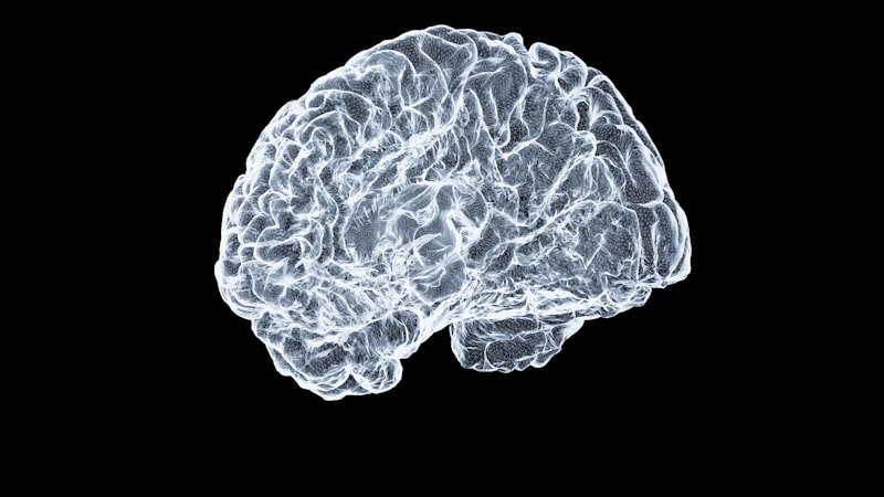
This visualization tool is essential for understanding dynamic biological events, linking morphology with molecular function. For instance, in neuroscience, autoradiography can map the precise locations of receptor populations or track the uptake of glucose (using 18F-FDG in certain variations) to infer metabolic activity in specific brain regions. The power of autoradiography lies in its sensitivity and its ability to maintain the structural integrity of the tissue while providing molecular localization data, bridging the gap between biochemistry and morphology, and allowing researchers to correlate molecular activity with anatomical structure.
Historical Context and Development
The origins of autoradiography predate the widespread availability of synthetic radioisotopes, tracing back to the late 19th century following the discovery of radioactivity by Henri Becquerel, who observed that uranium salts could expose photographic plates. Early experiments involved placing naturally radioactive mineral samples onto photographic plates, demonstrating the basic principle of self-exposure. However, the technique truly flourished after World War II, coinciding with advancements in nuclear technology that made radioisotopes and high-resolution photographic emulsions readily accessible for biological research. Scientists quickly recognized the potential of combining specific metabolic tracers with this detection method to study cellular dynamics.
Initial biological applications were relatively crude, often using large tissue samples placed against standard X-ray film, providing macro-level localization suitable for whole-organism studies or large tissue blocks. A significant breakthrough occurred with the development of techniques for applying liquid photographic emulsions directly onto histological sections, paving the way for microscopic autoradiography. This innovation, pioneered by researchers like L.F. Bélanger and C.P. Leblond in the 1950s, dramatically enhanced resolution, allowing researchers to localize radioactive materials within individual cells and even subcellular structures, such as nuclei or mitochondria, thereby enabling the study of DNA and protein synthesis sites.
Further refinements included the introduction of specialized high-sensitivity emulsions, improvements in tissue fixation methods to prevent the leaching or diffusion of soluble tracers, and the standardization of development protocols to achieve optimal signal-to-noise ratios. The transition from purely macroscopic film-based methods to highly detailed microscopic grain analysis marked the maturation of autoradiography into a rigorous quantitative scientific tool, indispensable across fields ranging from endocrinology to genetics. These historical developments laid the groundwork for modern methodologies used today, particularly in receptor binding studies and investigations of cell proliferation kinetics, fundamentally changing how biologists viewed molecular events in a structural context.
The Physicochemical Principles of Autoradiography
The efficacy of autoradiography relies heavily on the specific physicochemical interaction between emitted ionizing radiation and the silver halide crystals embedded within the photographic emulsion. When a radioisotope decays, it releases energy in the form of particles (e.g., beta particles, or electrons) or sometimes electromagnetic radiation (gamma rays). In biological applications, isotopes emitting low-energy beta particles, such as Tritium (3H), are overwhelmingly preferred because their short track length ensures that the exposure event occurs very close to the source of the radioactivity, thereby maximizing spatial resolution—a critical factor for subcellular localization.
The beta particle, upon entering the emulsion, possesses sufficient kinetic energy to transfer energy to the silver halide (AgBr) crystal lattice. This energy transfer causes ionization, leading to the liberation of free electrons within the crystal structure. These electrons subsequently reduce silver ions (Ag+) to neutral silver atoms (Ag0). These atoms accumulate at specific sensitivity sites on the crystal surface, forming a cluster, often termed a latent image speck, which is chemically unstable but physically detectable. The size and stability of this latent image are directly proportional to the amount of energy deposited, meaning the probability of forming a visible silver grain is directly related to the amount of radioactivity present in the underlying tissue section.
The final step involves chemical development, which serves as an amplification process. The developer solution selectively reduces the entire silver halide crystal containing a stable latent image speck into metallic silver grains, which appear black or dark brown when viewed microscopically. Crucially, crystals that have not been exposed to radiation remain largely unaffected and are subsequently dissolved and removed during the fixation process by agents like sodium thiosulfate. The resulting pattern of metallic silver grains, when viewed under darkfield or brightfield microscopy, serves as a permanent, high-resolution map of the original radioactive distribution, enabling quantitative data collection based on the density and spatial arrangement of the grains.
Detailed Methodologies: Film and Emulsion Application
Autoradiography methodologies are broadly categorized based on the type of detection medium used and the level of resolution required. Macro-autoradiography typically involves placing large tissue sections (such as whole-body sections from laboratory animals) or large analytical gels (from electrophoresis) in direct contact with standard X-ray film or specialized phosphor screens. This method is high-throughput, suitable for rapidly visualizing overall patterns of distribution, particularly in pharmacokinetic studies or analyzing results from DNA/RNA blotting procedures. While fast and scalable, the resolution achieved is generally low, offering localization only down to the millimetric or centimetric scale.
For high-resolution cellular and subcellular studies, microscopic autoradiography is mandatory, requiring the precise and delicate application of liquid photographic emulsion. The most common technique is the dipping technique (wet-mount). In this method, prepared tissue slides are carefully dipped into a molten photographic emulsion, often based on high-sensitivity nuclear track emulsions designed for fine grain size. The slides are then withdrawn slowly and dried vertically in a light-tight environment, resulting in a uniform, thin layer of emulsion covering the entire tissue section. This intimate and uniform contact is vital for capturing the weak emissions from isotopes like 3H, ensuring optimal spatial resolution.
The exposure process itself is highly sensitive to environmental factors and typically requires careful optimization. The duration can range from several days to many months, depending critically on the specific activity of the incorporated isotope, its concentration in the tissue, and the desired signal intensity. The slides must be stored in light-tight, dessicated containers, often refrigerated or frozen to minimize background fogging—the spurious formation of silver grains caused by cosmic rays, heat, or chemical contamination. Following adequate exposure time, the slides undergo strictly controlled developing, stopping, and fixing steps, followed by counterstaining of the tissue section to provide clear anatomical context necessary for accurate interpretation.
Microscopic Autoradiography Techniques
Microscopic autoradiography is further differentiated based on the required level of magnification and detail, leading to light microscopic and electron microscopic techniques. Light microscopic techniques are primarily used for cellular localization, such as identifying metabolically active cells or mapping receptor distribution within specific tissue layers. For instance, in situ hybridization often incorporates autoradiography by labeling oligonucleotide probes with 35S or 33P, allowing visualization of gene expression (mRNA) patterns within specific cell types or tissue compartments. The resulting silver grains are counted or measured relative to the underlying cell structure to quantify the expression level.
For the highest possible resolution, Electron Microscopic Autoradiography (EM Autoradiography) is employed. This technique allows researchers to localize radioactive molecules within subcellular organelles, such as mitochondria, endoplasmic reticulum, synaptic terminals, or nuclear pore complexes. The methodology requires ultrathin tissue sections (typically 50–100 nm thick) and specialized, extremely fine-grained emulsions applied as a monolayer onto the sections. Due to the requirement for ultra-high spatial resolution, researchers typically rely exclusively on Tritium (3H) because its very weak beta emission ensures that the resulting silver grain is deposited within nanometers of the radioactive source, minimizing the spread of the signal.
The challenges in EM autoradiography are substantial, involving ensuring minimal mechanical artifacts during the delicate emulsion application, achieving perfect physical contact between the emulsion layer and the ultrathin section, and compensating for the inherently low efficiency of detection due to the extreme thinness of the specimen. Despite these difficulties, the data provided—for example, the precise site of protein synthesis or membrane receptor insertion—is invaluable for understanding cellular ultrastructural machinery. Specialized statistical methods, such as grain density analysis and probability circle analysis, are typically necessary to accurately interpret the spatial relationship between the observed silver grains and the underlying ultrastructure.
Applications in Neuroscience and Receptor Mapping
Autoradiography holds a foundational and irreplaceable place in neuroscience research, particularly in the study of receptor pharmacology and brain mapping. Receptor autoradiography utilizes radiolabeled ligands—molecules designed to bind specifically and with high affinity to target receptors—to determine the density, distribution, and pharmacological properties of these receptors throughout the central and peripheral nervous systems. This technique allows for the precise mapping of receptor populations in different brain nuclei and cortical layers, providing crucial information regarding neural connectivity, signaling pathways, and functional segregation.
The methodology involves preparing fresh or fixed tissue sections and incubating them with the radiolabeled ligand, allowing it sufficient time to bind specifically to the target receptor sites. To ensure accurate measurement of specific binding, non-specific binding—ligand adhering to non-receptor sites or membrane lipids—is determined by incubating parallel sections in the presence of an excess concentration of a potent, non-labeled competing drug. The difference between the total binding signal and the non-specific binding signal yields the specific binding signal, which is directly proportional to the receptor density in that anatomical region. Following extensive washing and drying, the sections are exposed to film (usually tritium-sensitive film) or emulsion.
This approach is highly quantitative. By systematically varying the concentration of the radiolabeled ligand, researchers can generate saturation binding curves, which are then analyzed using classical pharmacological models. This analysis allows for the determination of crucial pharmacological parameters, including the maximum number of binding sites (Bmax, representing the absolute receptor density) and the equilibrium dissociation constant (Kd, representing the receptor affinity for the ligand). Autoradiography has been instrumental in identifying the spatial patterns of virtually every known neurotransmitter system, including dopaminergic, serotonergic, opioid, and GABAergic receptors, providing key insights into the etiology and progression of neurological disorders such as Parkinson’s disease, Alzheimer’s disease, and schizophrenia.
Applications in Molecular Biology and Genetics
Beyond anatomical and histological studies, autoradiography is a cornerstone technique in molecular biology, primarily for visualizing the results of separation techniques like gel electrophoresis. In genetics and biochemistry, it is indispensable for classic techniques such as DNA sequencing and various blotting procedures, where the goal is to detect and quantify specific molecular species based on size or charge separation.
- Blotting Techniques: Autoradiography is the standard method used to detect specific nucleic acid or protein fragments separated by gel electrophoresis. In Southern blotting (DNA), Northern blotting (RNA), and Western blotting (protein, when radiolabeled antibodies are used), target molecules are transferred from the gel matrix onto a solid support membrane. If the probe used to detect the target is radiolabeled (e.g., using high-energy 32P for nucleic acids), subsequent exposure of the membrane to X-ray film results in visible bands corresponding to the location and quantity of the target molecule.
- DNA Sequencing: The classical Sanger dideoxy chain termination method, foundational to modern genomics, relied heavily on autoradiography. DNA fragments, differentially terminated and labeled with isotopes like 35S or the higher-energy 32P, were separated with single-base resolution in high-resolution polyacrylamide gels. The resulting ladder of fragments, corresponding to the sequence, was visualized by exposing the dried gel to film, allowing researchers to read the sequence directly from the resulting band pattern.
- Metabolic Studies: Autoradiography is used extensively to track the incorporation of radiolabeled precursors (like 3H-thymidine for DNA, or 3H-uridine for RNA) into cellular machinery, thereby measuring the rate of synthesis and cellular proliferation.
The choice of isotope in molecular biology is often dictated by the need for speed and energy considerations. Phosphorus-32 (32P) is frequently used in blotting because its high-energy beta emission penetrates the gel and film efficiently, significantly reducing exposure times from weeks to hours compared to lower-energy emitters like 3H. However, this high energy comes at the significant cost of resolution, which is acceptable when analyzing discrete bands on a gel but utterly unsuitable for detailed microscopic localization within cells.
Quantitative Autoradiography and Data Analysis
The true scientific utility of autoradiography often lies in its capability for rigorous quantification, transforming visual patterns into absolute concentration data. Quantitative autoradiography (QAR) involves converting the visual signal—either the optical density of the exposed film or the silver grain count in microscopic preparations—into meaningful concentration data, typically expressed as femtomoles of ligand bound per unit mass of tissue (fmol/mg).
This quantification requires the meticulous use of standardized radioactive sources, or “standards,” which are co-exposed alongside the tissue sections. These standards, usually made from dried polymer or tissue pastes spiked with known concentrations of the same radioisotope, create a calibration curve relating radiation exposure to signal intensity. For macro-autoradiography using film, quantification involves densitometry. The exposed film is scanned using a high-resolution densitometer, and the optical density (darkness) of specific anatomical regions is measured using specialized image analysis software. These density values are then mathematically interpolated against the standard curve, allowing for the calculation of the absolute concentration of the radiolabel in the tissue.
For microscopic analysis, quantification is more localized and precise, typically performed by counting individual silver grains over defined cellular or subcellular areas (e.g., per cell nucleus or per square micrometer of cytoplasm). Sophisticated statistical analysis is crucial to correct for inherent variability, including background fogging (random grain deposition unrelated to the specimen) and to analyze the proximity of the grains to specific cellular structures using techniques like probability circle analysis. Modern specialized software assists in automating grain counting and analysis, enabling complex studies like determining the rate of DNA synthesis or the regional turnover rate of specific receptors with high objectivity.
Advantages, Limitations, and Technical Challenges
Autoradiography offers several distinct advantages over other contemporary localization and detection techniques. Its primary strength is its extraordinary high sensitivity; given a sufficiently long exposure time, even minute, picomolar quantities of radioactive material can be reliably detected. Furthermore, it provides unparalleled spatial resolution, particularly with low-energy isotopes like Tritium, allowing localization down to the subcellular level, which is critical for correlating molecular events with ultrastructure. Finally, it is a highly quantitative technique when appropriate standards and densitometry are employed, providing absolute concentration data rather than merely relative signal intensity, distinguishing it from purely qualitative immunohistochemical methods.
However, autoradiography is subject to several significant technical limitations and challenges that require careful methodological control.
- Resolution vs. Efficiency Trade-off: There is an inherent inverse relationship between resolution and detection efficiency. High-resolution isotopes (like 3H) require prohibitively long exposure times (low efficiency), while high-efficiency isotopes (like 32P) offer poor resolution due to their long particle track lengths, limiting their utility in histological applications.
- Artifacts and Chemography: Chemical interactions between residual tissue components, fixatives, or staining agents and the photographic emulsion can lead to artifacts known as chemography. This phenomenon can either enhance (positive chemography) or inhibit (negative chemography) the formation of the latent image, leading to inaccurate or false signals that complicate interpretation.
- Diffusion of Soluble Tracers: Maintaining the precise location of highly soluble radioactive tracers (e.g., certain neurotransmitters or ions) is technically demanding. Specialized and complex techniques, such as rapid freezing, freeze-drying, and dry-mounting of the emulsion under stringent temperature and humidity control, must be employed to prevent the diffusion or translocation of the tracer during tissue preparation and exposure.
- Protracted Time Commitment: The necessary exposure phase is often lengthy, frequently lasting weeks or even several months to accumulate enough signal for reliable detection and quantification, making the methodology time-consuming and inherently low-throughput.
Modern Variations and Future Directions
While classical film and emulsion techniques remain foundational, modern technology has introduced advanced variations that successfully address some of the limitations of traditional autoradiography, particularly concerning throughput, speed, and digital quantification.
One crucial modern development is the widespread use of Phosphor Imaging Screens, often termed computed autoradiography. Instead of relying on silver halide film, these systems utilize reusable screens containing phosphorescent crystals (e.g., barium fluorohalide) that absorb the energy from the emitted radiation, trapping electrons in metastable states. Upon subsequent exposure to a high-energy laser scanner, the trapped energy is released as visible light (photostimulated luminescence), generating a digital image. Phosphor imaging is significantly faster, requires shorter exposure times, and offers a much wider linear detection range than traditional film, enabling highly accurate quantification over a broader spectrum of sample activities.
Another major area of advancement is the convergence with sophisticated digital imaging and computed analysis technology. Digital image analysis systems now allow for automated, sophisticated processing, including complex background subtraction algorithms, and objective grain counting, dramatically improving the objectivity, accuracy, and speed of quantitative microscopic analysis. Furthermore, the integration of in vitro autoradiographic data with high-resolution anatomical maps derived from clinical imaging modalities (e.g., MRI or PET scans) through precise image registration techniques enhances the translational relevance of the findings. The future of autoradiography lies in the continued refinement of detection chemistries, the development of specialized probes, and the integration with automated, high-throughput robotic systems, ensuring its enduring importance in molecular imaging and histological research.