
Spindle Cells: The Neural Architects of Human Intuition
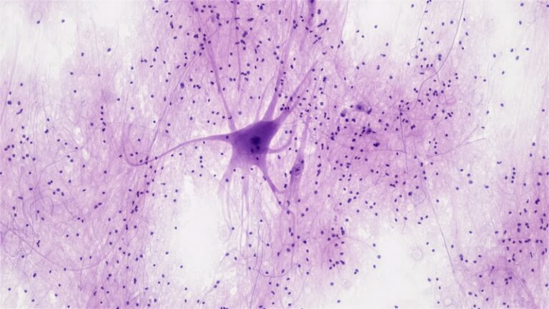
Introduction to Spindle Cells in Neuroscience Spindle cells, a unique class of neurons, are defined primarily by their distinctive morphology, characterized by a fusiform or spindle shape. These highly specialized cells are typically wider in the middle, housing the nucleus and major organelles, and feature narrow, tapering ends from which primary dendrites and the axon […]
Nissl Staining: Mapping the Architecture of the Mind
Introduction and Historical Context The Nissl method represents one of the most fundamental and enduring techniques in classical neurohistology, providing researchers with an essential tool for the microscopic examination of nervous tissue. This staining procedure was first introduced by the eminent German psychiatrist and neuropathologist, Franz Nissl, during the late nineteenth century. Specifically developed between […]
Neuroanatomy: Unfolding the Secrets of the Brain
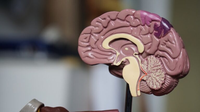
Introduction to the Folium and Etymology The term Folium, derived directly from the Latin word meaning “leaf,” is a foundational descriptor utilized within neuroanatomy and neuroscience to characterize a structure exhibiting a highly specific, leaf-like morphology. This anatomical term is most prominently and critically applied to describe the intricate, tightly packed folds of the cortex […]

Neural Imaging: Mapping the Mind Through Radioactivity
Introduction and Fundamental Definition Autoradiography refers to a sophisticated histological technique utilized primarily in biology, medicine, and chemistry to visualize the spatial distribution of radioactive compounds within complex samples. Fundamentally, it leverages the energy emitted by radioisotopes incorporated into a specimen to expose a photographic emulsion or film. This process creates a latent image that, […]
Schwann Cells: The Unsung Heroes of Neural Connectivity
Introduction and Definition of the Schwann Cell The **Schwann cell** represents a fundamental component of the peripheral nervous system (PNS), categorized as a type of glial cell, often referred to as neuroglia. These cells are distinguished by their vital role in supporting neuronal function, primarily through their interaction with axons, the long projections extending from […]
Biological Imaging: Slicing Through the Mind’s Layers
Introduction to the Microtome The microtome is an indispensable instrument in the fields of histology, pathology, and neuroscience, defined fundamentally as a mechanical device utilized for the preparation of ultra-thin sections of biological tissues or non-biological materials prior to microscopic examination. The term itself is derived from the Greek words “mikros,” meaning small, and “temnein,” […]
Spongioblasts: The Building Blocks of Your Brain
The Definitional Basis of the Spongioblast The term spongioblast refers to an essential, undifferentiated cell of ectodermal origin whose ultimate developmental destiny is the formation of the supporting cells of the central nervous system (CNS), collectively known as the neuroglia. Specifically, spongioblasts are precursors to the macroglia, which include astrocytes and oligodendrocytes, defining their foundational […]
Thalamic Nuclei: The Brain’s Secret Command Center
The Thalamic Nucleus: Central Hub of Brain Function The Core Definition of Thalamic Nuclei The term thalamic nucleus refers to any of the numerous distinct clusters of neuronal cell bodies, or nuclei, that constitute the Thalamus—a large, ovoid mass of gray matter situated deep within the forebrain. Positioned centrally in the Diencephalon, the thalamus acts […]
Cellular Psychology: Understanding the Mind Through Cells
Cytology: The Study of Cells The Core Definition of Cytology Cytology, often referred to as cell biology, is the fundamental branch of medicine and biology dedicated to the meticulous study of cells—the basic structural, functional, and biological units of all known living organisms. The core mission of cytology is to analyze the morphology, physiology, pathology, […]
PURKINJC-SANSON IMAGES
The Conceptual Framework of Purkinje-Sanson Images The Purkinje-Sanson image (PSI) represents a sophisticated optical imaging methodology primarily deployed to visualize the highly complex, microscopic architecture of diverse biological tissues. At its most fundamental level, this imaging modality relies on passing a controlled beam of light through a thin, carefully prepared tissue specimen. As the light […]
EPITHELIUM
Fundamental Definition and Structural Principles of Epithelium Epithelium, or epithelial tissue, represents one of the four primary types of animal tissue, serving as the essential biological interface between an organism and its environment. This tissue is composed of cells that are tightly packed into continuous sheets, forming the lining of internal cavities, the covering of […]
NISSL BODIES
Historical Discovery and Conceptual Foundation of Nissl Bodies The study of neuroanatomy was profoundly transformed in the late nineteenth and early twentieth centuries by the pioneering work of Franz Nissl, a distinguished German histologist and psychiatrist. In the early 1900s, Nissl developed specialized staining techniques that allowed for the unprecedented visualization of cellular structures within […]
CYTOARCHITECTURE
Introduction to Cytoarchitecture Cytoarchitecture, derived from the Greek terms meaning “cell structure” (kytos for cell, architektonia for architecture), is fundamentally the study of the internal and external organizational characteristics of cells within tissues. This discipline is a core branch of histology and cell biology, focusing intensely on the microscopic, three-dimensional arrangement of cellular components and […]
SECTIONING
Sectioning is a technique used in medical science to analyze tissue samples and gain a better understanding of the anatomy and physiology of the body. Sectioning involves cutting a sample of tissue into thin slices, which can then be viewed under a microscope. This technique is commonly used to study the structure of organs and […]