Schwann Cells: The Unsung Heroes of Neural Connectivity
- Introduction and Definition of the Schwann Cell
- Historical Context and Discovery
- Anatomy and Morphology of the Schwann Cell
- The Role of Myelination (Myelinating Schwann Cells)
- Non-Myelinating Schwann Cells (Remak Bundles)
- Regenerative Capacity and Role in Nerve Repair
- Clinical Significance and Associated Pathologies
Introduction and Definition of the Schwann Cell
The **Schwann cell** represents a fundamental component of the peripheral nervous system (PNS), categorized as a type of glial cell, often referred to as neuroglia. These cells are distinguished by their vital role in supporting neuronal function, primarily through their interaction with axons, the long projections extending from nerve cell bodies. They are indispensable for the efficient and rapid transmission of electrical impulses throughout the body outside of the brain and spinal cord. Specifically, the Schwann cell is responsible for insulating the vast majority of PNS axons by wrapping tightly around them, thereby forming the multilayered structure known as the **myelin sheath**. This critical insulation facilitates a specialized form of signal propagation called saltatory conduction, dramatically increasing nerve impulse velocity, which is essential for rapid motor responses and sensory processing.
Functionally, Schwann cells are highly versatile, exhibiting two primary phenotypes crucial for PNS homeostasis. The first and perhaps most recognized phenotype is the **myelinating Schwann cell**, which encases large-diameter axons with thick, segmented myelin sheaths. The second type, the **non-myelinating Schwann cell**, supports multiple smaller, unmyelinated axons by enveloping them in cytoplasmic folds, forming structures known as Remak bundles. Beyond their insulating and supportive roles, Schwann cells are unique among glial populations for their profound involvement in the regeneration and repair of damaged peripheral nerves. Following injury, they undergo remarkable phenotypic changes, guiding axonal regrowth and clearing cellular debris, highlighting their centrality not just in function but also in structural maintenance and recovery of the PNS.
Unlike the oligodendrocytes, which perform the analogous myelination function within the central nervous system (CNS), Schwann cells are unique in that a single cell is typically responsible for myelinating only one segment of a single axon. This characteristic contrasts sharply with oligodendrocytes, which can extend processes to myelinate multiple different axons simultaneously. This singular dedication to one axonal segment underlies the meticulous organization of the PNS myelin, forming distinct gaps between segments known as the **Nodes of Ranvier**, areas critical for maintaining the efficiency of signal transmission. Understanding the anatomical and functional attributes of the Schwann cell is paramount to comprehending both normal peripheral nerve physiology and the etiology of numerous debilitating neuropathies.
Historical Context and Discovery
The initial description and recognition of the cell that would eventually bear his name are credited to **Theodor Schwann** (1810–1882), a distinguished German physiologist and histologist. Schwann’s detailed microscopic investigations during the 1830s were instrumental in advancing cellular biology, culminating in his co-founding of the unified Cell Theory alongside Matthias Schleiden. While the existence of peripheral nerve sheaths was known prior to his work, Schwann provided crucial evidence detailing the cellular nature of this protective coating. His work established that the nerve fiber sheath was composed of distinct, living cellular units rather than simply being an amorphous, secreted substance. This groundbreaking observation shifted the paradigm of understanding how complex biological structures, like nerves, are built and maintained.
Schwann’s early research utilized nascent microscopic technology and staining methods, allowing him to visualize the intricate structures of animal tissues with unprecedented clarity. He observed the characteristic wrapping and spiral arrangement of the cellular layers around the nerve axons, deducing their primary role in forming the protective covering. Although the full physiological mechanism of **saltatory conduction** and the detailed molecular composition of myelin were elucidated much later, Schwann laid the fundamental groundwork by identifying the essential structural unit responsible for peripheral nerve insulation. His contributions were not confined solely to neurobiology; his comprehensive thesis on the conformity in the structure and growth of plants and animals firmly established the principle that all living organisms are composed of cells and cell products.
The period following Schwann’s initial description saw decades of intensive research aimed at clarifying the relationship between the axon and its surrounding cell. Key subsequent discoveries focused on determining how the myelin sheath was actually formed—a process now understood to involve the complex, progressive spiraling of the Schwann cell plasma membrane around the axonal cylinder. The designation of the cells as “Schwann cells” cemented his legacy in histology and neuroscience. These historical foundations underscore the enduring importance of anatomical observation in biological discovery, moving the field from a macroscopic view of nerve cables to a microscopic, cellular understanding of neuronal function and pathology.
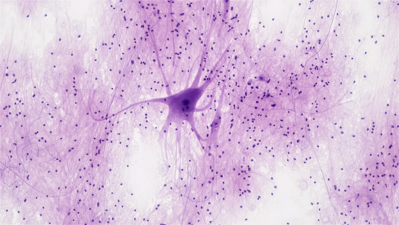
Anatomy and Morphology of the Schwann Cell
The morphology of the Schwann cell is highly specialized, directly reflecting its primary functions of insulation and support within the PNS. In its resting state, the cell possesses an ovoid nucleus and a cytoplasm rich in organelles necessary for the massive protein and lipid synthesis required for myelin production. The most striking anatomical feature of the myelinating Schwann cell is its capacity to flatten and extend its plasma membrane into a vast, ribbon-like sheet that wraps concentrically around a single, large-diameter axon. This wrapping process is highly ordered, resulting in dozens of compacted layers of membrane, which are devoid of cytoplasm in the compact region, forming the definitive **myelin sheath**. The thickness of this sheath is precisely regulated and is directly proportional to the diameter of the axon it covers, ensuring optimal conduction speed.
Crucially, the wrapping process is never fully complete along the entire length of the axon. Small, regular gaps exist between adjacent Schwann cell segments; these are the aforementioned **Nodes of Ranvier**. At the nodes, the axonal membrane is exposed to the extracellular fluid, and specialized clusters of voltage-gated sodium channels are localized. The junctional regions where the myelin ends and the node begins are known as the paranodal regions, characterized by intricate adhesion complexes (e.g., septate-like junctions) that securely anchor the Schwann cell to the axon and maintain the integrity of the myelin seal. These anatomical specializations—the compact myelin layers, the paranodal anchoring, and the nodal gaps—are all intrinsically necessary for the rapid, efficient mechanism of saltatory conduction.
The non-myelinating Schwann cell, while structurally distinct, also exhibits specialized morphology optimized for support. Instead of wrapping densely around a single axon, these cells envelop multiple smaller, usually unmyelinated, axons within deep invaginations or troughs of their cell surface. These formations are known as **Remak bundles**. Each axon within the bundle is suspended in the Schwann cell cytoplasm, but without the tight, multilayered compaction seen in myelin. This arrangement provides metabolic support and physical protection to these small axons, which often mediate functions like slow pain sensation or autonomic control. Thus, the overall morphological flexibility of the Schwann cell allows it to adapt its structure precisely to the diverse requirements of the various types of peripheral nerve fibers.
The Role of Myelination (Myelinating Schwann Cells)
Myelination by Schwann cells is arguably the most critical process ensuring the speed and fidelity of information transfer in the PNS. The initiation of myelination involves a complex molecular dialogue between the Schwann cell and the axon, often regulated by signaling molecules such as Neuregulin 1 (NRG1) expressed on the axon surface. Once initiated, the Schwann cell recognizes the appropriate axon and begins its characteristic spiral migration, sequentially wrapping its plasma membrane around the axon. As the inner layers of the membrane compact, the intervening cytoplasm is extruded, leaving behind a dense, lipid-rich structure that acts as a highly effective electrical insulator. This multilayered structure is stabilized by key myelin proteins, notably **Myelin Protein Zero (P0)**, which acts as a major adhesion molecule, effectively zippering the membrane layers together.
The primary physiological consequence of myelination is the facilitation of **saltatory conduction** (from the Latin saltare, meaning “to leap”). In unmyelinated axons, the action potential must be regenerated continuously along the entire length of the fiber, a slow and energetically costly process. Myelin, acting as a superb electrical insulator, prevents ion leakage across the axonal membrane in the internodal segments. As a result, the electrical signal leaps rapidly from one exposed region—the Node of Ranvier—to the next. At these nodes, the high concentration of voltage-gated sodium channels ensures that the incoming passive current rapidly triggers a new, full action potential, maintaining signal strength without requiring continuous regeneration along the insulated segment. This mechanism drastically reduces the required energy expenditure while increasing conduction velocity by up to fiftyfold compared to unmyelinated fibers.
Beyond simply accelerating conduction, the myelinating Schwann cell plays an indispensable role in the long-term metabolic and structural maintenance of the enclosed axon. The intimate relationship formed during myelination establishes a trophic dependence; the cell supplies essential nutrients and growth factors to the axon, particularly in the distal segments, which are far removed from the neuronal cell body. Specialized cytoplasmic channels and processes, such as the **Schmidt-Lanterman incisures** (pockets of remaining cytoplasm within the compact sheath), are thought to be important pathways for the transport of molecules necessary for myelin maintenance and axonal health. Therefore, the Schwann cell acts not merely as a passive insulator but as an active, metabolic partner crucial for the axon’s survival over the organism’s lifetime.
Non-Myelinating Schwann Cells (Remak Bundles)
While myelinating Schwann cells garner significant attention due to their role in fast conduction, the **non-myelinating Schwann cells** constitute the majority of Schwann cells in the PNS and perform equally crucial, albeit different, functions. These cells are associated with all small-diameter axons, which typically include Type C pain fibers, postganglionic autonomic fibers, and certain sensory afferents. Instead of tight, concentric wrapping, non-myelinating Schwann cells cradle multiple unmyelinated axons within deep longitudinal grooves or pockets in their cytoplasm, forming the characteristic **Remak bundles**. A single non-myelinating cell can support anywhere from two to twenty individual axons, providing physical separation and a controlled microenvironment for each.
The functions of the non-myelinating Schwann cell are primarily supportive and modulatory. They provide essential metabolic support and help regulate the ionic balance surrounding the unmyelinated axons, buffering the potassium concentration in the immediate vicinity of the neural membrane. Furthermore, they are involved in clearing neurotransmitters and other signaling molecules from the synaptic clefts and axonal surfaces, thereby modulating the excitability and communication efficiency of the supported fibers. Although these axons conduct impulses much slower than myelinated fibers, the close association with the non-myelinating Schwann cell ensures the continuous, sustained, and reliable conduction necessary for slow processes like chronic pain signaling and autonomic regulation of internal organs.
The non-myelinating phenotype is also highly significant in pathological contexts and in regeneration. In healthy nerves, they maintain a quiescent, supportive state. However, following nerve injury, non-myelinating cells, like their myelinating counterparts, rapidly proliferate and contribute to the regenerative process. They help define the pathway for potential axonal regrowth and participate in the clearance of debris. Their resilience and ubiquity throughout the peripheral nerve architecture highlight their role as primary caretakers and essential facilitators of nerve integrity, independent of the acceleration provided by myelin.
Regenerative Capacity and Role in Nerve Repair
One of the most remarkable features distinguishing the PNS from the CNS is its robust capacity for regeneration following injury, a process overwhelmingly dependent upon the adaptive capabilities of the **Schwann cell**. When a peripheral nerve axon is severed, the distal segment, disconnected from the neuronal cell body, rapidly degenerates in a process known as **Wallerian degeneration**. During this process, the myelin sheath fragments, and the axon breaks down. The Schwann cells surrounding the degenerating axon immediately activate a dramatic cellular transformation. They dedifferentiate, ceasing myelin production and initiating a proliferative phase.
These activated Schwann cells align themselves into organized, longitudinal columns known as **Büngner bands**. These bands serve as biological guides or conduits for the regenerating axonal sprouts originating from the proximal stump of the injured nerve. The activated Schwann cells express numerous cell adhesion molecules, neurotrophic factors (such as **Nerve Growth Factor, NGF**), and extracellular matrix components that are essential for promoting axonal elongation and survival. They also actively participate in phagocytosis, collaborating with macrophages to clear the debris of the degenerated myelin and axon fragments, thus preparing a clean pathway for the regrowth.
The success of nerve regeneration hinges entirely on the ability of the regenerating axon to navigate the environment created by the Schwann cell population and re-establish synaptic connections with its original target tissue. If the axon successfully traverses the gap and reaches the Bungner band, the Schwann cell guides it toward the target. Upon successful reinnervation, the Schwann cell redifferentiates, and if the axon is of sufficient diameter, it initiates remyelination, restoring the function of the nerve fiber. This profound plasticity makes the Schwann cell a central focus in therapeutic strategies aimed at improving functional recovery after peripheral nerve trauma.
Clinical Significance and Associated Pathologies
Given their crucial role in myelin integrity and nerve support, Schwann cells are implicated in a wide spectrum of peripheral neuropathies, ranging from acute autoimmune disorders to chronic hereditary conditions and neoplastic diseases. Dysfunction or destruction of the Schwann cell or its myelin product directly impairs signal conduction, leading to sensory deficits, muscle weakness, and autonomic failure. Understanding the mechanisms of Schwann cell pathology is essential for accurate diagnosis and the development of targeted treatments.
One of the most well-known acute disorders is **Guillain-Barré Syndrome (GBS)**, an acute inflammatory demyelinating polyneuropathy. In GBS, the immune system mistakenly attacks the Schwann cell membranes or the myelin proteins, leading to rapid, severe demyelination. This demyelination slows or blocks nerve conduction entirely, causing ascending paralysis that can be life-threatening if it affects the respiratory muscles. Conversely, **Chronic Inflammatory Demyelinating Polyneuropathy (CIDP)** presents as a similar but chronic and relapsing condition, also rooted in autoimmune damage to Schwann cell myelin.
Hereditary neuropathies frequently involve intrinsic defects in Schwann cell function or structure. The most common group of these disorders is **Charcot-Marie-Tooth Disease (CMT)**, which encompasses numerous genetic mutations affecting genes encoding myelin proteins (like P0 or connexins) or genes involved in the maintenance and signaling pathways of the Schwann cell itself. CMT typically leads to chronic, progressive demyelination or axonal loss, resulting in muscle atrophy and significant disability. Furthermore, Schwann cells are the origin of a common type of benign peripheral nerve sheath tumor known as a **Schwannoma** (or neurilemmoma). These tumors arise from the neoplastic transformation of Schwann cells and are often slow-growing, though they can cause pain and neurological deficits through compression of the nerve or surrounding structures.
The recognition that Schwann cell integrity is paramount to PNS health has driven extensive research into potential therapeutic interventions. Current research explores strategies such as gene therapy to correct CMT-related mutations in Schwann cells or the use of engineered cell scaffolds and transplanted Schwann cells to enhance the regenerative environment following severe nerve transection injuries. The Schwann cell thus remains a central target for both understanding neurological disease and developing restorative therapies.