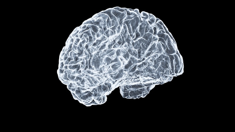
Neuroanatomy: Unfolding the Secrets of the Brain
Introduction to the Folium and Etymology
The term Folium, derived directly from the Latin word meaning “leaf,” is a foundational descriptor utilized within neuroanatomy and neuroscience to characterize a structure exhibiting a highly specific, leaf-like morphology. This anatomical term is most prominently and critically applied to describe the intricate, tightly packed folds of the cortex of the cerebellum. These repeated folds, known individually as folia (plural), represent the fundamental organizational units of the cerebellar gray matter, distinguishing its structure significantly from the broader gyri and sulci observed in the cerebral cortex. The primary function of this profound folding is the maximization of the cortical surface area, which is essential for accommodating the vast neuronal population necessary for the cerebellum’s complex computational roles.
The concept of the folium is indispensable for comprehending the gross and microscopic architecture of the cerebellum, a region often described as a massive, parallel array of highly ordered processing units. Each individual folium constitutes a narrow, transversely oriented ridge of gray matter, comprised of the standard three layers of the cerebellar cortex—the molecular, Purkinje cell, and granular layers—all enveloping a central core of white matter. This uniform, repetitive arrangement is structurally crucial, as it dictates the systematic distribution and flow of afferent and efferent neurological information across the cerebellar mantle, suggesting a profound modularity where specific functional operations may be segregated along these folds.
Historically, the adoption of the term folium highlights the early recognition by anatomists of the tissue’s remarkable physical resemblance to botanical leaves—thin, layered, and extensively convoluted. This nomenclature provides an immediate visual key to the structure’s physical properties: a thin planar sheet of cortex that has been repeatedly folded upon itself to achieve extreme compaction. The evolutionary necessity for such extensive foliation is directly linked to the immense computational load handled by the cerebellum, which houses more neurons than the rest of the brain combined. The resulting tight packing of the folia ensures that the requisite number of synapses, particularly those involving the numerous granule cells, can be efficiently maintained within the confined space of the posterior cranial fossa, optimizing neural processing efficiency.
Anatomical Context: The Cerebellar Cortex
The cerebellar cortex represents a remarkable instance of biological surface area optimization achieved through systematic folding, with the folia serving as the primary structural element dictating its organization. Although the cerebellum is conventionally subdivided into major lobes (anterior, posterior, and flocculonodular) and further into ten lobules, the underlying organizational pattern within these divisions is consistently defined by the folia. A typical folium extends across the width of a specific cerebellar lobule, often traversing the boundary between the medial vermis and the lateral hemispheres. This consistent transverse orientation plays a pivotal role in governing the flow of neural signals, which typically propagate perpendicular to the long axis of the folium, aligning with the specialized geometry of the cerebellar microcircuitry.
The massive surface area generated by the tight folial folding is staggering; estimates suggest the cerebellar cortex, when unfolded, approaches three-quarters the surface area of the entire cerebral cortex, despite the cerebellum accounting for only approximately ten percent of total brain volume. This extensive surface is imperative for housing the billions of neurons necessary for the cerebellum’s specialized role as a predictive engine and comparator for motor control, posture, and increasingly recognized cognitive functions. When the cerebellum is dissected, the white matter core within the folia is clearly visible, forming a distinctive, highly branched structure known as the arbor vitae, or “Tree of Life,” where each branch represents the central axonal core of a single folium.
Subtle variations in the precise morphology, depth, and spacing of the folia exist across the different functional zones of the cerebellum, reflecting specialized regional roles. For example, the folia within the vestibulocerebellum (flocculonodular lobe), crucial for balance, may differ morphologically from those in the cerebrocerebellum (lateral hemispheres), which are heavily involved in complex motor planning and cognitive tasks. Regardless of regional variation, the fundamental three-layered cortical structure is rigorously preserved within every fold. The close, parallel packing of adjacent folia facilitates complex interactions and highly parallel processing capabilities, enabling the cerebellum to rapidly and efficiently integrate vast quantities of sensory data and motor command signals to refine and coordinate output.
Microscopic Structure of the Folia
A high-magnification view of a cerebellar folium reveals the exquisite order of its microcircuitry, which is uniformly maintained across the entire convoluted surface. The structure is defined by three distinct cortical layers. The outermost layer is the molecular layer, characterized by its sparse cellular density but immense synaptic richness. It primarily contains the massive, highly branched dendritic arbors of the underlying Purkinje cells, the longitudinally oriented parallel fibers (axons of granule cells), and the cell bodies of inhibitory interneurons like basket and stellate cells. The molecular layer serves as the primary integration site where thousands of inputs converge to modulate the activity of the Purkinje cells.
The central layer, and arguably the most defining feature of the cerebellar cortex, is the Purkinje cell layer. This layer consists of a single, highly regular row of the largest neurons in the cerebellum, the Purkinje cells, which are the sole source of output from the cerebellar cortex, projecting inhibitory signals to the deep cerebellar nuclei. Crucially, the planar dendritic trees of these cells extend upward into the molecular layer, oriented strictly perpendicularly to the long axis of the folium. This precise anatomical alignment is pivotal to cerebellar function, as it determines how signals traveling along the parallel fibers (which run parallel to the folium’s axis) interact with the Purkinje cell dendrites, providing a mechanism for highly organized spatiotemporal summation of synaptic inputs.
The innermost layer, situated immediately adjacent to the white matter core of the folium, is the granular layer. This region possesses the highest neuronal density in the central nervous system, housing billions of tiny granule cells, along with inhibitory Golgi cells. The axons of the granule cells ascend into the molecular layer, where they bifurcate to form the parallel fibers, completing the circuit. The folium structure thus physically organizes this entire microcircuit: the white matter core supplies the necessary inputs and receives outputs; the granular layer performs initial processing of mossy fiber inputs; and the molecular layer integrates the signals before the final, precisely timed inhibitory output is generated by the planar Purkinje cells, ensuring efficiency and computational power.
Development and Formation of Cerebellar Folds
The formation of the cerebellar folia, a complex morphogenetic process termed foliation, is a meticulously controlled developmental event that spans late gestation and continues significantly into the postnatal period in humans. The process initiates with the formation of deep primary fissures, which delineate the major lobes and lobules, followed by the progressive generation of secondary and tertiary fissures that create the fine, leaf-like structure characteristic of the mature folium. This complex folding pattern is the result of differential growth rates and intricate cellular interactions.
The primary driving force behind foliation is the immense proliferation and subsequent inward migration of granule cell precursors, which initially reside in the external granular layer (EGL). As these cells proliferate rapidly and then begin their inward journey past the Purkinje cells to establish the internal granular layer, the resulting expansion of cellular volume necessitates the buckling and folding of the cerebellar surface. Molecular signaling pathways and mechanical tension must be tightly regulated to ensure the resulting folia are uniformly spaced and oriented in the characteristic parallel fashion seen in the mature brain.
Disruptions to the delicate timing or extent of granule cell genesis and migration can have severe consequences for the final folial architecture, leading to various forms of cerebellar dysgenesis or hypoplasia, where the folia may be poorly formed, excessively wide, or severely reduced in number. The successful establishment of the final, highly convoluted folial pattern is absolutely essential for the correct wiring and functional maturity of the cerebellar circuitry. The precise geometry and close packing of the folds are required to ensure the correct interaction between the parallel fibers and the Purkinje cell dendrites, which underpins the highly precise temporal coordination capabilities of the cerebellum.
Functional Significance of Folial Folding
The extreme extent of cortical folding into folia serves the crucial functional imperative of maximizing computational capability within restricted physical parameters. By packing the cortex so densely, the folial structure allows the cerebellum to house an exponentially large number of neurons and synapses, particularly the granule cells, which are hypothesized to function as pattern separators, converting diverse, often sparse input signals into complex, high-dimensional codes that are essential for accurate motor learning and predictive timing.
Moreover, the highly consistent, parallel orientation of the folia imposes a strict directionality on intracortical information flow. Input signals relayed via mossy fibers are processed across the granular layer, and the resulting activity propagates along the parallel fibers, which run longitudinally along the folium’s axis. This structure suggests that each small segment of the folium, or a small grouping of adjacent folia, functions as a highly specialized processing module, often referred to functionally as a microcomplex. These microcomplexes operate concurrently, processing distinct parameters of movement or specific aspects of cognitive tasks, thereby drastically enhancing the speed and efficiency of cerebellar operations.
The physical constraints inherent in the folial structure also guarantee that the topographical organization between the output cells—the Purkinje cells—and their targets in the deep cerebellar nuclei is maintained with high fidelity. This precise somatotopic and functional mapping is preserved across the folded surface, implying that specific, adjacent points on the cortex, located within neighboring folia, project to defined and adjacent domains within the deep nuclei. This strict reliance on ordered connectivity is entirely dependent upon the systematic, repetitive organization provided by the folial architecture, enabling the cerebellum to execute its role in highly refined, coordinated motor and non-motor control.
Clinical Relevance of Folial Morphology
Deviations from the normal morphology and organization of the cerebellar folia are often highly significant indicators of underlying neurological disease, making the careful assessment of folial pattern a standard component of clinical neuroimaging interpretation. Congenital conditions affecting cerebellar development, such as specific types of cerebellar hypoplasia, frequently present with visible abnormalities in folial structure, including a reduction in the number of folds, abnormally wide folial spacing (pachygyria), or complete disorganization of the cortical layers.
In the context of neurodegenerative disorders, including hereditary conditions like various spinocerebellar ataxias (SCAs) and acquired diseases like multiple system atrophy (MSA), the primary manifestation is often progressive atrophy. This atrophy is characterized radiologically by a perceptible thinning, widening of the fissures, and overall loss of definition in the folia, particularly within vulnerable lobules. Microscopically, this folial atrophy correlates directly with the pathological loss of crucial neurons, particularly Purkinje cells and granule cells, leading to the hallmark clinical symptoms of cerebellar dysfunction: ataxia, intention tremor, and dysmetria.
Furthermore, acute injuries, such as cerebellar strokes or localized trauma, often result in patterns of damage that reflect the vascular territories supplying the folia. Since specific groups of folia are supplied by defined arterial branches, focal ischemic lesions typically cause localized damage to particular functional microzones. Therefore, a precise understanding of the anatomical extent of folial damage is critical for accurately predicting the resulting functional deficits, whether they involve balance, coordination, or the increasingly recognized non-motor symptoms encompassed by the term cerebellar cognitive affective syndrome (CCAS).
Comparative Anatomy of the Folium
The existence of the folium structure is a deeply conserved anatomical feature across almost all vertebrate species, highlighting the essential role of surface area maximization in effective cerebellar function. However, the exact complexity, depth of folding, and overall extent of foliation demonstrate substantial variation, correlating significantly with the species’ brain size, overall body mass, and the complexity of their required motor and cognitive behaviors. Generally, species demanding high levels of sophisticated motor coordination, such as dolphins, large primates, and highly agile animals, exhibit significantly more elaborate and densely packed folia compared to species with simpler movement repertoires.
For instance, the human cerebellum displays an exceptionally high degree of folial convolution, which provides the necessary computational substrate for fine motor dexterity, complex articulated speech, and higher-order cognitive processing. Comparative neuroanatomical studies consistently reveal an evolutionary trend where an increase in behavioral complexity and reliance on advanced sensory-motor integration correlates positively with the overall cortical surface area generated by the folial architecture. This evidence strongly supports the hypothesis that the folium structure represents a highly successful evolutionary solution for maximizing synaptic capacity within constrained physical dimensions.
Despite the differences in overall folding complexity, the fundamental microscopic cellular organization within the folium—the tripartite layering, the strict planar orientation of the Purkinje cell dendrites, and the parallel fiber system—remains remarkably consistent across the vertebrate phylogeny, from teleost fish through to mammals. This structural uniformity underscores the universal efficiency of the cerebellar microcircuitry, which leverages the physical constraints and organizational principles of the folial structure to implement its core functional mechanism: the highly precise, inhibitory modulation of brain activity, driven by the massive parallel processing capacity facilitated by the dense cortical folding.