Spindle Cells: The Neural Architects of Human Intuition
- Introduction to Spindle Cells in Neuroscience
- Historical Context and Discovery of Von Economo Neurons
- Anatomical Location and Distribution
- Unique Morphology and Cytology
- Proposed Functions and Role in Cognition
- Clinical Significance and Pathology
- Clarification of Terminology: Spindle Cells vs. Muscle Spindles
Introduction to Spindle Cells in Neuroscience
Spindle cells, a unique class of neurons, are defined primarily by their distinctive morphology, characterized by a fusiform or spindle shape. These highly specialized cells are typically wider in the middle, housing the nucleus and major organelles, and feature narrow, tapering ends from which primary dendrites and the axon emerge. In the context of the human brain and comparative neuroanatomy, the term spindle cell is often used synonymously with Von Economo Neurons (VENs), acknowledging their discovery and subsequent detailed study. These neurons represent a highly evolved structural feature of the cortex, distinguishing themselves sharply from the more common pyramidal and stellate cells that dominate cortical architecture. Their morphology strongly suggests a functional adaptation for rapid signal transmission and integration across large cortical distances, linking systems critical for complex social behavior and emotional processing.
While relatively scarce compared to other neuronal subtypes, the presence and distribution of VENs are considered crucial indicators of advanced cognitive capacity, particularly within the domain of social cognition. These specialized neurons are overwhelmingly concentrated in areas related to the salience network, specifically the anterior cingulate cortex and the frontoinsular cortex. Their unique shape—a long, slender soma—provides a structural basis for efficient communication, facilitating quick, intuitive responses to emotionally or socially relevant stimuli. Understanding the spindle cell requires a detailed appreciation of both their cytological structure and their strategic anatomical placement within the cortical hierarchy, linking visceral experience with complex decision-making processes.
The study of spindle cells has provided profound insights into the neurological basis of human sociality and self-awareness. Their evolutionarily recent appearance and proliferation in species with intricate social structures, such as great apes, certain cetaceans, and elephants, underscores their importance in facilitating complex interactions, empathy, and rapid assessment of environmental threats or opportunities. The specific structure of the spindle cell allows for the integration of highly divergent sensory and internal signals into a singular output, hypothesized to be essential for generating the cohesive, immediate reactions necessary for adaptive social behavior. Thus, spindle cells are not merely morphological variants but are believed to be cornerstone elements in the neural circuitry governing advanced psychosocial functions.
Historical Context and Discovery of Von Economo Neurons
The initial identification and documentation of these unique neurons trace back to the early 20th century, primarily through the meticulous work of Austrian neurologist Constantin von Economo and his colleague Georg Koskinas. In their seminal 1925 atlas detailing the cytoarchitecture of the human cerebral cortex, they described these large, bipolar cells, noting their distinctive spindle shape and their concentration in specific forebrain regions. Although their initial findings categorized them based purely on morphology, distinguishing them from the prevalent pyramidal cells, the functional significance of these cells remained largely theoretical for many decades. This early recognition laid the groundwork for future research, establishing the morphological criteria by which these specialized neurons are still identified today.
For much of the mid-to-late 20th century, the spindle cell was often treated as an unusual variant of large layer V projection neurons rather than a distinct class. However, advancements in modern neuroscientific techniques, particularly immunohistochemistry and sophisticated quantitative analysis, enabled researchers to re-evaluate von Economo’s initial observations. These modern studies confirmed that VENs possess unique molecular markers and connections that differentiate them substantially from neighboring cell types, validating their classification as a distinct neuronal population. This resurgence of interest, particularly in the last two decades, has firmly established the spindle cell as a critical component of the advanced primate brain, linking their structure directly to evolutionary specialization in social and emotional cognition.
Further research into comparative neuroanatomy has significantly contextualized the importance of spindle cells. Studies have revealed that while rudimentary forms of these neurons may exist in some lower primates, their density and complexity dramatically increase in hominids, reaching peak development in the human brain. The presence of VENs in other highly intelligent, socially complex mammals, such as humpback whales, killer whales, and African elephants, strongly suggests convergent evolution—the independent development of this unique neuronal structure as an adaptation to the demands of navigating complex, long-term social networks. This evolutionary perspective underscores the hypothesis that spindle cells are integral to the cognitive machinery required for sophisticated, nuanced social intelligence.
Anatomical Location and Distribution
The anatomical distribution of spindle cells is highly specific and concentrated, which is a key factor in understanding their proposed functions. They are predominantly found within two major regions of the human brain: the Anterior Cingulate Cortex (ACC) and the Frontoinsular Cortex (FI). These two regions are functionally interconnected and form the core components of the brain’s salience network, which is responsible for detecting and filtering relevant internal and external stimuli, coordinating emotional responses, and regulating autonomic functions. The strategic placement of VENs in these integrating hubs suggests a crucial role in mediating the interactions between emotional states and cognitive control mechanisms.
Within the ACC, spindle cells are typically located in layer Vb, a principal output layer of the cortex. The ACC itself is critically involved in error detection, motivational control, anticipation of reward and punishment, and emotional processing. The presence of VENs in this region suggests they function as rapid conduits for transmitting integrated information about emotional context and potential consequences to other brain regions. This location highlights their role in linking visceral and affective states—originating from subcortical structures like the amygdala and hypothalamus—with high-level cognitive processes necessary for complex decision-making and self-regulation. Their positioning facilitates the rapid translation of internal feeling states into behavioral imperatives.
Similarly, the frontoinsular cortex (FI) is heavily populated by VENs. The FI is widely recognized as the primary site for interoception—the sense of the physiological condition of the body—and plays a pivotal role in conscious emotional awareness and subjective feelings. The spindle cells in the FI are believed to integrate information regarding internal bodily states (e.g., heart rate, hunger, pain) with external social cues, contributing significantly to the subjective experience of emotion and self-awareness. The highly localized concentration of these specialized neurons in the ACC and FI reinforces the notion that they are critical components in the brain’s immediate, intuitive system for assessing internal and environmental relevance, often referred to as the “gut feeling” mechanism.
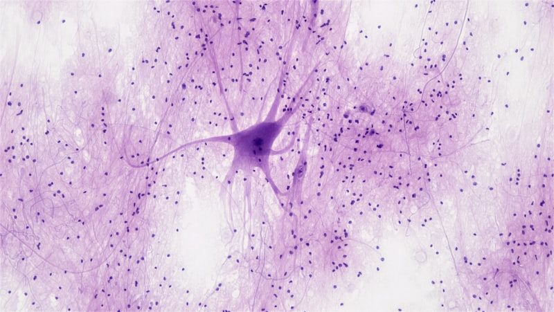
Unique Morphology and Cytology
The defining characteristic of the spindle cell is its fusiform morphology, which gives it its name. Unlike the ubiquitous pyramidal neurons, which possess a large triangular soma and multiple complex dendritic trees, the VEN soma is elongated, resulting in a narrow, bipolar configuration. This unique shape means that the VEN possesses only two primary dendrites that emerge from opposite poles of the soma, creating a streamlined, efficient structure. This simple bipolar organization contrasts sharply with the extensive arborization of other cortical neurons, suggesting a specialization for rapid, linear transmission rather than complex, localized integration of signals.
This specific structural architecture is hypothesized to confer a physiological advantage, particularly relating to speed. The elongated soma and restricted dendritic field likely enable the spindle cell to act as a fast-track transmission line between major cortical areas. By limiting the number of synaptic inputs it processes locally, the VEN may be optimized for relaying an integrated, urgent signal across the cortical mantle, specifically between the ACC and the FI. This rapid communication pathway is essential for the instantaneous and coordinated responses required for navigating dynamic social environments and responding to immediate emotional threats, lending credence to their role in the swift assessment of salience.
Cytologically, spindle cells are also notable for their large size, making them some of the largest neurons in the human brain. Although they have fewer primary dendrites, their axons are believed to be extensively and heavily myelinated, which further supports the hypothesis of highly efficient, long-distance signal relay. Histochemical studies have indicated that VENs express distinct neurochemical markers compared to nearby pyramidal cells, reinforcing their unique identity and physiological specialization. The combination of their large size, bipolar orientation, and strategic placement suggests that spindle cells are crucial output neurons designed to broadcast critical, high-level integrated information quickly throughout the neural network.
Proposed Functions and Role in Cognition
Functionally, spindle cells are intimately linked to higher-order cognitive capabilities, particularly those concerning social processing, rapid intuition, and emotional insight. They are hypothesized to serve as critical integrators within the salience network, performing a crucial role in the brain’s ability to instantaneously evaluate complex social and emotional situations. This rapid assessment is essential for behaviors such as empathy, recognizing social cues, and making quick decisions under emotionally charged conditions, forming the neural basis for what is often described as intuitive judgment or social intelligence.
One prominent theory suggests that VENs act as “gating” neurons, responsible for integrating diffuse inputs related to internal state, external threat, and social context, and then quickly determining which information is most salient and requires an immediate cognitive or behavioral response. Their bipolar morphology facilitates the efficient collection of integrated signals and the rapid projection of a unified output, bypassing the more complex, slower processing pathways of multi-polar neurons. This speed is vital for time-sensitive social interactions, enabling individuals to adapt their behavior fluidly based on subtle, rapidly changing social dynamics.
Furthermore, spindle cell activity is strongly implicated in self-awareness and consciousness. Given their concentration in the FI, the primary center for interoception, they are hypothesized to be instrumental in translating raw physiological sensations into subjective, felt emotions. This transformation is fundamental to the construction of a cohesive sense of self and the ability to distinguish internal states from external stimuli. Disruption to the function or integrity of these cells therefore profoundly impacts the core processes of social engagement and the internal mapping of emotional experience, linking them directly to complex psychiatric and neurological disorders.
Clinical Significance and Pathology
The selective vulnerability of spindle cells to specific diseases highlights their critical role in maintaining robust emotional and social functioning. Perhaps the most significant clinical correlation is their profound loss in the behavioral variant of Frontotemporal Dementia (FTD). FTD, particularly the subtype affecting social and emotional behavior, is characterized by early and severe atrophy in the anterior cingulate and frontoinsular cortices. Post-mortem studies consistently reveal a dramatic and preferential reduction in the density of VENs in these areas, often preceding the widespread neurofibrillary tangles or plaques associated with Alzheimer’s disease. This finding strongly suggests that the degeneration of spindle cells may be a primary driver of the hallmark symptoms of FTD, which include loss of empathy, social disinhibition, and impaired decision-making.
Beyond FTD, alterations in spindle cell morphology and density have been observed in other severe psychiatric conditions. Research suggests potential abnormalities in VEN counts or distribution in individuals with schizophrenia and autism spectrum disorders (ASD). In schizophrenia, disturbances in the salience network are central to symptoms like disorganized thought and inappropriate emotional responses, and these disturbances may be linked to abnormal spindle cell function or development. Similarly, in ASD, where difficulties in social interaction and emotional reciprocity are core features, altered VEN development could contribute to the impaired processing of complex social cues and theory of mind deficits.
The vulnerability of spindle cells is often attributed to their large size and high metabolic demand, potentially making them more susceptible to excitotoxicity, oxidative stress, or specific proteinopathies associated with neurodegenerative processes. The selective targeting of VENs across multiple distinct pathologies—ranging from neurodegeneration (FTD) to neurodevelopmental disorders (ASD)—underscores their importance as a biomarker for disease states affecting social and emotional intelligence. Consequently, research focused on the survival and function of spindle cells holds significant promise for developing therapeutic strategies aimed at preserving or restoring complex human social capabilities.
Clarification of Terminology: Spindle Cells vs. Muscle Spindles
A crucial point of clarification within biological terminology is the necessary distinction between the spindle cell, referring to the specialized neuron (VEN) in the cerebral cortex, and the muscle spindle, which is an entirely different structure located in the periphery. While both terms share the descriptor “spindle” due to the general fusiform shape of their respective components, their function, location, and physiological roles are completely disparate. Confusion between these terms is common, necessitating precise contextual use, particularly in interdisciplinary discussions encompassing neuroscience, anatomy, and physiology.
To ensure clarity, the following distinctions must be maintained:
- Spindle Cell (VEN): A unique, large bipolar neuron found exclusively in the layer Vb of the anterior cingulate and frontoinsular cortices. Its function is high-level cognitive integration, social cognition, and emotional processing.
- Muscle Spindle: An encapsulated sensory receptor found within the belly of skeletal muscle. Its function is entirely proprioceptive, detecting changes in muscle length and tension to facilitate motor control and reflex action.
Therefore, when discussing central nervous system function, particularly cognition, emotion, or pathology related to cortical development, the use of spindle cell refers explicitly to the Von Economo Neuron. Conversely, when discussing reflex arcs, motor control, or peripheral nervous system anatomy, the appropriate term is muscle spindle. Maintaining this strict contextual separation is vital for accurate scientific communication and interpretation of research findings regarding these fundamentally different biological structures.