Psychosomatic Health: The Hidden Link to Heart Disease
- Introduction and Definition of Bacterial Endocarditis
- Pathophysiology and Mechanisms of Valvular Damage
- Etiology: Bacterial Sources and Primary Risk Factors
- Clinical Presentation and Systemic Manifestations
- Major Complications: Embolism and Congestive Heart Failure
- Diagnosis and Therapeutic Strategies
- Psychosocial and Public Health Implications
Introduction and Definition of Bacterial Endocarditis
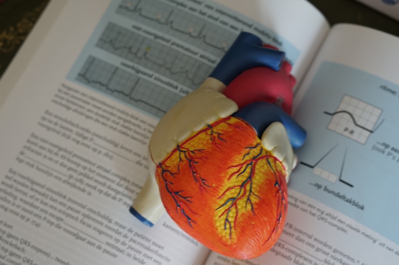
Bacterial Endocarditis (BE) represents a severe and potentially life-threatening condition characterized by the inflammation of the endocardium, which is the inner lining of the heart chambers and valves. This inflammatory process is invariably triggered by a bacterial infection that manages to colonize this otherwise sterile environment. The immediate consequence of this colonization is the formation of infectious masses, known as vegetations, primarily located on the heart valves. These vegetations are complex matrices composed of fibrin, platelets, inflammatory cells, and the causative microorganisms themselves, serving as a persistent source of infection within the circulatory system. The disease necessitates prompt recognition and aggressive therapeutic intervention due to its rapid destructive potential, often leading to irreversible structural cardiac damage and significant systemic complications if treatment is delayed or inadequate.
The impact of Bacterial Endocarditis extends far beyond localized inflammation; its defining characteristic is the progressive and often catastrophic damage inflicted upon the delicate structures of the heart valves. As the bacteria proliferate within the vegetations, they release enzymes and trigger intense inflammatory responses that physically erode the valve leaflets, chordae tendineae, or papillary muscles. This structural degradation results in impaired valvular function, most commonly manifesting as valvular insufficiency or regurgitation, where the valve fails to close properly, leading to backward flow of blood. Less commonly, but equally detrimental, the damage can cause stenosis, restricting forward blood flow. Ultimately, this compromised valvular integrity severely impairs the heart’s pumping action, demanding increased myocardial effort and precipitating a decline in overall cardiac efficiency, which is the cornerstone of subsequent heart failure development.
Clinically, BE often presents with a range of non-specific systemic symptoms, making early diagnosis challenging. The persistent bacterial presence in the bloodstream (bacteremia) drives systemic responses such as persistent or relapsing fever, chills, night sweats, and profound malaise. However, the true danger lies in the potential for life-threatening complications, particularly the risk of systemic embolism, where fragments of the infected vegetations detach and travel through the circulation, lodging in distant organs. Historically, BE was almost uniformly fatal prior to the advent of antibiotics, and while modern medicine offers effective treatments, it remains associated with high morbidity and mortality, especially in high-risk populations such as those with pre-existing valvular disease or those engaging in high-risk behaviors like unhygienic intravenous drug use.
Pathophysiology and Mechanisms of Valvular Damage
The pathogenesis of Bacterial Endocarditis requires a complex interplay between underlying cardiac pathology and transient or persistent bacteremia. Typically, the initial step involves damage or turbulence to the endocardial surface, which allows circulating bacteria to adhere. This initial non-bacterial thrombotic endocarditis (NBTE) lesion—a sterile aggregate of platelets and fibrin—forms primarily at sites of high-velocity blood flow or pre-existing structural abnormalities. Once bacteria enter the bloodstream, they preferentially adhere to this NBTE site, initiating the formation of the infectious vegetation. Specific bacterial surface components, known as adhesins, facilitate this binding process. Once attached, the bacteria rapidly multiply, becoming encased within a protective matrix of fibrin and platelets, which shields them effectively from host immune defenses, including phagocytes, and significantly reduces the penetration of systemically administered antibiotics.
The destructive mechanism is twofold: direct bacterial invasion and the massive inflammatory response elicited by the host. Direct invasion involves the release of lytic enzymes by the bacteria, such as proteases and collagenases, which actively dissolve the structural components of the valve leaflets, leading to perforation, rupture of the chordae tendineae, or tissue necrosis. Simultaneously, the persistent activation of the immune system results in an intense localized inflammatory reaction characterized by the infiltration of neutrophils and macrophages. While intended to clear the infection, this sustained inflammation contributes significantly to the bystander damage of the surrounding valve tissue. This combined assault rapidly compromises the mechanical integrity of the heart valve, creating large, friable vegetations that impair coaptation, leading directly to the hallmark functional impairment of the heart.
The location and size of the vegetation dictate the specific physiological consequences. In cases involving the mitral or aortic valves (the left side of the heart), resulting regurgitation leads to volume overload in the left atrium and ventricle, respectively, forcing the heart to work harder to maintain adequate cardiac output. This continuous volume and pressure stress initiates a cycle of ventricular remodeling and dilation, ultimately diminishing the heart’s intrinsic contractility. The resulting hemodynamic burden culminates in the clinical syndrome of congestive heart failure. In contrast, right-sided endocarditis, frequently observed in patients with a history of intravenous drug use, typically affects the tricuspid valve. While right-sided failure may occur, the more immediate threat involves septic pulmonary embolism, as vegetations dislodge and travel directly into the pulmonary circulation, causing pneumonia, abscesses, or infarction.
Etiology: Bacterial Sources and Primary Risk Factors
The causative agents of Bacterial Endocarditis are diverse, though a few bacterial species predominate, reflecting the source of the bacteremia and the host’s underlying risk factors. The most virulent and rapidly destructive form is often caused by Staphylococcus aureus, a bacterium commonly found on the skin and mucosal surfaces. Infections caused by S. aureus are notorious for their ability to colonize previously healthy valves and for their rapid destruction of cardiac tissue. Other common culprits include various species of Streptococcus, particularly the Viridans group (often associated with dental procedures or poor oral hygiene) and Enterococcus species (frequently linked to genitourinary or gastrointestinal sources). Less common but clinically significant are the HACEK organisms (Haemophilus, Actinobacillus, Cardiobacterium, Eikenella, Kingella), which typically cause subacute forms of the disease.
A critical and increasing risk factor in modern epidemiology is a history of unhygienic intravenous drug use (IVDU). Individuals who inject illicit substances intravenously often use non-sterile techniques, including licking needles, using contaminated injection solutions (diluents), or sharing equipment, which introduces high concentrations of bacteria directly into the bloodstream. Furthermore, the substances themselves, such as cocaine or heroin, may be cut with fillers that cause micro-trauma to the endocardium, providing optimal sites for bacterial adherence. This population predominantly suffers from right-sided endocarditis (tricuspid valve involvement) because the contaminated blood reaches the right side of the heart first. The organisms involved in IVDU-related endocarditis are often virulent skin flora, predominantly methicillin-sensitive or methicillin-resistant S. aureus (MSSA/MRSA), leading to highly destructive and recurrent infections that present substantial challenges for both medical and surgical management due to the underlying behavioral factors.
Beyond illicit drug use, several intrinsic factors predispose individuals to BE. Any condition that alters the normal flow dynamics or integrity of the endocardium increases risk. These risk factors can be categorized as follows:
- Pre-existing Valvular Disease: Rheumatic heart disease, bicuspid aortic valve, mitral valve prolapse with regurgitation, or degenerative calcific valve disease.
- Prosthetic Valves: Mechanical or bioprosthetic valves are foreign bodies that provide excellent surfaces for bacterial colonization. Infections in these patients carry exceptionally high mortality rates and often require surgical replacement.
- Congenital Heart Defects: Conditions like ventricular septal defects, patent ductus arteriosus, or tetralogy of Fallot often create turbulent blood flow patterns highly conducive to NBTE formation.
- Healthcare Exposure: Patients with long-term indwelling catheters, implanted cardiac devices (pacemakers, defibrillators), or those undergoing hemodialysis are at risk for nosocomial bacteremia, particularly with highly resistant organisms.
Understanding these underlying conditions is paramount for identifying high-risk patients who may benefit from prophylactic antibiotics prior to high-risk procedures, such as specific dental work.
Clinical Presentation and Systemic Manifestations
The clinical picture of Bacterial Endocarditis is notoriously heterogeneous, ranging from a fulminant, rapidly destructive acute illness to a more insidious, subacute presentation. In acute BE, often caused by highly virulent organisms like S. aureus, the onset is abrupt, characterized by high, spiking fever, rigors, and rapid deterioration of the patient’s condition, frequently mimicking severe sepsis. Subacute BE, conversely, typically caused by less virulent organisms (e.g., Viridans streptococci), may evolve over weeks or months with non-specific constitutional symptoms such as low-grade fever, unexplained weight loss, fatigue, arthralgias (joint pain), and myalgias (muscle aches). This often leads to significant diagnostic delay, as these symptoms can be easily attributed to common viral illnesses or chronic inflammatory conditions.
Beyond the generalized systemic signs resulting from persistent bacteremia, BE is characterized by several classic peripheral signs caused by the immunological response and the showering of micro-emboli. These manifestations, although less common today due to earlier diagnosis, are highly suggestive of the disease:
- Osler’s Nodes: Painful, palpable, reddish-purple nodules found on the pads of the fingers and toes, representing immune complex deposition.
- Janeway Lesions: Non-tender, hemorrhagic spots found on the palms and soles, caused by septic micro-emboli.
- Roth Spots: Retinal hemorrhages with pale centers, reflecting immune complex vasculitis in the eyes.
- Splinter Hemorrhages: Small, linear hemorrhages found under the nail beds, also indicative of micro-embolism.
The presence of a new or changing heart murmur is a crucial clinical finding, strongly suggesting acute valvular damage and prompting immediate investigation.
The systemic nature of the infection means that virtually any organ system can be affected. Neurological complications are particularly feared, resulting from septic emboli migrating to the cerebral arteries, causing ischemic stroke, transient ischemic attacks (TIAs), or the formation of mycotic aneurysms (infected arterial wall dilations), which carry a high risk of rupture and catastrophic hemorrhage. Renal involvement is also common, manifesting as glomerulonephritis due to immune complex deposition, potentially leading to renal failure. Furthermore, the persistent infection places immense strain on the hematological system, often resulting in anemia of chronic disease, splenomegaly (enlargement of the spleen), and characteristic laboratory findings such as an elevated erythrocyte sedimentation rate (ESR) and C-reactive protein (CRP). These widespread systemic consequences underscore why BE is not merely a cardiac infection but a complex, multi-system inflammatory and infectious disorder.
Major Complications: Embolism and Congestive Heart Failure
The two most critical and life-threatening complications of Bacterial Endocarditis are systemic embolism and the rapid deterioration into congestive heart failure (CHF). Embolic events occur when fragments of the friable vegetations break off and enter the systemic or pulmonary circulation. Left-sided endocarditis (aortic and mitral valves) poses the highest risk for systemic emboli, which travel to the brain, spleen, kidneys, and extremities. Cerebral embolism is the most devastating, often resulting in permanent neurological deficits or death. The risk of embolism is directly related to the size, mobility, and specific bacterial composition of the vegetation; for instance, large, highly mobile vegetations caused by S. aureus are associated with an extremely high embolic rate, particularly in the early phases of treatment before antibiotics stabilize the infectious process.
The consequences of septic embolism vary depending on the target organ. When lodged in the spleen or kidneys, emboli can cause infarction, abscess formation, and pain. Embolism to the coronary arteries is rare but can precipitate myocardial infarction. In patients with right-sided endocarditis (IDVU patients), the emboli are typically shed into the pulmonary circulation, causing septic pulmonary embolism. These often present as multiple nodular infiltrates on chest imaging, frequently cavitating and leading to pulmonary abscesses or infectious pneumonia, significantly impairing respiratory function and contributing to overall systemic deterioration. The management of embolic risk often requires careful risk stratification, potentially necessitating urgent surgical intervention if emboli recur despite appropriate antibiotic therapy or if the vegetation size exceeds established high-risk thresholds.
Congestive heart failure, resulting from acute, severe valvular regurgitation, is the leading cause of death in patients with BE, even when the infection itself is microbiologically cured. The acute destruction of a valve, particularly the aortic valve, imposes an overwhelming volume load on the ventricle, which cannot adapt quickly enough. This rapid hemodynamic decompensation necessitates swift surgical repair or replacement of the damaged valve. If the damage is profound—such as a torn aortic cusp or ruptured mitral chordae—the heart is unable to maintain adequate forward flow, leading to pulmonary edema and systemic hypoperfusion. Therefore, the presence of new or worsening heart failure is frequently the most crucial determinant for the timing of emergency cardiac surgery, superseding the completion of the standard antibiotic course, as surgical intervention is often required not only to remove the infectious source but primarily to restore hemodynamic stability and prevent fatal circulatory collapse.
Diagnosis and Therapeutic Strategies
Diagnosis of Bacterial Endocarditis relies on a combination of clinical findings, microbiological identification, and advanced imaging techniques, integrated primarily through the modified Duke Criteria. The initial diagnostic cornerstone involves drawing multiple sets of blood cultures before initiating antibiotic therapy. Isolating the causative bacterium is essential not only for confirmation but also for determining antibiotic sensitivities, which guide specific, targeted treatment. However, a significant minority of cases may be culture-negative, necessitating reliance on clinical and imaging evidence.
Imaging is critical for visualizing the vegetations and assessing the extent of cardiac damage. Echocardiography is the primary imaging modality:
- Transthoracic Echocardiography (TTE): Generally the initial test, useful for detecting large vegetations, assessing global cardiac function, and identifying significant valvular damage.
- Transesophageal Echocardiography (TEE): Offers superior resolution and clarity, particularly crucial for detecting small vegetations, assessing prosthetic valves, identifying abscesses or fistulae, and is considered mandatory when TTE results are non-diagnostic or when clinical suspicion remains high.
Additional imaging, such as computed tomography (CT) or magnetic resonance imaging (MRI), may be used to identify complications like septic emboli in the brain or mycotic aneurysms.
Therapeutic management of BE is invariably complex and typically requires a prolonged course of high-dose intravenous antibiotics, administered for four to six weeks, often in an inpatient setting initially. The selection of the antibiotic regimen is strictly guided by the susceptibility profile of the isolated organism. For highly destructive organisms like S. aureus, combination therapy may be employed. The prolonged duration is necessary because the bacteria are sequestered within the fibrin and platelet matrix of the vegetation, requiring sustained high serum concentrations of antibiotics to penetrate and sterilize the site. Despite effective antibiotic therapy, approximately 40% to 50% of patients will require cardiac surgery. Indications for surgery include moderate-to-severe heart failure due to valvular dysfunction, uncontrolled local infection (e.g., abscess, fistula formation), or evidence of recurrent systemic emboli despite optimal medical management. The surgical procedure involves debridement of infected tissue and often replacement of the damaged native valve with a prosthetic valve.
Psychosocial and Public Health Implications
The connection between Bacterial Endocarditis and unhygienic intravenous drug use highlights significant public health and psychosocial challenges. For patients with IVDU-associated BE, the cycle of infection and relapse is often tied directly to ongoing substance use disorder. Medical treatment of BE in this population is complicated by adherence issues, potential conflict between required inpatient care and drug dependence, and the high likelihood of recurrent infection if injecting habits continue post-discharge. Treating these patients requires an integrated approach that addresses both the acute medical crisis and the underlying addiction, often involving dedicated infectious disease specialists, cardiologists, social workers, and addiction counselors.
Prevention strategies are essential, particularly targeting the vulnerable population of active drug users. Public health interventions focus on harm reduction models, including the promotion of sterile injection practices, access to needle exchange programs, and providing education on the serious cardiac risks associated with unhygienic use. Furthermore, early access to treatment programs for substance use disorder can significantly reduce the incidence of primary and recurrent BE. The psychological burden on survivors is also substantial; they often face long recovery periods, the realization of permanent heart damage, and the need for lifelong medical follow-up, which can lead to anxiety, depression, and post-traumatic stress related to the severity of the illness.
From a broader perspective, BE serves as a sentinel indicator of underlying systemic health disparities. Patients with complex congenital heart disease or prosthetic valves require meticulous, lifelong dental and medical care to prevent bacteremia, often necessitating antibiotic prophylaxis prior to procedures known to introduce bacteria into the bloodstream (e.g., specific dental extractions). Failure in the healthcare system to adequately manage chronic conditions, coupled with the rising rates of opioid and injection drug abuse, ensures that Bacterial Endocarditis remains a persistent, high-acuity challenge in cardiology and infectious disease worldwide, demanding rigorous adherence to both medical protocols and comprehensive psychosocial support systems to improve long-term outcomes.