Bulbar Anatomy: The Science of Brainstem Function
- Definition and Etymology of Bulbar
- Anatomical Contexts: Bulb-like Structures
- Focus on the Medulla Oblongata: The Primary Medical Context
- Cranial Nerves Associated with Bulbar Function
- Symptoms and Clinical Manifestations of Bulbar Dysfunction
- Bulbar Paralysis: A Key Clinical Application
- Bulbar Poliomyelitis and Viral Impact
- Diagnostic Approaches and Differential Diagnosis
Definition and Etymology of Bulbar
The term bulbar is an adjective derived from the Latin term bulbus, meaning a bulb or an expanded, rounded structure. In its broadest anatomical application, bulbar pertains to any structure resembling or related to a bulb, reflecting a basic morphological description used across various biological fields. However, the medical and neurological usage has become significantly specialized, almost exclusively referring to structures associated with the lower brainstem. Initially, the adjective was employed universally to describe various anatomical “bulbs,” such as the bulbus oculi (the eyeball) or the expanded base of hair follicles. Over time, particularly within clinical neuroscience, the semantic range narrowed substantially, focusing on the most critical bulb-like structure within the central nervous system: the medulla oblongata. This narrowing of definition is essential for understanding clinical syndromes, as the presence of bulbar symptoms immediately directs diagnostic inquiry toward specific nuclei located within this vital region of the brainstem, indicating profound dysfunction in areas controlling essential, life-sustaining functions such as respiration, speech, and deglutition.
Understanding the etymological precision is crucial because while the term technically applies to any bulb-like structure, modern clinical medicine has codified its meaning to denote conditions originating specifically from the lower cranial nerve nuclei situated in the medulla. This anatomical focus underscores the severity of bulbar conditions, as the medulla oblongata is a critical nexus for automatic regulatory functions. The formal, precise usage ensures that when a clinician refers to a bulbar condition, they are communicating a specific, high-stakes neurological localization. This localization involves the motor neurons responsible for controlling muscles of the pharynx, larynx, palate, and tongue, which are innervated by cranial nerves IX (Glossopharyngeal), X (Vagus), XI (Accessory), and XII (Hypoglossal). The implications of damage in this region are always serious, necessitating immediate and specialized medical attention due to the direct threat posed to airway maintenance and nutritional intake.
The transition from a general descriptive adjective to a specific clinical descriptor highlights the evolutionary process of medical terminology. While anatomical texts might still use bulbar in reference to the ocular or urethral bulb, the overwhelming clinical context demands the association with the brainstem. This linguistic prioritization reflects the extreme clinical significance of medullary pathology compared to other bulb-like structures. When assessing a patient presenting with symptoms like difficulty swallowing or speaking, the adjective bulbar serves as an immediate neurological shorthand, signaling a potential lesion affecting the lower motor neurons housed within the brainstem’s central “bulb.” The subsequent exploration of related conditions, such as bulbar paralysis or bulbar poliomyelitis, reinforces this specialized anatomical focus, cementing the medulla oblongata as the primary referent for the term in high-level neurological discourse.
Anatomical Contexts: Bulb-like Structures
Anatomically, the concept of a “bulb” refers to any rounded, expanded, or thickened part of an organ or structure, and the adjective bulbar therefore describes something pertaining to such a structure. Beyond the highly specific neurological context, several structures throughout the human body possess this bulbous morphology. For instance, the eye is anatomically referred to as the bulbus oculi, and the term bulbar conjunctiva refers to the thin membrane covering the visible part of the eyeball. Similarly, the initial segment of the duodenum, just distal to the stomach, is often termed the duodenal bulb due to its dilated appearance on imaging. This diversity in general anatomical usage contrasts sharply with the clinical specialization, where these peripheral references are typically secondary to the primary neurological meaning. It is the central location and critical function of the brainstem’s enlargement that overwhelmingly dominates the clinical application of the term bulbar.
The key anatomical structure that defines the modern clinical term is the caudal portion of the brainstem, specifically the medulla oblongata. The medulla is situated superior to the spinal cord and inferior to the pons, forming a slightly expanded, tapered structure often likened to a bulb or stalk where the central nervous system enters the skull. This region is densely packed with crucial nuclei and ascending and descending tracts, making its integrity essential for maintaining life. The concentration of motor nuclei for the lower cranial nerves (specifically IX, X, XI, and XII) within this medullary bulb establishes the anatomical basis for bulbar syndromes. Damage to these motor neuron pools disrupts the efferent signals necessary for performing complex, coordinated muscular activities such as phonation (voice production), articulation (speech clarity), and deglutition (swallowing), leading to the specific constellation of symptoms known as bulbar palsy.
It is important to differentiate between general anatomical usage and specific pathological localization. While the term may technically apply to structures like the base of the hair follicle or the vestibule of the vagina, these instances lack the clinical weight carried by the neurological application. When a physician refers to a bulbar condition, they are highlighting a direct impact on the cranial nerve nuclei located within the medulla oblongata. This precise localization is critical for diagnosis and prognosis because the structures housed within this bulb control automatic and reflexive functions that are indispensable for survival. Therefore, while multiple structures are bulb-like, the medical definition of bulbar pathology has functionally become synonymous with pathology of the lower brainstem motor nuclei, serving as a powerful indicator of severe neurological compromise.
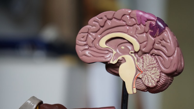
Focus on the Medulla Oblongata: The Primary Medical Context
Medically, bulbar denotes a condition pertaining directly to the medulla oblongata, which is the anatomical “bulb” of the brainstem. This structure is arguably the most vital center of the entire central nervous system, housing regulatory centers for autonomic functions that sustain life without conscious effort. Key among these are the cardiovascular centers, which regulate heart rate and blood pressure, and the respiratory centers, which control the rhythm and depth of breathing. When a disease process, whether infectious, degenerative, or ischemic, affects the medulla, the resulting syndrome is termed bulbar, emphasizing the critical nature of the compromised region. The severity of bulbar dysfunction stems directly from the fact that damage to this area threatens the fundamental ability to maintain homeostasis and execute basic motor skills required for daily survival, such as clearing the throat and managing secretions.
The medulla oblongata acts as the relay station between the brain and the spinal cord, and critically, it contains the motor neuron cell bodies (nuclei) for the last four cranial nerves. These nerves—Glossopharyngeal (IX), Vagus (X), Accessory (XI), and Hypoglossal (XII)—are collectively responsible for the intricate movements of the tongue, soft palate, pharynx, and larynx. These movements facilitate speech, chewing, and the coordinated reflex of swallowing. Pathological processes targeting these motor nuclei result in muscle weakness and atrophy in the territories supplied by these nerves, manifesting clinically as dysphagia (difficulty swallowing) and dysarthria (difficulty speaking). These specific symptoms are the hallmarks of a true bulbar syndrome, distinguishing it from general neurological weakness. The precise anatomical localization within the medulla ensures that the resulting clinical picture is highly recognizable and carries a grave prognosis if the underlying cause is progressive.
Moreover, the concentration of vital centers within this small bulbar region explains the rapid and often fatal progression of severe bulbar syndromes. Unlike peripheral nerve damage, which might affect a limb, medullary damage immediately impacts essential regulatory systems and the ability to protect the airway. Respiratory failure is a common complication in advanced bulbar pathology, resulting either from the direct involvement of respiratory centers within the medulla or from the inability of the patient to clear the airways due to paralysis of laryngeal and pharyngeal muscles. Therefore, the term bulbar, when used clinically, signifies not just an anatomical location, but a functional threat to the patient’s most basic physiological processes, necessitating intensive monitoring and often requiring supportive measures such as tracheostomy or nutritional support via gastrostomy tube.
Cranial Nerves Associated with Bulbar Function
The core components of bulbar function are mediated by the cranial nerves whose nuclei are situated within the medulla oblongata. These are predominantly the lower cranial nerves, specifically Cranial Nerves IX (Glossopharyngeal), X (Vagus), XI (Accessory), and XII (Hypoglossal). The functions of these nerves are tightly integrated, controlling the motor output to the muscles of the pharynx, larynx, soft palate, and tongue, which are collectively responsible for the complex actions of speech and swallowing. Dysfunction in the motor components of these nerves’ nuclei is the definitive cause of true bulbar palsy. For example, damage to the nucleus ambiguous, which contains the motor neurons for IX and X, results in severe weakness of the pharyngeal constrictors and vocal cords, leading to nasal regurgitation and a weak, breathy voice (dysphonia).
Detailed examination of the role of these nerves clarifies the clinical presentation of bulbar syndromes. The Vagus nerve (X) is perhaps the most critical, controlling the elevation of the soft palate and the movement of the vocal cords; its paralysis leads to a characteristic lack of gag reflex, deviation of the uvula toward the intact side, and severe dysphagia. The Hypoglossal nerve (XII) controls all intrinsic and most extrinsic muscles of the tongue; when its nucleus is affected, the tongue becomes weak, fasciculating (twitching), and atrophied, resulting in profound difficulty with articulation and pushing food back for swallowing. The coordination required for a successful swallow involves sequential, precise activation of muscles controlled by all four of these bulbar nerves, meaning that even partial damage to the medullary nuclei can severely compromise nutritional intake and place the patient at high risk for aspiration pneumonia.
The precise involvement of these cranial nerve nuclei allows clinicians to distinguish between bulbar syndromes and higher-level motor disorders. The presence of lower motor neuron signs—such as muscle atrophy, weakness, and fasciculations—in the tongue or facial muscles is a strong indicator that the pathology lies directly within the medullary nuclei or the nerve roots exiting the brainstem. These signs contrast significantly with the findings in pseudobulbar palsy, where the damage occurs to the corticobulbar tracts above the medulla, sparing the motor neuron cell bodies themselves. Therefore, understanding which specific cranial nerves are housed in the bulbar region provides the anatomical framework for diagnosing and classifying diseases such as Progressive Bulbar Palsy, where the degeneration is confined specifically to these vulnerable motor neuron pools.
Symptoms and Clinical Manifestations of Bulbar Dysfunction
The clinical manifestations of bulbar dysfunction are characterized by a specific triad of symptoms related to impaired function of the muscles controlled by the lower cranial nerves: dysphagia, dysarthria, and dysphonia. Dysphagia, or difficulty swallowing, is often the most life-threatening symptom. It results from the weakness of the pharyngeal constrictors and the failure of the soft palate to elevate properly, leading to the inability to move a food bolus from the mouth to the esophagus. Patients often report choking, coughing after swallowing, and nasal regurgitation of liquids. This impaired swallowing mechanism significantly increases the risk of aspiration, where food or liquid enters the trachea and lungs, invariably leading to aspiration pneumonia, a frequent cause of morbidity and mortality in bulbar syndromes.
Dysarthria, which is difficulty in articulating speech, arises from weakness and incoordination of the muscles of the tongue and lips. The speech of a patient with bulbar palsy is typically slow, slurred, and often nasal in quality due to the paralysis of the soft palate, preventing the closure of the nasopharynx during speaking. When the tongue is severely affected, the patient struggles to form consonants that require precise tongue movements, leading to unintelligibility. Concurrently, dysphonia, or a change in voice quality, often manifests as a weak, breathy, or low-pitched voice due to paralysis of the vocal cords (controlled by the Vagus nerve). These speech impairments severely restrict communication, further isolating the patient and contributing to the psychological burden of the disease.
In addition to the primary triad, bulbar dysfunction is often characterized by the presence of lower motor neuron signs specific to the affected musculature. Clinicians look for fasciculations (visible muscle twitching) and atrophy (wasting) of the tongue, which are highly suggestive signs of motor neuron disease affecting the Hypoglossal nucleus. The jaw jerk reflex is typically diminished or absent in true bulbar palsy, contrasting with the exaggerated jaw jerk seen in pseudobulbar palsy. Furthermore, patients may exhibit difficulty managing oral secretions due to impaired pharyngeal clearing mechanisms, leading to drooling and pooling of saliva. The combination of these specific cranial nerve deficits provides a clear clinical signature, compelling neurologists to investigate degenerative processes like Amyotrophic Lateral Sclerosis or specific infectious etiologies such as bulbar poliomyelitis.
Bulbar Paralysis: A Key Clinical Application
The term bulbar paralysis (or bulbar palsy) is a clinical descriptor signifying weakness or paralysis of the muscles innervated by the motor nuclei of the lower cranial nerves (IX, X, XI, XII), reflecting a lower motor neuron lesion in the medulla oblongata. This condition is most often associated with rapidly progressive and catastrophic neurodegenerative disorders, predominantly Amyotrophic Lateral Sclerosis (ALS), where the disease presentation is specifically termed Progressive Bulbar Palsy (PBP) when the bulbar symptoms are the first or most prominent signs. In PBP, the motor neurons in the medulla degenerate selectively, leading to profound and progressive difficulty with swallowing and speech before significant limb weakness occurs. This localized degeneration highlights the vulnerability of the bulbar motor neuron pool to the pathological processes characteristic of ALS.
Bulbar paralysis must be carefully differentiated from other forms of paralysis due to its severe implications for survival. In its classic presentation, bulbar paralysis involves flaccid weakness, muscle atrophy, and fasciculations in the tongue, palate, and facial muscles. The underlying etiology determines the prognosis, but the immediate threat remains the loss of airway protection and subsequent aspiration. While ALS is the most frequent cause in the adult population, bulbar paralysis can also result from other conditions that damage the medulla, including brainstem stroke (infarction or hemorrhage), tumors, or inflammatory processes such as Guillain-Barré syndrome (specifically the Bickerstaff’s brainstem encephalitis variant). The hallmark of true bulbar paralysis is the presence of signs indicating direct damage to the lower motor neuron cell body, such as the aforementioned atrophy and fasciculations, confirming the medullary localization.
Management of bulbar paralysis is primarily supportive, focusing intensely on maintaining nutrition and respiratory function. Due to the progressive nature of diseases like ALS, patients often require early intervention measures. These interventions include percutaneous endoscopic gastrostomy (PEG) tube placement to ensure adequate caloric intake without the risk of aspiration, and non-invasive positive pressure ventilation (NIPPV) to support weakened respiratory muscles. The progression of bulbar symptoms is frequently rapid, sometimes leading to complete loss of speech (anarthria) and total dependence on assisted feeding and breathing within a year or two of onset. Therefore, the diagnosis of bulbar paralysis necessitates prompt multidisciplinary care involving neurology, speech pathology, respiratory therapy, and nutritionists to manage the complex physical and psychological challenges facing the patient.
Bulbar Poliomyelitis and Viral Impact
Historically, one of the most devastating and feared causes of bulbar dysfunction was bulbar poliomyelitis. Poliomyelitis, caused by the poliovirus, is a highly contagious viral infection that exhibits a pronounced neurotropism, selectively invading and destroying motor neurons within the central nervous system. While the spinal form of polio caused paralysis in the limbs, the bulbar form specifically targeted the motor nuclei located within the medulla oblongata, the brainstem bulb. This form of the disease was particularly lethal because the viral destruction of these bulbar nuclei directly incapacitated the vital centers controlling breathing and swallowing, leading to rapid mortality even in young, otherwise healthy individuals.
In bulbar poliomyelitis, the virus caused inflammation and necrosis of the neurons in the nucleus ambiguous (affecting IX and X) and the Hypoglossal nucleus (XII). The resulting paralysis led to severe, acute bulbar symptoms: complete inability to swallow, resulting in pooled secretions and choking, and paralysis of the pharyngeal and laryngeal muscles. Crucially, the bulbar form could also involve the respiratory centers in the medulla itself, disrupting the automatic rhythm of breathing. During the epidemics of the mid-20th century, patients suffering from bulbar polio often required immediate placement in negative-pressure ventilators, famously known as “iron lungs,” to mechanically sustain respiration until the acute phase of the infection subsided, if they survived the initial neuronal destruction.
Although widespread vaccination has dramatically reduced the incidence of wild poliovirus globally, the historical context of bulbar poliomyelitis remains critical for understanding the catastrophic potential of medullary damage. The disease serves as a stark example of a non-degenerative etiology causing acute bulbar paralysis. The clinical presentation of acute bulbar polio—rapid onset of dysphagia, dysarthria, and respiratory compromise—is a benchmark against which other acute bulbar syndromes, such as those caused by stroke or certain autoimmune conditions, are measured. The residual weakness and post-polio syndrome experienced by survivors further underscore the permanent damage inflicted upon the motor neuron pools housed within the delicate bulbar region of the brainstem.
Diagnostic Approaches and Differential Diagnosis
The diagnostic process for bulbar dysfunction is complex, requiring precise localization of the pathology and differentiation from conditions that mimic true bulbar palsy. The primary challenge lies in distinguishing true bulbar palsy (a lower motor neuron lesion in the medulla) from pseudobulbar palsy (an upper motor neuron lesion affecting the corticobulbar tracts above the medulla). Clinical examination is paramount; true bulbar palsy presents with signs of LMN damage, including muscle atrophy, fasciculations, and a weak or absent gag reflex, whereas pseudobulbar palsy presents with UMN signs, such as spasticity, hyperreflexia (an exaggerated gag reflex and jaw jerk), and the characteristic emotional lability known as pseudobulbar affect.
A comprehensive diagnostic workup for suspected bulbar syndrome involves several sophisticated techniques. Electromyography (EMG) and nerve conduction studies are crucial, as they can reveal denervation and fasciculation potentials specifically in the bulbar muscles (tongue, soft palate), confirming lower motor neuron damage localized to the medulla. Imaging studies, particularly Magnetic Resonance Imaging (MRI) of the brainstem, are essential to rule out structural causes such as tumors, vascular malformations, or brainstem strokes, which would represent acute or subacute, non-degenerative etiologies. In cases where an infectious or autoimmune cause is suspected, cerebrospinal fluid (CSF) analysis may be necessary to look for elevated protein or inflammatory markers.
The differential diagnosis is extensive and requires careful consideration of both acute and chronic conditions. Acute causes might include brainstem stroke, Bickerstaff’s encephalitis, or diphtheria. Chronic, progressive causes are most often motor neuron diseases like ALS (Progressive Bulbar Palsy). Furthermore, certain myopathies or neuromuscular junction disorders, such as myasthenia gravis, can cause fluctuating bulbar weakness. Since the prognosis and treatment pathways vary dramatically depending on the underlying cause—from immunosuppression for autoimmune disorders to symptom management for degenerative diseases—the accurate classification of bulbar symptoms through careful clinical assessment, electrophysiology, and imaging is the cornerstone of effective neurological management.