Biological Cannula: Pathways to Healing and Human Insight
- Introduction and Definition of the Cannula
- Historical Context and Evolution of Internal Access Devices
- Basic Design and Essential Components
- Primary Medical Applications and Clinical Utility
- Specialized Applications in Neuroscience and Behavioral Research
- Insertion Techniques and Associated Procedural Risks
- Materials Science, Biocompatibility, and Gauge Standardization
- Future Directions and Technological Advancements
Introduction and Definition of the Cannula
The term cannula (plural: cannulae or cannulas) refers to a highly specialized, slender tube designed for safe and temporary or semi-permanent insertion into the body. Fundamentally, this instrument serves as a conduit, creating a controlled pathway between the external environment and a targeted internal structure, typically a body cavity, duct, or blood vessel. Its utility spans two critical, often complementary, functions: facilitating the controlled removal of fluids, gases, or pathological materials from a specific anatomical location, and conversely, enabling the precise introduction of therapeutic agents, diagnostic contrast media, or nutritional solutions into the circulatory system or targeted tissue. This dual capability establishes the cannula as an indispensable tool in modern medical and biological sciences, underpinning procedures ranging from routine intravenous access to highly complex neurosurgical interventions.
Unlike the standard hypodermic needle, which is primarily a sharp, solid shaft used for rapid injection or aspiration, the cannula is characteristically hollow and often incorporates a retractable sharp component, known as a trocar or stylet, used solely for the initial penetration. Once the device is correctly positioned within the desired lumen or space, the sharp component is withdrawn, leaving the flexible or semi-rigid plastic tube in place. This design philosophy significantly enhances patient safety by minimizing the risk of vessel wall perforation or tissue damage once access is secured. The strategic deployment of cannulae ensures sustained, reliable access for prolonged treatments, thereby overcoming the limitations associated with repeated puncturing required by traditional needle injection methods.
The conceptual simplicity of the cannula belies its profound impact on clinical practice, serving as the cornerstone for minimally invasive procedures across nearly every medical specialty. Whether employed in cardiology to deliver stents, in anesthesiology for continuous pain management, or in laboratory settings for precise chemical delivery in research models, the core principle remains consistent: to establish a stable, safe, and controlled interface with the internal physiology. Understanding the structure, material composition, and application profiles of various cannulae is paramount for professionals seeking to provide effective care while mitigating procedural risks associated with internal access.
Historical Context and Evolution of Internal Access Devices
The necessity of draining harmful accumulations of fluid, such as pus or edema, from the human body dates back to antiquity, suggesting that early, rudimentary forms of drainage tubes were likely employed by ancient civilizations. However, the development of the modern, sophisticated cannula as we know it today is intrinsically linked to the advancements in surgical technique and, particularly, the rise of intravenous therapy in the 17th and 18th centuries. Early attempts at blood transfusion, though often unsuccessful due to poor understanding of blood compatibility and asepsis, highlighted the critical need for reliable, non-collapsing access points into the vascular system. These initial devices were crude, often constructed of rigid materials like silver or brass, and lacked the necessary biocompatibility and flexibility for long-term placement.
Significant leaps occurred in the mid-19th century with the burgeoning acceptance of antiseptic principles championed by figures like Joseph Lister. The realization that materials inserted into the body must be sterile and inert spurred innovation in material science. The transition from purely metallic instruments to those incorporating rubber, glass, and eventually synthetic polymers marked a pivotal moment. The materials had to be non-thrombogenic—that is, they must not provoke the formation of blood clots—and sufficiently smooth to minimize trauma upon insertion and residence within the delicate vascular lining, known as the endothelium. This period saw the introduction of early designs that attempted to separate the puncturing element from the indwelling tube.
The mid-20th century revolutionized cannula design through the widespread adoption of plastics, such as polytetrafluoroethylene (PTFE, or Teflon) and polyethylene. These polymers offered superior flexibility, reduced friction, and enhanced biocompatibility compared to earlier materials. The development of the over-the-needle catheter design, where a plastic sheath surrounds a penetrating needle, became the industry standard for peripheral intravenous access. This innovation allowed for the immediate removal of the sharp needle after insertion, dramatically reducing the risk of accidental needle-stick injuries to healthcare workers and increasing patient comfort during prolonged infusion therapies.
Furthermore, the evolution of specialized cannulae, such as those designed for arterial blood gas sampling or central venous pressure monitoring, followed the general trend toward miniaturization and material refinement. The continuous drive to reduce invasiveness and improve the duration of safe internal access has led directly to the highly specialized instruments utilized today, including those equipped with anti-microbial coatings and integrated sensing capabilities, which represent a significant technological distance from their metallic predecessors.
Basic Design and Essential Components
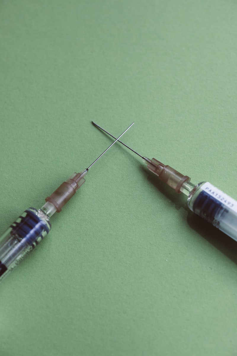
A modern cannula system is typically composed of several integrated components, each serving a specific mechanical or functional purpose, ensuring the device operates reliably and safely. The primary component is the cannula tube itself, which is the hollow, flexible or semi-rigid sheath that remains inside the body. This tube must possess specific mechanical properties, including sufficient tensile strength to prevent breakage and flexibility to conform to the curvature of vessels or anatomical structures without kinking. The internal diameter, or lumen, dictates the flow rate of fluids, making lumen size a critical specification chosen based on the intended application, such as high-volume resuscitation versus slow medication infusion.
Crucial to the insertion process is the trocar or stylet. This is a sharp, solid, or semi-hollow metallic core that extends through the lumen of the cannula tube. Its pointed tip facilitates the clean, rapid penetration of skin and underlying tissue layers. The stylet is designed to be slightly longer than the cannula sheath, ensuring that the sharp tip leads the way. Once successful vascular or cavity access is confirmed, typically through a visual flashback of blood or fluid into the hub, the stylet is immediately and carefully withdrawn, leaving only the soft cannula tube in position. This mechanism is central to the safety profile of the device, mitigating the trauma associated with indwelling sharp objects.
At the proximal end of the device, remaining outside the body, is the hub, which serves as the interface between the indwelling tube and external equipment. The hub is usually color-coded according to a standardized gauge system (e.g., green for 18-gauge, pink for 20-gauge) to allow for quick identification of the cannula size and flow capacity. The hub typically incorporates Luer lock or Luer slip fittings, standardized connectors that ensure a secure, leak-proof attachment to infusion lines, syringes, or stopcocks. Furthermore, many hubs include wings or fixation plates that assist in securing the cannula to the patient’s skin with tape or dressings, preventing accidental dislodgement and subsequent infiltration or infection.
A final, critical component is the flashback chamber, often integrated into the hub or the proximal end of the stylet. This transparent reservoir allows the clinician to visually confirm successful placement. As the needle enters the target vessel, the internal pressure causes blood or fluid to flow back into this chamber. This visual confirmation is instantaneous and vital, ensuring that the clinician does not advance the device unnecessarily, which could lead to complications or failed placement. The integration of these components—the flexible tube, the sharp stylet, the secure hub, and the visual confirmation system—defines the modern, reliable cannula delivery system.
Primary Medical Applications and Clinical Utility
The clinical utility of cannulae is vast, touching nearly every aspect of patient care where internal fluid management or access is required. Perhaps the most common application is in establishing peripheral intravenous (IV) access, essential for administering hydration fluids, electrolytes, and medications directly into the venous circulation. These procedures utilize smaller gauge cannulae (typically 18 to 24 gauge) and are vital in emergency medicine, surgical preparation, and long-term inpatient care. The reliability of peripheral IV access is foundational to modern pharmacology, ensuring therapeutic agents reach the systemic circulation rapidly and predictably.
Beyond peripheral access, cannulae are critical for central venous access, where larger, longer catheters are threaded into major veins, such as the subclavian or internal jugular, leading toward the heart. Central lines are necessary for the administration of caustic medications (like certain chemotherapy agents), for total parenteral nutrition (TPN), or for monitoring central venous pressure (CVP). These procedures require meticulous technique, often guided by ultrasound or fluoroscopy, due to the proximity of vital structures. Central venous cannulation provides a durable, high-flow access point crucial for critically ill patients requiring intensive support.
In surgical contexts, specialized cannulae are employed for aspiration and drainage. For example, in plastic surgery, large-bore cannulae are used during liposuction to safely aspirate subcutaneous adipose tissue. In thoracic and abdominal surgery, drainage cannulae, often referred to as drains, are inserted post-operatively to evacuate accumulated blood, serum, or air (as in a chest tube following lung collapse), thereby preventing compression and promoting healing of internal organs. These drainage devices are structurally rigid enough to maintain patency but flexible enough to minimize irritation to surrounding tissues.
Furthermore, diagnostic procedures heavily rely on cannulae. Angiography, the visualization of blood vessels, requires the insertion of a catheter—a form of cannula—into an artery (often in the groin or wrist) to deliver radiopaque contrast dye. Similarly, in hemodialysis, specialized, high-flow cannulae are temporarily or permanently placed into shunts or fistulas to allow for the efficient exchange of blood between the patient and the dialysis machine. The selection of the appropriate cannula type, size, and material is meticulously managed based on the specific fluid dynamics and anatomical constraints of the procedure being performed.
Finally, in the realm of anesthesia, epidural and spinal cannulae are extremely thin, flexible tubes inserted into the spinal column area to provide continuous regional analgesia. These specialized cannulae allow for the slow, sustained delivery of anesthetic agents directly to the nerve roots, offering highly effective pain management during labor, major surgery, and chronic pain treatment. The precision required for these placements underscores the sophisticated nature of these medical instruments.
Specialized Applications in Neuroscience and Behavioral Research
Although the cannula is fundamentally a medical device, its application extends deeply into experimental psychology, behavioral neuroscience, and neuropharmacology, where it serves as a critical tool for investigating the complex dynamics of the central nervous system. In this research context, cannulae are often miniaturized for intracerebral implantation, allowing for the chronic, targeted access to specific brain regions in animal models, such as the hippocampus, amygdala, or specific nuclei. This capability is essential for studying the neural substrates of learning, memory, addiction, and psychiatric disorders.
One of the most powerful applications is microdialysis, a technique that uses a specialized, fine-gauge cannula with a semi-permeable membrane at its tip. Once implanted, this cannula is perfused with a solution that mimics the extracellular fluid. Small molecules, including neurotransmitters (like dopamine, serotonin, or glutamate), hormones, and metabolites, diffuse across the membrane and into the perfusate, which is then collected and analyzed using high-performance liquid chromatography (HPLC). This allows researchers to monitor the release and metabolism of critical neurochemicals in real-time while the animal is engaged in a behavioral task, providing crucial insights into the neural correlates of behavior and motivation.
Moreover, intracerebral cannulation enables the highly localized delivery of pharmacological agents, a technique known as intracranial drug infusion. Researchers can administer specific receptor agonists or antagonists directly into a targeted brain region to transiently inhibit or excite neural activity. This approach circumvents the blood-brain barrier and avoids systemic side effects, allowing for the precise mapping of neurocircuitry involved in specific behaviors. For instance, a researcher might infuse a dopamine antagonist into the nucleus accumbens to study its effect on reward-seeking behavior, establishing causality between neurotransmitter function in that specific region and the resulting observable behavior.
In clinical neurosurgery, specialized guidance cannulae are sometimes used during procedures such as Deep Brain Stimulation (DBS). While the final electrode placement is precise, temporary cannulae may be used for microelectrode recording (MER) or for the targeted introduction of small amounts of substances to confirm physiological responses before permanent device implantation. These neurosurgical applications demand extreme precision, often requiring cannulae with sub-millimeter tolerances, emphasizing the role of this instrument in both basic science discovery and advanced clinical treatment for neurological and psychiatric conditions.
Insertion Techniques and Associated Procedural Risks
The success and safety of cannulation depend heavily on the proper execution of specific insertion techniques, which vary according to the target vessel or cavity. For peripheral venous access, the standard technique involves stabilizing the vein, penetrating the skin and vessel wall with the stylet-cannula assembly, confirming flashback, and then advancing the plastic cannula sheath while simultaneously withdrawing the sharp stylet. Meticulous adherence to aseptic technique is paramount throughout this process to prevent the introduction of pathogens.
For accessing deeper or more critical vessels, particularly in central venous or arterial cannulation, the Seldinger technique is often employed. This method involves several sequential steps: first, puncturing the vessel with a thin access needle; second, threading a flexible guidewire through the needle and into the vessel; third, removing the access needle while the guidewire remains in place; fourth, passing a dilator and then the final cannula over the guidewire; and finally, removing the guidewire. The use of the guidewire ensures that the vessel lumen is maintained and that the potentially damaging cannula tip does not blindly search for the pathway, significantly reducing the risk of vessel trauma.
Despite stringent procedures, cannulation carries inherent risks that must be managed. The most pervasive complication is infection. Any breach of the skin barrier introduces a risk of bacterial entry, which can lead to localized phlebitis (inflammation of the vein) or, in severe cases, systemic sepsis, particularly with central venous lines. To mitigate this, strict skin preparation (using chlorhexidine or iodine solutions) and sterile dressing techniques are mandatory protocols.
Other significant risks include thrombosis (blood clot formation) around the catheter tip, which can obstruct blood flow and potentially lead to embolism; infiltration or extravasation, where the cannula punctures through the far wall of the vessel, causing infused fluids to leak into surrounding tissues; and mechanical complications such as nerve damage, hematoma formation, or accidental pneumothorax (lung collapse) if central lines are placed near the apex of the lung. Continuous monitoring and timely removal or repositioning of the cannula are essential steps in minimizing these potentially serious complications.
Materials Science, Biocompatibility, and Gauge Standardization
The material composition of a cannula is a fundamental determinant of its function, longevity, and safety within the human body. Materials must exhibit high biocompatibility, meaning they should not provoke adverse immunological, toxicological, or inflammatory reactions when in contact with tissues or blood. Early cannulae made of rigid metals often caused significant mechanical trauma and inflammation; modern devices utilize sophisticated polymers engineered specifically for medical use.
Common polymer materials include Polytetrafluoroethylene (PTFE), known for its extremely low coefficient of friction, which aids in smooth insertion and reduces tissue drag. Polyurethane (PU) is another popular choice, valued for its superior flexibility and kink resistance, making it ideal for central venous catheters that must navigate curved vessel pathways. Some specialized cannulae, particularly those used in high-pressure procedures or for structural support, still incorporate medical-grade stainless steel for the stylet and portions of the hub. The internal surface of the tube is often treated or coated to minimize protein adsorption and platelet aggregation, thereby reducing the risk of catheter-related thrombosis.
The size of the cannula is internationally standardized using the French scale (Fr), particularly for larger catheters (where 1 Fr is approximately 0.33 mm of outer diameter), and the Birmingham gauge (G) for smaller, peripheral devices. It is counter-intuitive but crucial to note that the gauge system is inverse: a smaller gauge number indicates a larger external diameter and thus a higher flow rate. For instance, a 14-gauge cannula is significantly larger than a 22-gauge cannula. The choice of gauge directly affects the clinical scenario; for rapid fluid administration during trauma, a large-bore 14G or 16G cannula is required, while for routine medication delivery in pediatric or geriatric patients, a smaller 22G or 24G might be used to protect fragile veins.
Future Directions and Technological Advancements
The evolution of cannula technology is currently focused on integrating smart features and enhancing material performance to address the persistent challenges of infection and thrombosis. A major area of research involves the development of antimicrobial coatings. These coatings, often incorporating silver compounds or chlorhexidine, are designed to slowly release antiseptic agents onto the cannula surface, dramatically reducing the colonization of bacteria and lowering the incidence of catheter-related bloodstream infections (CRBSIs), which are a significant cause of morbidity and mortality in hospitalized patients.
Another promising area is the creation of sensing cannulae. Future iterations may integrate microscopic sensors capable of monitoring physiological parameters in real-time, such as local tissue pH, oxygen saturation, or even specific analyte concentrations (e.g., glucose or lactate). This capability would transform the cannula from a simple conduit into a continuous diagnostic tool, providing clinicians with immediate feedback on the efficacy of treatment or the onset of localized complications. For example, an arterial cannula could monitor blood gas levels continuously, replacing the need for intermittent blood draws.
Furthermore, improvements in polymer science are leading to the development of novel materials that are not only more biocompatible but also feature active anti-thrombogenic surfaces. These materials mimic the natural properties of the vascular endothelium, actively repelling platelets and clotting factors rather than simply being passively inert. Coupled with advancements in insertion techniques, such as increasingly precise ultrasound and robotic guidance systems, the future of cannulation aims toward devices that are safer, smaller, longer-lasting, and capable of providing integrated diagnostic information directly from the internal access site.