Cell Proliferation: The Biological Engine of Human Growth
- Defining Cell Proliferation and its Biological Imperative
- The Mechanics of Mitosis and the Cell Cycle
- Differentiation: The Path from Proliferation to Specialization
- Regulation of Proliferation: Checkpoints and Signaling Pathways
- Role in Development, Growth, and Homeostasis
- Cell Proliferation in Tissue Repair and Regeneration
- Dysregulation: Proliferation and Pathological States
Defining Cell Proliferation and its Biological Imperative
Cell proliferation is fundamentally defined as the continuous, integrated process of cellular multiplication and subsequent differentiation, representing one of the most essential biological activities necessary for life. It encompasses the methodical increase in the total number of cells within a tissue or organism. This increase in cellular mass and quantity is primarily achieved through the mechanism of `mitosis`, or somatic cell division, which ensures the precise replication of genetic material and the equitable distribution of cellular components into two daughter cells. Proliferation is not merely about increasing cell numbers; it is intrinsically linked to the organism’s capacity for growth, maintenance, and complex adaptation. Without controlled proliferation, an organism cannot mature from a zygote, replace dying cells, heal wounds, or respond physiologically to environmental demands.
The core requirement of proliferation is to produce progeny cells that are genetically identical to the parent cell, thereby preserving the integrity of the tissue or organ system. This process ensures that organs can increase in size, often correlating directly with the overall growth of the organism, but it also allows specialized populations, such as hematopoietic stem cells in the bone marrow, to continuously replenish highly transient cell lines like erythrocytes and leukocytes. The rate of proliferation varies dramatically across cell types; some, like epithelial cells of the gut lining, exhibit extremely high turnover rates, while others, such as mature neurons, cease proliferating shortly after development, entering a quiescent state known as G0.
Furthermore, cell proliferation is inextricably tied to the concept of structural and functional adaptation. As an organism develops, cells must not only multiply but must also differentiate to acquire specialized roles, allowing for the formation of complex tissues like muscular, nervous, or connective tissue. This adaptive proliferation and differentiation enables the organism to achieve a more intricate structure and function, transitioning from simple, undifferentiated clusters of cells to highly organized, functional biological systems. The coordination between the speed of multiplication and the timing of specialization is a hallmark of healthy developmental biology and is governed by vast networks of genetic and molecular signals.
The Mechanics of Mitosis and the Cell Cycle
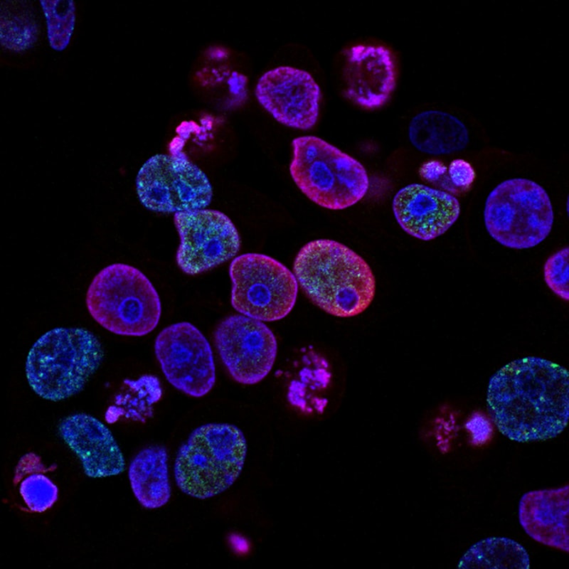
The physical realization of cell proliferation is managed by the highly conserved mechanism known as the `cell cycle`, a precise sequence of events that results in cell division. The cell cycle is traditionally divided into two major phases: Interphase, which consists of G1 (Gap 1), S (Synthesis), and G2 (Gap 2); and the M phase (Mitosis). Interphase is the preparatory period where the cell grows, duplicates its organelles, and, most critically, replicates its DNA. The duration and fidelity of Interphase dictate the success of the resulting division and the health of the daughter cells, making it a critical regulatory hub for proliferation.
During the G1 phase, the cell monitors its internal and external environment, ensuring conditions are suitable for division and accumulating the necessary enzymes and building blocks. The transition into the S phase marks the commitment to proliferation, as this is when DNA synthesis occurs, leading to the duplication of the entire genome. This process must be flawless, as errors introduced during the `S phase` can lead to mutations, chromosomal instability, or cell death. Following S phase, the G2 phase provides a final safety gap where the cell synthesizes proteins required for mitosis and checks the integrity of the newly synthesized DNA before proceeding to division.
The M phase, or mitotic phase, represents the actual physical separation of the duplicated cellular components. Mitosis itself is subdivided into prophase, metaphase, anaphase, and telophase, culminating in cytokinesis, the division of the cytoplasm. During prophase, the condensed chromosomes become visible; in metaphase, they align centrally along the metaphase plate; during anaphase, sister chromatids separate and move to opposite poles; and finally, in telophase and cytokinesis, the two new nuclei form, and the cell physically splits into two distinct daughter cells. This entire orchestrated sequence ensures that each daughter cell receives a complete and identical set of chromosomes, thereby completing the proliferative event.
Differentiation: The Path from Proliferation to Specialization
While proliferation generates the necessary quantity of cells, differentiation provides the crucial quality, converting newly born cells into functional components of complex tissues. Cellular differentiation is the process by which a less specialized cell, often a proliferative stem cell or progenitor cell, becomes a more specialized cell type, such as a cardiomyocyte, hepatocyte, or osteoblast. This process is essential because mere multiplication without specialization would result in a disorganized mass of identical cells, incapable of sustaining complex physiological functions. Proliferation and differentiation are often viewed as opposing forces, but in developing and regenerating tissues, they are tightly coupled and temporally regulated.
The initial stages of development rely heavily on pluripotent stem cells, which possess both immense proliferative capacity and the potential to differentiate into any cell type. As development progresses, these cells produce multipotent or unipotent `progenitor cells`. Progenitor cells are still highly proliferative but are lineage-restricted, meaning their differentiation potential is limited to specific tissue types. For instance, a myeloid progenitor cell can proliferate rapidly but can only differentiate into various blood cells (macrophages, neutrophils, etc.), not into neural cells or muscle cells. This controlled restriction ensures the organized formation of distinct organs and systems.
The signals that drive a cell to cease rapid proliferation and commit to differentiation are complex, involving changes in gene expression dictated by transcription factors, external microenvironmental cues, and cell-to-cell contact signals. Once a cell commits fully to terminal differentiation, it often loses its proliferative capacity, entering a permanent or semi-permanent G0 state. Classic examples include highly specialized cells like mature skeletal muscle fibers, which permanently exit the cell cycle. The balance between maintaining a pool of highly proliferative stem cells (for replenishment) and producing terminally differentiated, functional cells (for performance) is a critical axis of tissue `homeostasis`.
Regulation of Proliferation: Checkpoints and Signaling Pathways
The high stakes associated with cell proliferation necessitate incredibly stringent regulatory mechanisms. Uncontrolled proliferation is synonymous with disease, while insufficient proliferation leads to developmental defects or poor healing. Regulation occurs primarily through intracellular checkpoints and external signaling pathways mediated by growth factors. The cell cycle checkpoints function as molecular surveillance systems, pausing the cycle if necessary until crucial events, such as DNA repair or chromosome alignment, are successfully completed. The most critical checkpoint is the G1 checkpoint (or Restriction point), where the cell decides whether to commit to division or enter G0.
Internal regulation relies heavily on the interaction between cyclins and `Cyclin-Dependent Kinases (Cdks)`. Cyclins are regulatory proteins whose concentration fluctuates throughout the cell cycle, binding to and activating Cdks. These Cdk-cyclin complexes phosphorylate target proteins that drive the cell from one phase to the next. For instance, the activation of Cyclin E/Cdk2 is necessary for entry into S phase. If DNA damage is detected, regulatory proteins like p53 can trigger the production of Cdk inhibitors (CKIs), halting the cycle at the G1 checkpoint, allowing time for repair, or initiating apoptosis if the damage is irreparable.
External control is exerted by a vast array of signaling molecules, primarily polypeptide `growth factors`. These factors (such as Epidermal Growth Factor or Platelet-Derived Growth Factor) bind to specific receptors on the cell surface, initiating intracellular signaling cascades (like the Ras-MAPK pathway) that ultimately lead to the activation of transcription factors necessary for initiating proliferation. Conversely, inhibitory signals, such as transforming growth factor beta (TGF-β), actively suppress proliferation, ensuring that cells only divide when and where they are required. The interplay between these promoting and inhibiting signals dictates the precise rate of proliferation within any given tissue environment.
Role in Development, Growth, and Homeostasis
In the context of development, cell proliferation is the driving force behind embryogenesis and organogenesis. Starting from a single fertilized egg, massive waves of rapid, yet tightly coordinated, cell division produce the billions of cells required to form a complete organism. During the embryonic phase, proliferation rates are maximal, and the resulting cells are guided by complex morphogenic gradients and transcription factors to form the primary germ layers and ultimately, all differentiated tissues. Errors in the timing or location of proliferation during this phase can lead to severe congenital defects, highlighting the exquisite sensitivity of developmental programming.
Postnatally, proliferation continues to be essential for somatic growth, particularly during childhood and adolescence. Organs and tissues increase in size through a combination of increased cell number (hyperplasia, driven by proliferation) and increased cell size (hypertrophy). Once adult size is reached, proliferation transitions from a growth mechanism to a maintenance mechanism, focusing on tissue homeostasis. Homeostatic proliferation involves replacing cells lost through normal wear and tear, senescence, or programmed cell death (`apoptosis`).
Tissues with high turnover rates, such as the blood system (hematopoiesis), the skin (epidermis), and the gut lining, rely on constant, regulated proliferation of specific stem cell niches to maintain functional capacity. For example, the bone marrow produces billions of new blood cells daily through continuous proliferation and differentiation of hematopoietic stem cells. If this proliferative capacity were compromised (e.g., due to chemotherapy or radiation), the organism would quickly succumb to deficiencies in oxygen transport, immunity, and clotting, underscoring the vital, ongoing nature of homeostatic cell multiplication.
Cell Proliferation in Tissue Repair and Regeneration
One of the most immediate and critical roles of cell proliferation in the adult organism is its contribution to tissue repair following injury. When tissue integrity is compromised, the body initiates a complex wound healing cascade that relies heavily on controlled, localized cell division to restore the structural barrier and functional capacity. The repair process typically moves through three phases: inflammation, proliferation, and remodeling. The proliferative phase specifically involves the rapid mobilization and multiplication of several cell types at the injury site.
Key cell populations involved in reparative proliferation include fibroblasts, endothelial cells, and epithelial cells. `Fibroblasts` proliferate rapidly to synthesize new extracellular matrix components, forming granulation tissue that acts as a temporary scaffold. Simultaneously, endothelial cells proliferate to form new blood vessels (angiogenesis), which are crucial for supplying oxygen and nutrients to the rapidly growing tissue mass. Finally, epithelial cells at the wound margins proliferate and migrate to cover the denuded surface, restoring the protective epidermal barrier. This localized, temporary surge in proliferation is highly regulated by signals released by platelets and immune cells present in the wound bed.
In instances of true regeneration, such as liver partial hepatectomy or certain tissue damage in lower vertebrates, the proliferative response is even more robust, aiming not just for repair (scar formation) but for the complete restoration of the original tissue architecture. For example, the mammalian liver exhibits remarkable regenerative capacity, responding to tissue loss by re-entering the cell cycle en masse. Hepatocytes, normally quiescent, are stimulated by specific growth factors to proliferate until the original organ mass is restored, demonstrating the latent proliferative potential retained by many terminally differentiated cells when the appropriate signals are presented.
Dysregulation: Proliferation and Pathological States
While essential for life, defects in the control mechanisms governing cell proliferation are the molecular basis of numerous pathological states, most notably `cancer`. Cancer is fundamentally characterized by uncontrolled, excessive cell proliferation that disregards normal inhibitory signals and checkpoints. This dysregulation results from the accumulation of genetic mutations or epigenetic changes that affect key regulatory genes, tipping the delicate balance away from stasis and toward relentless multiplication.
The mutations typically target two classes of genes: proto-oncogenes and tumor suppressor genes. Proto-oncogenes, when mutated, become hyperactive oncogenes that constitutively promote cell division, essentially putting the foot down on the cell cycle accelerator. Conversely, tumor suppressor genes, such such as `p53` or Rb (retinoblastoma protein), normally function to inhibit inappropriate proliferation or trigger apoptosis. When these genes are inactivated or lost, the brakes on the cell cycle are removed, allowing damaged cells to proliferate unchecked and bypass crucial checkpoints, often leading to genomic instability.
The clinical management of proliferative diseases, particularly cancer, often focuses on therapeutically targeting the proliferative machinery. Many chemotherapeutic agents function as antimetabolites or DNA-damaging agents designed to interfere with the S phase or M phase of the cell cycle, preferentially killing rapidly dividing cells. Modern targeted therapies, however, focus on inhibiting specific components of the signaling pathways (like tyrosine kinases) that drive uncontrolled proliferation. Understanding the molecular intricacies of cell proliferation—from the internal Cdk controls to the external growth factor signaling—remains central to both developmental biology and the ongoing search for cures for proliferative disorders.