Neuroanatomy: How Brain Structures Shape Human Behavior
- Introduction
- Corpus Cavernosum Urethrae: Core Definition
- Corpus Cavernosum Urethrae: Historical Context
- Corpus Cavernosum Urethrae: Practical Example
- Corpus Cavernosum Urethrae: Significance and Impact
- Corpus Cavernosum Urethrae: Connections and Relations
- Corpus Striatum: Core Definition
- Corpus Striatum: Historical Context
- Corpus Striatum: Practical Example
- Corpus Striatum: Significance and Impact
- Corpus Striatum: Connections and Relations
- Interrelation and Clinical Relevance
- Conclusion
Introduction
The human body is an intricate network of specialized structures, each performing vital roles in maintaining physiological function and supporting complex behaviors. Among these, the corpus cavernosum urethrae and the corpus striatum represent two distinct anatomical entities located in vastly different regions, yet both are fundamental to specific bodily processes. The corpus cavernosum urethrae, an integral part of the male urinary and reproductive systems, plays a crucial role in maintaining urethral patency and supporting pelvic floor integrity. Conversely, the corpus striatum, a pivotal subcortical structure within the brain, is central to motor control, learning, and various cognitive functions. This encyclopedia entry will delve into the core definitions, historical contexts, practical implications, and broader significance of both structures, ultimately exploring their distinct contributions to human physiology and their clinical relevance, particularly where the original text suggests a potential (though indirect for the striatum) overlap in relation to bladder and pelvic floor muscle control.
While the initial pairing of these two structures might seem unusual given their disparate locations – one in the pelvis and the other in the brain – their combined consideration highlights the interconnectedness of bodily systems. Understanding the detailed anatomy and physiology of each, from the micro-level cellular composition to their macro-level functional contributions, is essential for appreciating their importance in both healthy functioning and various pathological conditions. This comprehensive overview aims to bridge complex scientific information with accessible explanations, providing a foundational understanding for a general audience interested in human biology and its psychological implications.
Corpus Cavernosum Urethrae: Core Definition
The corpus cavernosum urethrae, also widely known as the corpus spongiosum penis in males, is a singular, cylindrical mass of erectile tissue that surrounds the urethra within the penis. Its primary function is to prevent the compression of the urethra during erection, thereby ensuring the patency of the urethral lumen for the passage of semen and urine. Unlike the paired corpora cavernosa, which are primarily responsible for the rigidity of the penis during erection, the corpus cavernosum urethrae remains relatively less rigid, preventing the urethra from collapsing under pressure and allowing fluid expulsion.
This specialized tissue is characterized by a dense network of vascular spaces, or sinusoids, lined by endothelium and surrounded by smooth muscle and connective tissue, collectively known as the tunica albuginea. During sexual arousal, parasympathetic nerve impulses trigger the release of neurotransmitters, leading to the relaxation of the smooth muscle within the sinusoidal spaces. This relaxation allows for a rapid influx of blood, causing the tissue to engorge and expand. While it contributes to the overall volume of the erect penis, its distinct physiological role lies in safeguarding urethral function, making it a critical component of both urinary and reproductive health.
Corpus Cavernosum Urethrae: Historical Context
The anatomical understanding of the corpus cavernosum urethrae, alongside other structures of the male reproductive and urinary systems, emerged gradually through centuries of anatomical dissection and observation. Early anatomists, such as those from ancient Greece, possessed a rudimentary knowledge of human internal structures, though often limited by cultural taboos and technological constraints. It was during the Renaissance, with figures like Andreas Vesalius in the 16th century, that systematic human dissection became more widespread and detailed anatomical descriptions began to be compiled. Vesalius’s “De humani corporis fabrica” (On the Fabric of the Human Body) provided groundbreaking illustrations and text that meticulously detailed structures previously unknown or misunderstood.
Further refinements in anatomical knowledge continued through the subsequent centuries, with numerous physicians and anatomists contributing to a more precise understanding of the pelvic region. The specific nomenclature and functional roles of structures like the corpus cavernosum urethrae were elucidated as microscopy advanced and physiological experiments became possible. The appreciation of its unique role in maintaining urethral patency during erection, distinct from the primary erectile function of the corpora cavernosa, represents a culmination of these detailed anatomical investigations and physiological deductions over many generations of scientific inquiry.
Corpus Cavernosum Urethrae: Practical Example
A practical example illustrating the function of the corpus cavernosum urethrae can be observed during the process of urination or ejaculation in males. Consider a scenario where an individual needs to urinate. The bladder contracts, and the urethral sphincters relax, allowing urine to flow out through the urethra. During this process, the corpus cavernosum urethrae ensures that the urethra remains open and uncompressed, even as surrounding muscles might exert pressure. This is a passive role in urination, maintaining a clear conduit.
More dynamically, during sexual arousal and erection, the penis undergoes significant engorgement. Without the specialized structure of the corpus cavernosum urethrae, the immense pressure generated by the engorgement of the paired corpora cavernosa could potentially compress the urethra, thereby impeding the flow of semen during ejaculation. The inherent design of the corpus cavernosum urethrae, which swells but does not become as rigid as the corpora cavernosa, acts as a protective sheath around the urethra. This “how-to” of its function demonstrates a critical physiological safeguard, preventing mechanical obstruction and ensuring that reproductive fluid can be expelled effectively, highlighting its indispensable contribution to normal sexual and urinary function.
Corpus Cavernosum Urethrae: Significance and Impact
The significance of the corpus cavernosum urethrae extends deeply into the fields of urology and andrology, underscoring its importance for both male urinary and reproductive health. Its anatomical integrity and physiological function are paramount for normal micturition and successful reproduction. When this structure is compromised, either through injury, disease, or congenital abnormalities, it can lead to significant clinical issues. For instance, damage to the corpus cavernosum urethrae can result in urethral strictures, making urination difficult and painful, or even leading to urinary retention.
Furthermore, its role in preventing urethral compression during erection means that dysfunction can indirectly impact erectile dysfunction, though it is more commonly associated with issues directly affecting the corpora cavernosa. In surgical procedures involving the urethra or penis, understanding the precise anatomy and vascularization of the corpus cavernosum urethrae is critical for minimizing complications and preserving function. Its application in medicine, therefore, lies in guiding surgical interventions, diagnosing conditions related to urethral patency, and developing treatments that maintain or restore its crucial role in the genitourinary system.
Corpus Cavernosum Urethrae: Connections and Relations
The corpus cavernosum urethrae is intimately connected with several other structures within the male pelvis, forming a complex functional unit. It is directly related to the urethra, which it completely encircles, providing both structural support and functional protection. It is also anatomically associated with the paired corpora cavernosa, which lie dorsally to it and are primarily responsible for penile rigidity during erection. The intricate interplay between these three erectile bodies facilitates the complex process of penile erection and ejaculation.
More broadly, the corpus cavernosum urethrae is part of the larger pelvic floor musculature and structures, which collectively support pelvic organs and control urinary and fecal continence. Its function is influenced by the innervation of the autonomic nervous system, highlighting its connection to neuroanatomy and neurophysiology. Therefore, this concept belongs to the broader category of human anatomy, specifically within urology and andrology, which are medical subfields focusing on the urinary tract and male reproductive system, respectively. While not directly a psychological concept, its dysfunction can significantly impact an individual’s quality of life and psychological well-being due to issues like incontinence or sexual dysfunction.
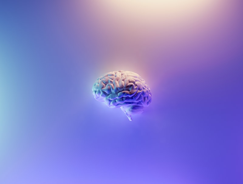
Corpus Striatum: Core Definition
The corpus striatum is a major subcortical component of the basal ganglia system in the brain, playing a critical role in motor control, learning, and reward-motivated behaviors. It derives its name from the Latin “corpus” (body) and “striatum” (striped), referring to its distinctive striped appearance due to the bundles of nerve fibers, primarily myelinated axons, that traverse it. Structurally, the corpus striatum is composed of three primary nuclei: the caudate nucleus, the putamen, and the globus pallidus (though sometimes the globus pallidus is considered a separate but closely related component of the basal ganglia, with the caudate and putamen forming the “neostriatum” or simply “striatum”).
The key idea behind the corpus striatum’s function is its role as the primary input nucleus of the basal ganglia, receiving extensive excitatory glutamatergic projections from the entire cerebral cortex. This vast array of cortical inputs carries information about sensory, motor, and cognitive processes. The striatum then processes this information, integrating it with modulatory input from the substantia nigra (specifically, dopamine) and other brain regions. This intricate processing is fundamental to the initiation and regulation of voluntary movement, the formation of habits, and the evaluation of rewards, making it a cornerstone of behavioral control and learning.
Corpus Striatum: Historical Context
The discovery and understanding of the corpus striatum have evolved over centuries, intertwined with the broader history of neuroanatomy. Early anatomists, using gross dissection techniques, identified distinct masses within the brain, though their functions remained largely speculative. The term “corpus striatum” itself gained prominence as the internal structure, marked by fiber bundles, became clearer. In the 19th century, with advances in microscopy and staining techniques, a more detailed cellular architecture of the brain began to emerge.
The true significance of the corpus striatum in motor control began to be appreciated in the late 19th and early 20th centuries through clinical observations of patients with movement disorders. Physicians noted that damage to specific deep brain nuclei, including the striatum, led to characteristic symptoms such as tremors, rigidity, and involuntary movements. This clinical-pathological correlation formed the foundation for understanding its role in neurological conditions like Parkinson’s disease. Later, in the mid-20th century, neurophysiological experiments and the discovery of neurotransmitters, particularly dopamine, provided a deeper understanding of the striatum’s intricate circuitry and its role in mediating behavior.
Corpus Striatum: Practical Example
A relatable practical example illustrating the function of the corpus striatum is the process of learning a new motor skill, such as riding a bicycle or playing a musical instrument. Initially, these actions require intense conscious effort and concentration, engaging cortical areas directly. However, with repeated practice, the movements become smoother, more automatic, and require less conscious thought – they become habitual. This transition from conscious effort to automaticity is largely mediated by the corpus striatum.
In the “how-to” of learning to ride a bicycle, for instance, the brain continuously receives sensory feedback about balance and movement. The corpus striatum, particularly its input nuclei (caudate and putamen), processes this information and, through its connections within the basal ganglia, refines the motor commands. It learns to associate specific sensory cues with successful movements and, importantly, with the reward of staying upright. Over time, the striatum forms strong neural pathways that execute the complex sequence of balancing, pedaling, and steering without the need for high-level cortical oversight, transforming a challenging task into a fluid, almost unconscious skill. This illustrates its crucial role in procedural learning and habit formation.
Corpus Striatum: Significance and Impact
The significance of the corpus striatum in neuroscience and psychology is profound, particularly concerning its role in motor control, cognition, and motivation. Its dysfunction is implicated in a wide array of neurological and psychiatric disorders, making it a focal point for research and clinical intervention. For example, the degeneration of dopaminergic neurons projecting to the striatum from the substantia nigra is the hallmark pathology of Parkinson’s disease, leading to characteristic motor symptoms like tremor, rigidity, and bradykinesia. Conversely, hyperactivity in certain striatal pathways is associated with hyperkinetic disorders such as Huntington’s disease and Tourette syndrome.
Beyond motor disorders, the corpus striatum is also critical in understanding addiction, reward processing, and decision-making, which are central themes in cognitive psychology and behavioral neuroscience. Its application in modern medicine extends from the diagnosis and treatment of movement disorders through pharmacological interventions and deep brain stimulation, to informing therapeutic strategies for addiction and impulse control disorders. Understanding the striatum’s intricate circuitry provides invaluable insights into the neurological basis of complex behaviors and mental health conditions.
Corpus Striatum: Connections and Relations
The corpus striatum is a highly interconnected structure, forming the core of the basal ganglia, a group of subcortical nuclei vital for motor control, procedural learning, and executive functions. It receives massive input from virtually all areas of the cerebral cortex and from the substantia nigra (dopaminergic projections), and in turn, projects to other basal ganglia nuclei, primarily the globus pallidus and substantia nigra pars reticulata. These connections form complex feedback loops that regulate cortical activity.
Related concepts include dopamine, a key neurotransmitter whose imbalance in the striatum is central to Parkinson’s disease and addiction; motor learning, which heavily relies on striatal plasticity; and reward pathways, where the striatum plays a crucial role in processing pleasurable stimuli. The corpus striatum belongs to the broader category of neuroscience, specifically within neuroanatomy, neurophysiology, and its functional aspects are deeply explored in cognitive psychology, behavioral neuroscience, and psychopharmacology, as its activity underlies learning, motivation, and the development of habitual behaviors.
Interrelation and Clinical Relevance
While the corpus cavernosum urethrae and the corpus striatum are anatomically distinct and primarily serve different physiological systems, the original context of their joint mention suggests an intriguing, albeit mostly indirect, connection through their roles in the control of bladder and pelvic floor muscles, and their vulnerability to neurological disorders. The corpus cavernosum urethrae directly supports the urethra and contributes to pelvic floor integrity. Its function is crucial for normal urinary continence and sexual function, which can be directly impacted by localized damage or dysfunction.
The corpus striatum, by contrast, is a central brain structure involved in complex motor planning and execution. Its influence on bladder and pelvic floor muscle control is not direct but occurs through its modulation of descending motor pathways that ultimately innervate these muscles. Conditions like Parkinson’s disease, multiple sclerosis, and stroke, which primarily affect the central nervous system, can disrupt the striatum’s function. This disruption can then lead to secondary effects on urinary function, resulting in symptoms such as urinary incontinence or pelvic floor dysfunction, illustrating how neurological disorders impacting brain structures like the striatum can have far-reaching consequences for peripheral bodily functions.
Therefore, while the corpus cavernosum urethrae has a direct, local role in pelvic function, the corpus striatum’s influence is mediated through the complex neural circuitry of the brain and spinal cord. Abnormalities in either structure, or the pathways connecting them, can lead to a variety of debilitating conditions. Understanding these distinct yet sometimes functionally convergent roles is paramount for accurate diagnosis and effective management of patients presenting with symptoms affecting either motor control or urogenital function, especially in the context of broader neurological illnesses.
Conclusion
In summary, the corpus cavernosum urethrae and the corpus striatum represent two fundamentally important, albeit anatomically disparate, structures within the human body. The corpus cavernosum urethrae, located in the male pelvis, is an essential component of the urinary and reproductive systems, ensuring urethral patency and contributing to the integrity of the pelvic floor. Its primary role is to facilitate the unimpeded flow of urine and semen, a function critical for daily physiological needs and reproductive success.
Conversely, the corpus striatum, a key subcortical structure of the brain’s basal ganglia, is central to motor control, habit formation, and reward-motivated behaviors. Its complex neural circuitry integrates cortical inputs and dopaminergic modulation to orchestrate voluntary movements and facilitate learning. While one operates at the periphery, maintaining local structural and functional integrity, the other directs sophisticated central nervous system processes. Both structures, however, are susceptible to various pathologies, with their dysfunction leading to significant clinical challenges, ranging from urogenital issues to severe neurological movement disorders, underscoring their vital and distinct contributions to overall human health and psychological well-being.