Cranial Anomalies: Understanding Deviations in Head Shape
- Understanding Cranial Anomalies: A Core Definition
- Historical Perspectives and Early Recognition
- Prevalence and Demographic Insights: The Epidemiology of Cranial Anomalies
- Unraveling the Causes: Etiology of Cranial Anomalies
- Diagnostic Approaches and Modern Techniques
- Illustrating the Impact: A Practical Example
- Comprehensive Management Strategies and Interventions
- Profound Significance and Broad Impact in Medicine
- Interdisciplinary Connections and Related Concepts
Understanding Cranial Anomalies: A Core Definition
A cranial anomaly is a medical term that refers to any structural abnormality of the skull or head, deviating from typical anatomical development. These conditions encompass a broad spectrum of deformities, ranging from subtle variations in shape that may have primarily cosmetic implications to severe malformations that profoundly impact neurological function and overall health. Such anomalies can either be present at birth, termed congenital anomalies, or develop later in life due to various factors. The core mechanism behind these conditions often involves disruptions in the intricate processes of embryonic and fetal development, particularly concerning the formation and growth of the craniofacial skeleton and its relationship with the developing brain.
The fundamental principle underlying cranial anomalies is a deviation from the precisely orchestrated sequence of events that govern craniofacial development. During early embryogenesis, specialized cells known as neural crest cells migrate and differentiate to form the bones and cartilages of the face and skull. Simultaneously, the developing brain undergoes rapid growth and differentiation, requiring the skull to expand in a coordinated manner. Any interference with these genetic programs or external influences during critical developmental windows can lead to abnormal skull shape, size, or structure, which in turn can impede normal brain development or cause complications due to restricted intracranial space.
The severity and clinical presentation of cranial anomalies are highly variable. Some individuals may present with only minor skull asymmetries that require no intervention, while others may suffer from life-threatening conditions such as anencephaly, where a significant portion of the brain and skull fails to develop. The functional consequences can include developmental delays, neurological deficits, sensory impairments, and breathing difficulties, underscoring the critical protective and supportive roles of the skull for the central nervous system and sensory organs. Understanding these variations is paramount for accurate diagnosis, prognosis, and the formulation of appropriate management strategies.
Historical Perspectives and Early Recognition
The recognition of cranial anomalies, particularly those visible at birth, dates back to ancient civilizations. Historical records and archaeological findings suggest that various cultures observed and sometimes even depicted individuals with distinct head shapes. However, the understanding of these conditions was often rooted in superstition or rudimentary observations, with explanations frequently involving divine intervention, curses, or environmental phenomena that were poorly understood. Early medical texts from civilizations such as ancient Egypt, Greece, and Rome contain descriptions of birth defects, though systematic classification and scientific inquiry into their etiology were largely absent.
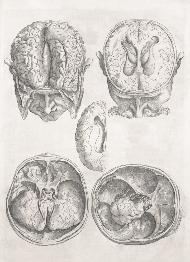
The scientific study of human anatomy and embryology, which began to flourish during the Renaissance and intensified in the 19th century, laid the groundwork for a more structured understanding of congenital malformations. Anatomists like Andreas Vesalius provided detailed illustrations of the human skeleton, including the skull, while early embryologists began to observe and document developmental processes. The late 19th and early 20th centuries saw the emergence of pathology as a distinct field, allowing for post-mortem examinations that linked macroscopic abnormalities to underlying structural changes. This period marked a shift from mere observation to attempts at categorizing and hypothesizing about the origins of such conditions.
Significant advancements in the understanding and diagnosis of cranial anomalies truly began in the mid-20th century with the advent of advanced imaging technologies. Before techniques like X-rays, and later, Computed Tomography (CT) and Magnetic Resonance Imaging (MRI), internal skull and brain structures could not be visualized non-invasively. These innovations revolutionized the ability of clinicians to precisely identify the nature and extent of cranial malformations, enabling more accurate diagnoses, better prognostication, and more effective surgical planning. This technological leap transformed the field from descriptive pathology to a dynamic area of medical intervention and genetic research.
Prevalence and Demographic Insights: The Epidemiology of Cranial Anomalies
The epidemiology of cranial anomalies reveals them to be relatively rare occurrences within the general population. Current estimates suggest an incidence rate of approximately 0.5 to 1.0 per 1,000 live births in developed countries. This seemingly low figure belies the profound impact these conditions have on affected individuals and their families, often requiring extensive medical intervention and long-term care. The precise prevalence can vary slightly based on geographical region, ethnic background, and the comprehensiveness of diagnostic screening programs, highlighting the importance of standardized reporting for accurate epidemiological data.
A critical epidemiological insight is that approximately 70% of cranial anomalies do not occur in isolation but are found in the context of multiple congenital anomalies. This observation suggests that many skull malformations are not isolated developmental errors but rather manifestations of broader systemic developmental issues or complex genetic syndromes affecting various organ systems. When a cranial anomaly is part of a larger pattern of birth defects, it often necessitates a comprehensive evaluation to identify all associated conditions and to understand the underlying genetic or environmental cause, which is crucial for multidisciplinary management and genetic counseling.
Furthermore, epidemiological studies have identified a noticeable sex disparity in the incidence of cranial anomalies, with males being more frequently affected than females. The male-to-female ratio is approximately 2:1, indicating that male infants are twice as likely to be diagnosed with these conditions. The exact reasons for this disparity are not fully understood but may involve complex interactions between sex-linked genetic factors, differential susceptibility to environmental teratogens, or hormonal influences during critical developmental stages. Further research is ongoing to elucidate the biological mechanisms behind this observed sex difference, which could provide valuable insights into the etiology of these conditions.
Unraveling the Causes: Etiology of Cranial Anomalies
The etiology of cranial anomalies is multifaceted, stemming from a complex interplay of genetic and environmental factors. Genetic defects represent the most common cause, accounting for approximately 70% of all reported cases. These genetic alterations can be inherited from parents, following Mendelian patterns of inheritance such as autosomal dominant or recessive traits, or they can arise as spontaneous, *de novo* mutations in the developing embryo. Such genetic anomalies can disrupt critical pathways involved in bone formation, cell proliferation, or tissue patterning, leading to specific syndromes like Apert syndrome, Crouzon syndrome, or Pfeiffer syndrome, each characterized by distinct craniofacial malformations.
Beyond genetic predispositions, a significant proportion of cranial anomalies can be attributed to environmental factors, particularly exposures during the prenatal period. These environmental influences, often referred to as teratogens, can directly interfere with fetal development. Notable examples include prenatal alcohol exposure, which can lead to Fetal Alcohol Spectrum Disorders characterized by distinctive facial features and brain abnormalities, and certain maternal drug uses, such as anticonvulsants or retinoids, which are known to have teratogenic effects. Other environmental contributors might include exposure to certain chemicals, radiation, or extreme maternal nutrient deficiencies during critical stages of gestation.
Maternal infections during pregnancy also represent a significant etiological factor for cranial anomalies. Specific pathogens have been definitively linked to developmental disruptions that can manifest in the skull and brain. Infections such as rubella (German measles), cytomegalovirus (CMV), and toxoplasmosis are particularly concerning. These infectious agents can cross the placental barrier and directly infect the developing fetus, causing inflammation, cell damage, and interference with neural migration and craniofacial bone development. The resulting damage can range from subtle microcephaly to severe structural defects, depending on the timing of the infection during gestation and the virulence of the pathogen.
Diagnostic Approaches and Modern Techniques
The diagnosis of cranial anomalies typically begins with prenatal screening, primarily through routine prenatal ultrasound examinations. Advanced ultrasound technology allows for detailed visualization of fetal anatomy, enabling the detection of abnormalities in skull shape, size, and the presence of associated brain malformations as early as the second trimester. Sonographers and obstetricians look for indicators such as an unusual head circumference, abnormal cranial contours, or signs of hydrocephalus, which can prompt further investigation and specialized fetal imaging. Early detection through these methods is crucial for enabling timely counseling for expectant parents and planning for postnatal management.
Upon birth, a thorough physical examination is paramount. Pediatricians and neonatologists meticulously assess the infant’s head for any asymmetry, unusual fontanelle size or tension, palpable ridges indicating premature suture fusion, or distinctive facial features that might suggest a syndromic condition. If a cranial anomaly is suspected or confirmed, advanced imaging techniques become indispensable. Computed Tomography (CT) scans provide exquisitely detailed images of bone structures, invaluable for assessing skull morphology, suture patency, and bone defects. Magnetic Resonance Imaging (MRI), on the other hand, excels at visualizing soft tissues, offering superior detail of the brain, its development, and any associated intracranial abnormalities, such as malformations of cortical development or hydrocephalus.
In cases where the etiology remains unclear or a genetic predisposition is suspected, genetic testing plays a pivotal role in confirming the diagnosis and guiding management. Techniques such as karyotyping, chromosomal microarray analysis, and whole exome or genome sequencing can identify specific genetic mutations or chromosomal aberrations responsible for the cranial anomaly. This genetic information is not only critical for understanding the prognosis and potential associated conditions but also provides crucial data for genetic counseling, allowing families to understand recurrence risks and make informed reproductive decisions. The integration of clinical examination, advanced imaging, and genetic analysis ensures a comprehensive diagnostic approach.
Illustrating the Impact: A Practical Example
To illustrate the practical implications of a cranial anomaly, let’s consider a common condition known as craniosynostosis. This condition involves the premature fusion of one or more of the fibrous sutures that connect the bones of the skull. Normally, these sutures remain open during infancy and childhood, allowing the brain to grow and the skull to expand in a flexible manner. When one or more sutures fuse prematurely, the skull’s growth is restricted in the direction perpendicular to the fused suture, leading to compensatory growth in other directions, resulting in an abnormally shaped head.
For example, if the sagittal suture, which runs along the top of the head from front to back, fuses prematurely, the skull cannot grow laterally. Instead, it grows longer and narrower, a condition known as scaphocephaly. This physical manifestation is not merely a cosmetic concern; it demonstrates the fundamental principle that the skull’s ability to grow is intrinsically linked to the brain’s developmental needs. The “how-to” of this principle is evident: the prematurely fused suture acts as a rigid barrier, preventing the natural expansion that accommodates the rapidly growing brain. The brain, seeking space, will push the remaining unfused sutures to expand, causing the characteristic elongated head shape.
The practical impact extends beyond the visible head shape. In severe or untreated cases of craniosynostosis, the restricted cranial volume can lead to increased intracranial pressure, which can have serious consequences for brain development. This pressure can manifest as headaches, developmental delays, visual impairment, or even cognitive deficits. Thus, the timely diagnosis and intervention, often involving surgical correction, are crucial to relieve pressure on the brain, allow for normal growth, and improve the child’s developmental trajectory and quality of life. This example vividly demonstrates how a structural anomaly of the skull directly impacts neurological function and necessitates a proactive medical approach.
Comprehensive Management Strategies and Interventions
The management of cranial anomalies is highly individualized, depending largely on the specific type, severity, and associated symptoms of the condition. In cases where the anomaly is mild, such as positional plagiocephaly (a flattened head shape due to external pressure, not suture fusion), and does not affect brain function or development, a conservative approach involving observation and repositioning techniques may suffice. Close monitoring by a pediatric specialist ensures that the condition does not worsen and that developmental milestones are met. For some mild cosmetic concerns, especially those related to external molding, no active treatment beyond watchful waiting might be necessary.
For more severe or functionally significant anomalies, surgical correction often becomes the primary mode of intervention. The goals of surgery are multifaceted: to improve the aesthetic appearance of the skull, to relieve pressure on the brain (especially in cases of craniosynostosis or hydrocephalus), and to create adequate space for future brain growth. Surgical procedures can range from minimally invasive endoscopic techniques for certain types of craniosynostosis to extensive cranial vault remodeling, which involves reshaping and repositioning multiple skull bones. These complex operations are typically performed by multidisciplinary teams comprising neurosurgeons, craniofacial surgeons, and other specialists, emphasizing a collaborative approach to optimize outcomes.
Beyond surgical interventions, other therapeutic modalities play a vital role in comprehensive management. The use of corrective helmets or orthotics is commonly recommended for infants with moderate positional plagiocephaly or as an adjunct therapy post-surgery to help mold the skull into a more typical shape. These custom-fitted helmets apply gentle, consistent pressure to redirect skull growth over several months. Furthermore, long-term follow-up with developmental pediatricians, physical therapists, occupational therapists, and speech therapists is often crucial, particularly when cranial anomalies are associated with developmental delays or neurological deficits. This holistic approach addresses not only the structural issue but also the functional development of the child.
Finally, genetic counseling is an indispensable component of managing cranial anomalies, especially when a genetic etiology is identified or suspected. Genetic counselors provide families with detailed information about the specific condition, its inheritance patterns, the risk of recurrence in future pregnancies, and available reproductive options. They also offer psychological support and connect families with support groups and resources, empowering them to make informed decisions and cope with the challenges associated with a child’s diagnosis. This comprehensive framework of medical, surgical, and supportive care aims to maximize the child’s health, development, and quality of life.
Profound Significance and Broad Impact in Medicine
The study and management of cranial anomalies hold profound significance across multiple medical disciplines. For pediatric neurology, understanding these conditions is critical for assessing brain development, identifying potential neurological deficits, and managing associated conditions like seizures or hydrocephalus. In neurosurgery and craniofacial surgery, cranial anomalies represent a significant portion of their clinical practice, driving innovations in surgical techniques, imaging, and biomaterials. Furthermore, these conditions provide crucial insights into fundamental processes of human embryology, bone biology, and brain development, making them a focal point for basic and translational research.
The impact of cranial anomaly research extends directly into clinical applications, revolutionizing diagnostic protocols and therapeutic interventions. Advanced prenatal screening technologies have improved early detection, allowing for better preparation and intervention planning. Surgical techniques have become increasingly sophisticated, minimizing invasiveness and improving outcomes, particularly in complex cases requiring extensive reconstruction. Ongoing research continues to explore genetic therapies, tissue engineering, and advanced imaging modalities to further enhance diagnostic accuracy, predict developmental trajectories, and refine treatment strategies, continually pushing the boundaries of what is possible in reconstructive and developmental medicine.
Beyond direct medical care, the understanding of cranial anomalies has broader public health implications. It highlights the importance of maternal health during pregnancy, emphasizing factors like proper nutrition (e.g., folic acid supplementation for neural tube defects, a related category of birth defects), avoidance of teratogenic substances, and timely vaccination against infectious diseases like rubella. Public health campaigns aimed at promoting healthy pregnancies and raising awareness about preventable risk factors contribute to reducing the incidence of certain congenital anomalies, underscoring the interconnectedness of medical knowledge, clinical practice, and community well-being.
Interdisciplinary Connections and Related Concepts
Cranial anomalies are deeply interconnected with a wide array of other psychological and medical concepts, underscoring the interdisciplinary nature of modern healthcare. Related concepts include specific conditions such as craniosynostosis, which is a common type of cranial anomaly involving premature suture fusion. Other related conditions include hydrocephalus, an accumulation of cerebrospinal fluid that can cause macrocephaly (an abnormally large head) and exert pressure on the brain, and microcephaly, where the head is abnormally small, often indicative of impaired brain development. Conditions like anencephaly and spina bifida fall under the broader category of neural tube defects, which are also developmental anomalies affecting the central nervous system and its protective bony structures.
These conditions primarily fall within the broader categories of Developmental Biology, Pediatric Neurology, Medical Genetics, and Craniofacial Surgery. Developmental biology provides the foundational understanding of how these anomalies arise during embryogenesis. Pediatric neurology focuses on the neurological consequences and developmental outcomes. Medical genetics plays a crucial role in identifying the genetic underpinnings and providing counseling. Craniofacial surgery and neurosurgery are central to the surgical management and reconstruction of affected structures. The collaborative efforts among these specializations are essential for providing comprehensive care, from diagnosis to long-term follow-up and rehabilitation.
Furthermore, understanding cranial anomalies contributes to broader scientific inquiries into human development, genetic expression, and the intricate interactions between genotype and phenotype. It sheds light on how environmental factors can modulate genetic predispositions, influencing the manifestation and severity of developmental conditions. Research in this area often intersects with fields like epigenetics, maternal-fetal medicine, and developmental psychology, as the physical anomalies can also influence cognitive and behavioral development. This complex web of connections highlights that cranial anomalies are not isolated medical phenomena but integral components of a larger understanding of human health and development.