Cranial Nerves: The Biological Blueprint of Human Behavior
The Core Definition of Cranial Nerves
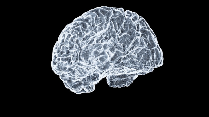
Cranial nerves are a distinct set of twelve pairs of nerves that emerge directly from the brain, primarily the brainstem, rather than from the spinal cord. Unlike spinal nerves which are organized segmentally along the vertebral column, cranial nerves are specialized for specific functions related to the head, neck, and, in one notable case, the thoracic and abdominal cavities. They serve as crucial conduits for both sensory information entering the central nervous system and motor commands exiting it, thereby orchestrating a vast array of vital functions from our most basic senses to complex facial expressions and autonomic regulation. This unique origin and distribution distinguish them as a critical component of the peripheral nervous system, directly interfacing with the central processing unit of the brain.
The fundamental mechanism behind the operation of cranial nerves involves their direct connection to the brain, allowing for rapid and precise communication between the central nervous system and the sensory organs and muscles of the head and neck. Each pair of cranial nerves is assigned a Roman numeral from I to XII, typically based on their anatomical position from front to back of the brain. Some cranial nerves are purely sensory nerves, transmitting information such as smell, sight, or hearing. Others are purely motor nerves, controlling muscle movements like eye movements or facial expressions. A significant number are, in fact, mixed nerves, containing both sensory and motor fibers, enabling them to perform a dual role, such as the trigeminal nerve which handles both facial sensation and chewing. This intricate organization allows for highly specialized control over the sensory input and motor output governing the cephalic region.
The intricate network formed by these nerves is responsible for an astonishing array of functions that define our daily interactions with the world. From the moment we perceive a scent or taste a flavor, to the complex coordination required to speak, swallow, or maintain balance, cranial nerves are actively engaged. They are not merely simple conduits but are integral to the sophisticated processing that occurs within the brain, relaying critical data and executing commands with remarkable efficiency. Understanding their individual roles and collective synergy is paramount to comprehending the overall functionality of the human nervous system and diagnosing neurological conditions affecting these pathways.
Historical Context and Discovery
The study of cranial nerves dates back to ancient times, with early anatomists and physicians making observations about nerves emerging from the head. Galen of Pergamon, a prominent Roman physician and philosopher in the 2nd century AD, made significant contributions to the understanding of anatomy, including distinguishing between sensory and motor nerves. He identified seven pairs of cranial nerves, though his numbering and exact definitions differed from the modern classification. His work, based largely on animal dissection, laid foundational groundwork for centuries, emphasizing the importance of nerves in transmitting sensations and controlling movement, even if the precise origins and functions of all cranial nerves were not yet fully elucidated.
Over the centuries, particularly during the Renaissance and the Enlightenment, detailed anatomical dissections became more common, leading to a refined understanding of the nervous system. Key figures like Thomas Willis in the 17th century, known for his seminal work “Cerebri Anatome” (Anatomy of the Brain), further advanced the classification and description of cranial nerves. Willis, along with Christopher Wren, provided detailed illustrations and descriptions, contributing to the establishment of the modern twelve-pair classification. This period marked a shift from purely observational anatomy to a more systematic and detailed investigation, driven by advancements in dissection techniques and a growing scientific curiosity about the intricate structures of the human body.
The 19th century brought further clarity, as neurologists and physiologists began to correlate specific nerve damage with observable deficits in sensory perception or motor function. Researchers like Charles Bell and François Magendie contributed significantly to differentiating sensory and motor nerve functions, a principle known as the Bell-Magendie law, which also applied to understanding the mixed nature of some cranial nerves. This era cemented the functional understanding of each cranial nerve, moving beyond mere anatomical description to a more dynamic comprehension of their physiological roles in mediating the complex interplay between the brain and the body’s sensory and motor apparatus of the head and neck.
Anatomy and Classification of the Twelve Cranial Nerves
The twelve pairs of cranial nerves are systematically identified by Roman numerals (I-XII) and by descriptive names that often reflect their primary function or anatomical location. Their points of emergence from the brain are primarily from the brainstem, with the exception of the olfactory (I) and optic (II) nerves, which arise from the cerebrum and diencephalon, respectively. These nerves can be broadly categorized based on their primary fiber type: purely sensory, purely motor, or mixed (containing both sensory and motor fibers). This classification is crucial for understanding their diverse roles in the human body.
The purely sensory cranial nerves are responsible for conveying specialized sensory information to the brain. This category includes the Olfactory Nerve (I) for the sense of smell, the Optic Nerve (II) for vision, and the Vestibulocochlear Nerve (VIII) for hearing and balance. These nerves transmit raw sensory data from receptors in specialized organs directly to specific processing centers within the brain, forming the initial steps of our perception of the external world. Damage to any of these nerves can result in significant sensory deficits, highlighting their irreplaceable roles.
The purely motor cranial nerves are dedicated to controlling muscle movements. This group comprises the Oculomotor Nerve (III), Trochlear Nerve (IV), and Abducens Nerve (VI), which collectively govern eye movements. Additionally, the Accessory Nerve (XI) controls specific neck and shoulder muscles, and the Hypoglossal Nerve (XII) is responsible for tongue movements critical for speech and swallowing. Lastly, the mixed cranial nerves, such as the Trigeminal Nerve (V), Facial Nerve (VII), Glossopharyngeal Nerve (IX), and Vagus Nerve (X), combine both sensory and motor functions, enabling them to handle complex tasks like facial sensation and chewing, facial expression and taste, or taste, swallowing, and autonomic regulation of internal organs.
- Olfactory Nerve (I): Primarily responsible for the sense of smell, carrying sensory information from the olfactory epithelium in the nasal cavity to the brain.
- Optic Nerve (II): Responsible for transmitting visual information from the retina of the eye to the brain, forming the basis of sight.
- Oculomotor Nerve (III): Controls most extrinsic eye muscles, enabling various eye movements, and also innervates the iris (pupil constriction) and ciliary body (lens accommodation).
- Trochlear Nerve (IV): Innervates the superior oblique muscle of the eye, responsible for downward and inward eye rotation.
- Trigeminal Nerve (V): A large mixed nerve with three main branches. It conveys sensory information from the face, scalp, teeth, and oral cavity, and provides motor innervation to the muscles of mastication (chewing).
- Abducens Nerve (VI): Controls the lateral rectus muscle, responsible for abducting the eye (moving it laterally away from the midline).
- Facial Nerve (VII): A mixed nerve that controls muscles of facial expression, conveys taste sensation from the anterior two-thirds of the tongue, and innervates lacrimal (tear) and salivary glands.
- Vestibulocochlear Nerve (VIII): Purely sensory, consisting of two parts: the vestibular component for balance and head position, and the cochlear component for hearing.
- Glossopharyngeal Nerve (IX): A mixed nerve involved in taste sensation from the posterior one-third of the tongue, innervating the stylopharyngeus muscle for swallowing, and regulating salivary glands.
- Vagus Nerve (X): The most extensive cranial nerve, with wide-ranging autonomic functions. It regulates heart rate, digestion, respiration, and provides motor innervation to muscles of the larynx and pharynx for speech and swallowing, as well as visceral sensation from the thoracic and abdominal organs.
- Accessory Nerve (XI): Primarily motor, controlling the sternocleidomastoid and trapezius muscles, which are involved in head and shoulder movement.
- Hypoglossal Nerve (XII): Purely motor, controlling the intrinsic and extrinsic muscles of the tongue, crucial for speech articulation and swallowing.
A Practical Example: The Act of Eating an Apple
To illustrate the complex and coordinated functions of cranial nerves in everyday life, consider the seemingly simple act of eating an apple. This routine activity involves a harmonious interplay of multiple cranial nerves, each contributing a specialized role from the initial perception of the apple to its final mastication and swallowing. This example highlights how these nerves work in concert to facilitate a fundamental human behavior.
The process begins even before the apple reaches the mouth. The Olfactory Nerve (I) detects the apple’s sweet aroma, signaling to the brain that food is present. Simultaneously, the Optic Nerve (II) processes the visual information, allowing us to see the apple’s color, shape, and identify it. As the hand reaches for the apple, the Oculomotor (III), Trochlear (IV), and Abducens (VI) nerves coordinate eye movements to track the apple’s position, ensuring accurate grasping. Once the apple is brought closer, the Facial Nerve (VII) might cause a subtle smile of anticipation, and the Glossopharyngeal (IX) and Vagus (X) nerves begin to stimulate salivary glands, preparing the mouth for consumption.
Upon taking a bite, the Trigeminal Nerve (V) becomes highly active, conveying sensory information about the apple’s texture and temperature from the teeth and mouth to the brain. Concurrently, its motor component powers the muscles of mastication, enabling strong chewing. As chewing proceeds, the Facial Nerve (VII) continues to provide taste sensations from the front of the tongue, while the Glossopharyngeal Nerve (IX) handles taste from the back, and both contribute to managing saliva. The Hypoglossal Nerve (XII) meticulously controls the tongue’s movements, manipulating the food bolus within the mouth, pushing it against the teeth for grinding, and ultimately preparing it for swallowing. Finally, the coordinated actions of the Glossopharyngeal (IX), Vagus (X), and some voluntary muscles controlled by the Accessory Nerve (XI) facilitate the complex process of swallowing, moving the food safely down the pharynx and esophagus, completing the cycle of consumption. This intricate dance of neural activity is performed effortlessly, yet it underscores the essential role of cranial nerves in our daily existence.
Significance and Impact in Psychology and Medicine
The comprehensive understanding of cranial nerves is of immense significance across various scientific and medical disciplines, particularly in neurology, psychology, and general medicine. Their direct connection to the brain makes them invaluable indicators of neurological health. Damage or dysfunction in any of these nerves can lead to a wide spectrum of symptoms, ranging from sensory deficits like loss of smell or vision, to motor impairments affecting facial movement, speech, or swallowing. Therefore, the assessment of cranial nerve function is a cornerstone of neurological examination, providing critical diagnostic clues for conditions such as stroke, tumors, multiple sclerosis, or Bell’s palsy.
From a psychological perspective, cranial nerves underpin many fundamental aspects of perception and expression that are central to human experience and social interaction. For instance, the integrity of the olfactory and optic nerves is vital for our sensory experience of the world, influencing mood, memory, and spatial awareness. The facial nerve’s role in facial expressions is critical for non-verbal communication, empathy, and social bonding, making its impairment (e.g., in facial paralysis) profoundly impactful on psychological well-being. Furthermore, the vagus nerve’s extensive autonomic functions link directly to stress response, emotional regulation, and gut-brain axis interactions, increasingly recognized as crucial in psychological disorders like anxiety and depression.
Beyond diagnosis, the knowledge of cranial nerve anatomy and physiology is pivotal for various medical interventions and therapeutic strategies. Neurosurgeons rely on precise anatomical knowledge to avoid nerve damage during intricate brain and skull base surgeries. Therapists, including speech-language pathologists and occupational therapists, design rehabilitation programs targeting specific cranial nerve deficits to restore functions like speech, swallowing, and facial control. Moreover, advancements in understanding cranial nerve pathways have opened avenues for novel treatments, such as vagus nerve stimulation for epilepsy and depression, demonstrating their enduring importance in both clinical practice and cutting-edge research aimed at improving human health and quality of life.
Connections and Relations to Broader Concepts
Cranial nerves are intricately linked to a broader network of psychological and neuroscientific concepts, forming essential bridges between the central nervous system and the external world, as well as critical internal regulatory systems. They represent a specialized component of the peripheral nervous system (PNS), specifically the somatic and autonomic divisions. While spinal nerves primarily serve the trunk and limbs, cranial nerves extend the reach of the brain’s direct control and sensory input to the head, neck, and vital organs, ensuring a comprehensive integration of bodily functions. Their study falls squarely within the subfields of Neuroanatomy, Neurophysiology, and clinical Neurology, contributing to our understanding of the structural and functional organization of the nervous system.
They are fundamentally related to the concepts of Sensory Perception and Motor Control. Each sensory cranial nerve (Olfactory, Optic, Vestibulocochlear) is a dedicated pathway for specific sensory modalities, directly feeding into the brain’s respective cortical areas for processing and interpretation. This direct input is crucial for forming our sensory experience of reality. On the motor side, cranial nerves are effector pathways for movements crucial for survival and interaction, such as chewing, swallowing, speaking, and expressive facial movements. These motor functions are often under the voluntary control of the cerebral cortex, demonstrating the hierarchical organization of motor control within the nervous system.
Furthermore, cranial nerves, particularly the Vagus Nerve (X), play a significant role in the Autonomic Nervous System, influencing involuntary bodily functions. The vagus nerve is a major component of the parasympathetic nervous system, responsible for the “rest and digest” response, regulating heart rate, digestion, and respiration. Its widespread influence connects cranial nerve function to broader physiological and psychological states, including stress regulation, emotional processing, and the gut-brain axis. This highlights that cranial nerves are not isolated entities but are deeply integrated into the overarching functional architecture of the entire nervous system, contributing to both conscious experience and subconscious physiological regulation.