Cranial Anatomy: The Biological Foundation of the Mind
- Definition, Scope, and Etymology
- Detailed Anatomy and Structure of the Cranium
- Functions and Protective Role of the Cranium
- The System of Cranial Nerves
- Clinical Significance and Pathologies
- Diagnostic and Imaging Modalities
- Cranial Surgical Interventions
- Developmental and Lifespan Aspects of Cranial Structure
Definition, Scope, and Etymology
The term cranial functions strictly as an adjective, utilized within medical, anatomical, and psychological contexts to denote anything pertaining to, relating to, or situated within the cranium—the bony structure that encases and protects the brain. This structure is often referred to interchangeably as the braincase or, less precisely in common parlance, the skull, though the latter technically includes the facial skeleton. The conceptual understanding of the cranial space is fundamental to neuroscience, as it defines the immediate physical environment of the central nervous system, influencing everything from fluid dynamics to neuronal protection.
Etymologically, the word traces its roots directly back to the Greek term kranion, meaning “skull,” which was adopted into Latin and subsequently into modern scientific nomenclature. The adjective form is essential for specificity, allowing clinicians and researchers to clearly delineate structures that are housed within the skull (e.g., cranial nerves, cranial arteries) from those that are merely cephalic (relating to the head in general). This distinction is vital when discussing pathologies or procedures, as the integrity of the cranial vault is paramount to survival and neurological function.
A classic example illustrating the function and relevance of the cranial surface involves surgical intervention designed to mitigate pressure, such as the historical procedure of trephination. As noted in clinical texts, “The tool would drill a small hole through the cranial surface to alleviate pressure on the brain,” demonstrating the necessary manipulation of the bony container to address underlying neurological emergencies. Such procedures underscore the rigid, protective nature of the cranium, which, while offering substantial defense against external forces, can become detrimental when internal swelling or hemorrhage occurs, demanding precise knowledge of cranial landmarks and structure.
The scope of the term is broad, encompassing not just the bone itself, but also the meningeal layers, the cerebrospinal fluid (CSF) system, and the complex vascular network that supports brain metabolism. Understanding cranial morphology is crucial for anthropologists studying human evolution, for forensic scientists identifying remains, and most importantly, for neurosurgeons planning complex operations, highlighting the term’s indispensable role across various scientific disciplines.
Detailed Anatomy and Structure of the Cranium
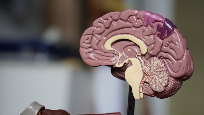
The cranium is not a single, monolithic bone but a composite structure formed by multiple bony plates that articulate through fixed, fibrous joints known as sutures, which provide both rigidity and slight elasticity, particularly important during trauma absorption. Anatomically, the cranium is typically divided into two main sections: the neurocranium, which forms the protective vault around the brain, and the viscerocranium (or facial skeleton), which supports the facial structures and houses the sensory organs. The neurocranium itself consists of eight bones: the unpaired frontal, occipital, sphenoid, and ethmoid bones, and the paired parietal and temporal bones, all intricately interlocking to form a protective shell.
The internal floor of the neurocranium is known as the cranial base, a complex, uneven surface divided into three fossae—the anterior, middle, and posterior cranial fossae—each corresponding to and supporting different lobes of the brain (frontal, temporal/parietal, and cerebellum/brainstem, respectively). These fossae contain numerous foramina and canals, which serve as conduits for the passage of the cranial nerves and major blood vessels, including the carotid arteries and jugular veins, connecting the contents of the skull to the rest of the body. The precise location and orientation of these openings are critically important in clinical settings, as they are common sites for nerve compression or vascular injury.
The sutures, such as the coronal, sagittal, and lambdoid sutures, are particularly noteworthy components of the cranial structure. While they remain flexible in infancy to allow for brain growth, they typically fuse in adulthood, creating a single, robust structure. The mechanical properties of the cranial bones, which are composed of outer and inner tables of compact bone separated by a layer of spongy bone known as the diploë, provide significant shock absorption. This layered architecture is a key element in protecting the delicate neural tissue from external kinetic forces, distributing impact over a wider area.
Furthermore, the inner surface of the cranium is lined by the meninges—the dura mater, arachnoid mater, and pia mater—which further cushion and compartmentalize the brain. The dura mater, the thickest and outermost layer, adheres closely to the inner table of the cranial vault, forming sinuses that drain venous blood from the brain. Understanding this complex three-dimensional relationship between the bone, the meningeal layers, and the underlying brain parenchyma is absolutely essential for diagnosing and treating conditions like epidural or subdural hematomas, which involve bleeding into the potential spaces defined by these layers.
Functions and Protective Role of the Cranium
The primary and most vital function of the cranial structure is the absolute protection of the highly sensitive and irreplaceable neural tissues of the brain, the command center of the entire organism. The rigid, arched vault dissipates kinetic energy from impact, serving as a biomechanical helmet that shields the brain from direct injury. This protective role extends beyond physical trauma; the cranial cavity also serves to maintain a highly controlled, stable internal chemical and pressure environment necessary for optimal neuronal signaling.
A critical function related to this stability is the maintenance of Intracranial Pressure (ICP) within narrow physiological limits, governed by the Monro-Kellie doctrine. This principle dictates that the sum of the volumes of brain tissue, cerebrospinal fluid (CSF), and blood within the non-expandable cranial vault must remain constant. If the volume of one component increases—for example, due to hemorrhage or swelling—the volume of the others, usually CSF or venous blood, must decrease to compensate. Failure of this compensation mechanism leads to elevated ICP, a life-threatening condition that can result in brain herniation and irreversible damage.
Beyond the brain itself, the cranial structure provides crucial bony protection and support for the major sensory organs of the head. The orbits, formed partially by cranial bones, shield the eyes, while the temporal bones house the delicate structures of the middle and inner ear, which are responsible for hearing and balance. This integration of sensory apparatus within the cranial boundaries demonstrates an evolutionary efficiency, concentrating vital components within a secure, robust housing.
The cranial bones also serve as essential attachment points for the muscles involved in mastication (chewing), facial expression, and head movement. While these functions are often attributed primarily to the viscerocranium, the temporal and occipital bones of the neurocranium provide critical anchors for powerful neck muscles that stabilize the head and allow for complex movement. Thus, the integrity of the cranial vault is linked not only to neurological health but also to fundamental motor and communicative functions.
The System of Cranial Nerves
The term cranial is intrinsically linked to the twelve pairs of cranial nerves (CN I through CN XII), which emerge directly from the brain or brainstem, rather than the spinal cord, and exit the cranium through specific foramina in the skull base. These nerves are pivotal components of the peripheral nervous system, responsible for sensory perception, motor control, and autonomic regulation within the head, neck, and torso, linking the central command structure to the external world and internal systems.
The functions of these nerves are diverse and highly specialized. For instance, CN I (Olfactory) and CN II (Optic) are dedicated solely to the special senses of smell and vision, respectively. CN III, IV, and VI (Oculomotor, Trochlear, and Abducens) are primary motor nerves governing eye movement, demonstrating the complexity required for precise visual tracking. Damage to any of these nerves, often due to compression within the cranial space resulting from trauma or tumors, can lead to distinct and debilitating neurological deficits, making their assessment a cornerstone of neurological examination.
Other nerves, such as CN V (Trigeminal), CN VII (Facial), CN IX (Glossopharyngeal), and CN X (Vagus), are mixed nerves, carrying both motor and sensory fibers. The Vagus nerve (CN X), in particular, is unique among the cranial nerves as its influence extends far beyond the head and neck, playing a crucial role in regulating heart rate, digestion, and respiratory function, thereby linking the cranial control center directly to vital visceral processes and autonomic stability throughout the thoracic and abdominal cavities.
Clinical evaluation of the cranial nerves provides invaluable insight into the localized function and health of the brainstem and surrounding cranial structures. A careful assessment of reflexes, sensation, and motor control—such as testing facial symmetry, tongue movement, or pupillary response—allows practitioners to localize lesions within the brainstem or along the nerve pathways. Therefore, the phrase “cranial nerve assessment” is synonymous with a rapid, detailed evaluation of the integrity of the brain’s foundational outputs and inputs.
Clinical Significance and Pathologies
The cranial cavity is the site of numerous significant pathologies, ranging from acute traumatic injuries to chronic neurodegenerative conditions, all of which fall under the scope of cranial medicine. Traumatic Brain Injury (TBI), resulting from blunt force trauma to the cranial vault, represents a major clinical challenge. Fractures of the cranial bones, particularly depressed fractures, can directly compromise the meninges and brain tissue, requiring immediate surgical stabilization to prevent infection and further neurological damage.
Vascular events are another critical category of cranial pathology. Intracranial hemorrhages, classified based on their location relative to the meninges (e.g., epidural, subdural, or subarachnoid hematomas), are often rapidly fatal due to the resulting mass effect and sharp increase in intracranial pressure. Furthermore, conditions such as aneurysms and arteriovenous malformations (AVMs) within the cranial circulation pose a constant risk of rupture, leading to hemorrhagic stroke, demanding specialized diagnostic techniques like cerebral angiography.
Beyond acute trauma, the cranial space is susceptible to various chronic and progressive disorders. Neoplasms, or brain tumors, whether primary (originating from glial cells or meninges) or metastatic (spreading from distant sites), exert pressure on surrounding neural structures, leading to a wide array of symptoms depending on the location, including seizures, cognitive deficits, and focal weakness. Hydrocephalus, the abnormal accumulation of cerebrospinal fluid (CSF) within the cranial ventricles, is another significant condition, often requiring surgical shunting to divert excess fluid and normalize ICP.
Infections, such as meningitis (inflammation of the meninges) or encephalitis (inflammation of the brain parenchyma), represent serious cranial emergencies. While these are often systemic diseases, their effects are localized within the rigid cranial confines, leading to severe inflammation, headache, and altered mental status. The management of all these conditions relies fundamentally on recognizing the spatial limitations imposed by the cranial structure and addressing any compromise to the contained neural tissue quickly and decisively.
Diagnostic and Imaging Modalities
Accurate assessment of the cranial structures and their contents relies heavily on sophisticated neuroimaging techniques, which allow physicians to visualize internal structures without invasive exploration. The initial diagnostic tool in acute trauma is often Computed Tomography (CT) scanning, which is rapid, widely available, and highly effective for identifying bony fractures, acute hemorrhages, and midline shifts indicative of mass effect or herniation within the cranial vault. CT scans provide excellent detail regarding the density and architecture of the cranial bones.
For detailed examination of the soft tissues of the brain, including tumors, ischemic stroke, and white matter diseases, Magnetic Resonance Imaging (MRI) is the superior modality. MRI provides higher spatial resolution and contrast differentiation between gray matter, white matter, and fluid spaces, enabling precise localization of neurological lesions that might not be visible on CT. Specialized MRI techniques, such as Functional MRI (fMRI) and Diffusion Tensor Imaging (DTI), further enhance the ability to map functional areas and structural connectivity within the cranial space.
Vascular assessment within the cranium frequently utilizes angiography, either conventional digital subtraction angiography (DSA) or CT/MR angiography (CTA/MRA). These methods are essential for diagnosing conditions affecting the cerebral vasculature, such as aneurysms, stenosis, or vasculitis, providing critical pre-operative mapping for neurosurgical procedures. The flow dynamics within the cranial arteries and veins are crucial indicators of brain perfusion and oxygenation status.
Furthermore, electrophysiological studies, such as Electroencephalography (EEG), although not imaging modalities in the structural sense, provide essential functional data regarding electrical activity emanating from the cerebral cortex within the cranium. EEG is indispensable for diagnosing seizure disorders and assessing levels of consciousness. The combined use of structural imaging (CT/MRI) and functional studies (EEG) allows for a comprehensive understanding of both the physical state and the operational status of the contents of the cranial cavity.
Cranial Surgical Interventions
Surgical management of cranial pathologies is often highly complex and requires meticulous planning due to the critical nature of the underlying neural structures. Procedures are typically categorized based on their relationship to the cranial bones. A craniotomy involves temporarily removing a section of the cranial bone (a bone flap), performing the necessary intracerebral surgery (e.g., tumor resection, aneurysm clipping), and then securely replacing the bone flap at the conclusion of the procedure. This technique is designed to minimize long-term structural deficits.
In contrast, a craniectomy involves the removal of a section of the skull which is not immediately replaced. This is most commonly performed as a decompressive craniectomy, a life-saving procedure executed when severe brain swelling (edema) is causing dangerously elevated intracranial pressure. By opening the rigid cranial vault, the surgeon creates space for the swelling brain, thereby preventing herniation and potentially irreversible brain damage, though the bone must later be replaced in a subsequent procedure called cranioplasty.
The historical reference to drilling a small hole in the cranial surface pertains to trephination, or burr hole placement, which remains a valid, though often preliminary, surgical technique. Burr holes are used for rapid evacuation of superficial hematomas (like chronic subdural hematomas) or for the placement of monitoring devices, such as intracranial pressure (ICP) monitors or external ventricular drains (EVDs) used to manage hydrocephalus. These small openings provide crucial access with minimal disruption to the cranial structure.
Modern neurosurgery increasingly utilizes minimally invasive techniques, including stereotactic surgery, which employs sophisticated imaging guidance (CT or MRI) to precisely target lesions deep within the cranial space, minimizing the need for large craniotomies. Endoscopic techniques also allow surgeons to navigate the cranial base and ventricular system through small incisions. The constant evolution of these surgical approaches aims to preserve the structural and functional integrity of the cranium while effectively treating critical neurological diseases.
Developmental and Lifespan Aspects of Cranial Structure
The development of the cranium begins early in gestation, and its structure undergoes significant changes throughout the lifespan, starting with the formation of the fetal skull. Unlike the adult cranium, the infant skull is composed of distinct bony plates separated by wide areas of fibrous tissue known as fontanelles, or “soft spots,” and unossified sutures. These fontanelles—the anterior fontanelle being the largest—serve two critical functions: they allow the skull to mold during passage through the birth canal, and they accommodate the rapid, tremendous growth of the brain during the first year of life.
The timing of suture closure is a key developmental milestone. If one or more sutures fuse prematurely, a condition known as craniosynostosis occurs. This premature fusion restricts brain growth perpendicular to the fused suture, leading to abnormal and often severe cranial deformities (plagiocephaly, scaphocephaly, etc.) and potentially elevated intracranial pressure. Correction of craniosynostosis typically requires complex pediatric neurosurgery to reshape the cranial vault and provide adequate space for the growing brain.
As an individual ages, the cranial bones generally undergo a process of increasing ossification and density, and the sutures fully fuse, creating a highly rigid structure. However, in advanced age, bone density may decrease, and the integrity of the bony shell can be compromised by conditions like osteoporosis, potentially increasing vulnerability to fracture following minor trauma. The inherent biomechanical properties of the cranium thus change dynamically throughout the lifespan, impacting both protective capacity and surgical feasibility.
Anthropological studies heavily rely on analysis of the cranial structure, particularly measurements related to cranial capacity, which provides insight into brain size evolution in hominids. Furthermore, the detailed morphology of the cranial bones and sutures can be used in forensic science to estimate age, sex, and ancestry of skeletal remains. Therefore, the study of the cranial structure spans not only medicine but also disciplines focused on human development and population history.