Dendrodendritic Synapses: How Neurons Talk to Each Other
- The Core Definition and Mechanism
- Historical Discovery and Context
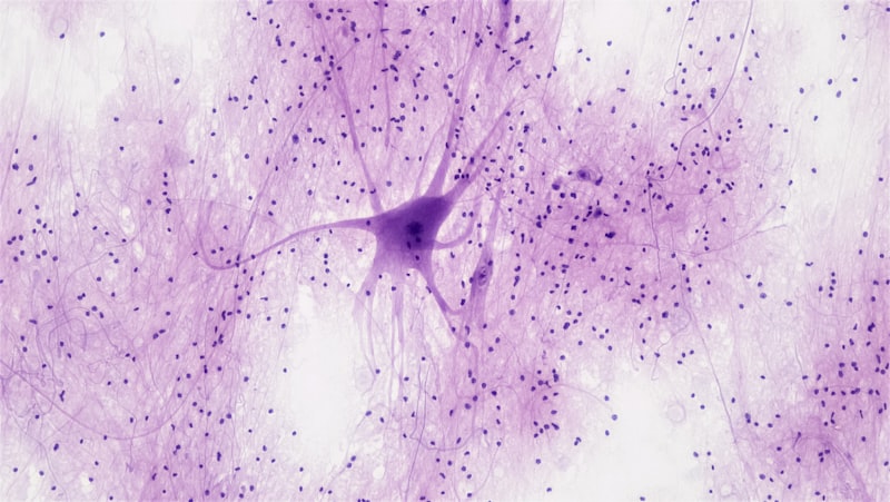
- Anatomy and Distinctive Structure
- Physiology and Neurotransmission
- Functional Roles in Neuronal Processing
- A Practical Example of Modulatory Function
- Significance in Neuroscience and Clinical Impact
- Related Concepts and Broader Classification
The Core Definition and Mechanism
The dendrodendritic synapse represents a highly specialized and unique form of connection within the central nervous system, fundamentally defined by the direct point of contact between the dendrite of one neuron and the dendrite of another neuron. Unlike the vast majority of neuronal connections—known as axodendritic or axosomatic synapses—which rely on the axon terminal as the presynaptic element, the dendrodendritic synapse utilizes the dendritic arbor itself for both sending and receiving signals. This connection challenges the traditional view of neuronal polarity, where dendrites were strictly considered passive receivers of information. The core mechanism involves a dendritic segment acting as a release site for neurotransmitters, thereby facilitating localized communication and modulation of signaling within small, interconnected neuronal circuits.
The key idea behind this specialized structure is not necessarily the transmission of massive, long-distance electrical signals, but rather the facilitation of complex, localized interactions, particularly within areas dedicated to sensory processing. This unique configuration allows neurons to engage in reciprocal or serial signaling loops that are confined to specific regions of the dendritic tree. This process often involves graded potential changes rather than the all-or-nothing firing characteristic of axonal transmission, enabling a fine-tuned level of integration and adjustment of incoming data before it reaches the cell body. Consequently, the dendrodendritic synapse functions primarily as a powerful modulator of local circuit activity, allowing for rapid adaptation and refinement of information processing.
While these synapses are less numerous than conventional axodendritic connections, their impact is disproportionately large in the regions where they reside, often defining the computational properties of those areas. The specialized architecture, where both terminals are structurally similar and often lack the extensive supporting machinery found at large axonal terminals, suggests a role focused on immediate, short-range influence. The function is highly dependent on the location and the specific types of receptors and neurotransmitters involved, contributing significantly to processes like lateral inhibition and gain control within local networks.
Historical Discovery and Context
The existence of synapses that deviate from the classic axo-dendritic model was first definitively established in the 1970s, marking a significant refinement of our understanding of neuronal circuitry. Prior to this period, the prevailing doctrine, largely influenced by Santiago Ramón y Cajal, held that the flow of information was strictly unidirectional: from axon to dendrite or soma. The discovery of the dendrodendritic connection, particularly in the mammalian olfactory bulb and the retina, forced neuroscientists to acknowledge a far more complex and versatile communication architecture within the nervous system. Key researchers utilized electron microscopy to visualize these unconventional contacts, providing irrefutable morphological evidence of direct dendritic-to-dendritic communication.
The initial research focused heavily on structures containing highly organized local circuits, such as the glomerular layer of the olfactory bulb, where dendrodendritic synapses form reciprocal connections between mitral cells and granule cells. This historical context reveals that the existence of these synapses was not theorized first but discovered through careful observation of specific, specialized brain regions. These initial findings demonstrated that dendrites were not merely passive input structures but were capable of serving an active, presynaptic role, dramatically expanding the functional repertoire attributed to these neuronal components.
This discovery was crucial because it provided a structural basis for understanding complex computational functions like lateral inhibition—the process by which the excitation of one neuron leads to the suppression of its neighbors. In the olfactory bulb, for instance, the reciprocal dendrodendritic connections facilitate the sharpening of odor discrimination by inhibiting the signaling of less-activated neighboring neurons. The historical realization that dendrites could act as both input and output elements opened up entirely new avenues in neurophysiology, leading to a deeper appreciation for the heterogeneity and adaptability of synaptic communication.
Anatomy and Distinctive Structure
The anatomy of the dendrodendritic synapse is distinct primarily due to the origin of the presynaptic terminal. In a conventional synapse, the presynaptic terminal is a specialized structure at the end of an axon, equipped with active zones optimized for rapid, large-scale release. In the dendrodendritic context, the presynaptic element is a segment of a dendrite, often housing a cluster of synaptic vesicles and a discernible active zone, though these features may be less pronounced or more dispersed than in typical axonal terminals. The postsynaptic element is usually a spine or a shaft of a neighboring dendrite, or occasionally the soma of the receiving cell.
A particularly intriguing anatomical arrangement frequently observed is the reciprocal dendrodendritic synapse. This occurs when two dendrites communicate bidirectionally at the same point of contact. Dendrite A acts as presynaptic to Dendrite B, while simultaneously, Dendrite B acts as presynaptic to Dendrite A. This tight coupling allows for extremely rapid, localized feedback loops, providing a powerful mechanism for synchronizing or regulating the excitability of the interconnected neurons. This structure highlights the efficiency of local microcircuitry, bypassing the need for signals to travel long distances down an axon to reach the target cell.
Furthermore, the morphology of these synapses often suggests their role in modulation. They are typically smaller and sometimes lack the robust scaffolding proteins and extensive mitochondrial support characteristic of high-throughput axonal synapses. The location of the dendrodendritic synapse—often proximal to the cell body or confined to specific dendritic branches—positions it perfectly to influence the integration of multiple inputs converging on the neuron. The structural characteristics underscore their function as integrators and local circuit modulators, rather than primary drivers of action potential generation.
Physiology and Neurotransmission
The physiological process of signal transmission at a dendrodendritic synapse follows the fundamental principles of chemical synaptic transmission but is adapted to the dendritic environment. When an electrical signal—usually a local graded potential rather than a full action potential—reaches the presynaptic dendritic terminal, it initiates the release of neurotransmitters. This release is typically triggered by the influx of calcium ions, leading to the fusion of small synaptic vesicles with the presynaptic membrane and the subsequent expulsion of chemical messengers into the synaptic cleft.
A key physiological distinction lies in the input requirements for release. Since the presynaptic element is a dendrite, which historically transmits graded potentials (changes in voltage that decay over distance) rather than all-or-nothing spikes, the release of neurotransmitters is often more finely graded in response to the amplitude and duration of the local depolarization. This allows the synapse to operate with a high degree of precision, capable of releasing small, modulated amounts of neurotransmitter based on the immediate local electrical state of the presynaptic dendrite.
Once released, the neurotransmitters diffuse across the synaptic cleft and bind to receptors on the postsynaptic dendrite. This binding induces an electrical signal—either excitatory (depolarizing) or inhibitory (hyperpolarizing)—in the receiving dendrite. Because these synapses often use inhibitory neurotransmitters like GABA (gamma-aminobutyric acid), their primary physiological role is frequently inhibition and dampening of excitatory signals, ensuring that the local circuit does not become overstimulated and allowing for sharper, more contrasted signaling among neighboring cells.
Functional Roles in Neuronal Processing
Dendrodendritic synapses play critical functional roles that extend beyond simple signal relay, acting instead as sophisticated regulators of neuronal activity. One of their most significant functions is facilitating short-term plasticity. Short-term plasticity refers to rapid, transient changes in synaptic strength—either facilitation (strengthening) or depression (weakening)—that occur in response to recent activity. Dendrodendritic connections are highly sensitive to recent firing patterns, allowing neurons to quickly adjust their communication strength in milliseconds. This rapid adaptability is crucial for processing dynamic input, such as quickly shifting sensory stimuli.
Beyond short-term changes, these synapses are also deeply involved in long-term plasticity, the mechanism underlying learning and memory. The ability of the nervous system to modify its connections over extended periods, known as Long-Term Potentiation (LTP) or Long-Term Depression (LTD), has been observed at dendrodendritic sites. By modifying the efficacy of these connections, local circuits can permanently alter their processing characteristics based on experience, thereby encoding complex information within the localized network structure.
A third vital functional role is the precise control of spike transmission and timing. While they may not initiate the primary spike, dendrodendritic synapses are highly efficient at modulating the likelihood or timing of an action potential in the postsynaptic cell. In circuits where rapid, precise processing is necessary—like those governing rhythmicity or sensory contrast—the inhibitory influence of a dendrodendritic connection can ensure that only the strongest, most relevant signals pass through to the main output pathway, effectively acting as a highly localized gatekeeper for neural information flow.
A Practical Example of Modulatory Function
To illustrate the powerful modulatory role of the dendrodendritic synapse, we can examine its function within the mammalian olfactory bulb, the primary structure responsible for processing scent information. When an animal inhales air, various odorants activate specific populations of sensory neurons, which in turn excite the main output neurons of the bulb, called mitral cells. However, simply transmitting all signals would lead to poor odor discrimination, especially between highly similar smells.
The critical processing occurs through granule cells, interneurons that lack axons and communicate almost exclusively via reciprocal dendrodendritic synapses with the mitral cell dendrites. The mitral cell (Dendrite A) excites the granule cell (Dendrite B), and in return, the activated granule cell immediately inhibits the mitral cell (Dendrite B inhibits Dendrite A). This forms a classic inhibitory feedback loop.
- Initial Excitation: A specific odor molecule causes a mitral cell (M1) to depolarize strongly. This depolarization spreads to the M1 dendrite.
- Presynaptic Release: The M1 dendrite, acting as the presynaptic element, releases an excitatory neurotransmitter onto the neighboring granule cell (G1) dendrite.
- Reciprocal Inhibition: The G1 dendrite is excited by M1, and immediately, G1’s own dendrite (now acting presynaptically) releases GABA, an inhibitory neurotransmitter, back onto the M1 dendrite.
- Signal Refinement: This immediate feedback inhibition dampens the original signal in M1, ensuring that the cell fires only briefly and strongly, enhancing the temporal precision of the signal. Crucially, the inhibitory effect often spreads laterally to neighboring mitral cells (M2), suppressing their activity even if they were weakly excited. This process, known as lateral inhibition, sharpens the neural representation of the odor, allowing the brain to clearly distinguish between similar scents.
Significance in Neuroscience and Clinical Impact
The significance of the dendrodendritic synapse in neuroscience is profound, as its discovery mandated a reassessment of fundamental neuroanatomical principles. It provided the structural evidence necessary to move beyond the simplistic, linear model of neural communication and embrace the reality of complex, highly interconnected local microcircuits. These synapses highlight the computational power vested in the dendritic tree itself, demonstrating that dendrites are not merely passive receivers but active, integrated components capable of sophisticated information processing, release, and feedback control. They are vital for localized computations that require immediate comparison and contrast of inputs, such as in sensory filtering mechanisms.
In terms of clinical impact, research increasingly suggests that dysfunction in these specialized local circuits may contribute to the etiology of various neurological and psychiatric disorders. Given their critical role in modulating local inhibitory tone and maintaining synaptic balance, alterations in the structure or function of dendrodendritic synapses have been implicated in conditions characterized by sensory processing deficits or circuit hyperexcitability. For example, some studies point toward potential irregularities in dendrodendritic organization or signaling within regions associated with sensory integration in individuals with Autism spectrum disorder (ASD).
Furthermore, understanding the mechanisms governing the long-term plasticity of these synapses could offer new targets for therapeutic interventions aimed at restoring balance in compromised neural circuits. If localized learning mechanisms are disrupted at the dendrodendritic level, it could explain specific deficits in sensory adaptation or fine motor control. The ongoing study of these unique connections continues to reveal sophisticated mechanisms of control that are essential for cognitive function and sensory discrimination, making them a crucial area of focus in modern neuropharmacology and clinical neuroscience.
Related Concepts and Broader Classification
The dendrodendritic synapse belongs broadly to the field of Cellular and Synaptic Neuroscience, specifically within the study of neurophysiology and microcircuitry. It is categorized as an unconventional chemical synapse due to its presynaptic element. Understanding its function requires comparison with other major types of synaptic connections.
Related concepts include:
- Axodendritic Synapse: The most common type, where an axon terminal connects to a dendrite. This is the classic, unidirectional model of signal transmission. The dendrodendritic synapse is functionally similar in its postsynaptic role but unique in its presynaptic role.
- Axoaxonic Synapse: A connection where an axon terminal contacts another axon terminal. These are crucial for presynaptic inhibition, where the activity of the primary axon is modulated, often reducing the amount of neurotransmitter it releases. This shares the dendrodendritic synapse’s role as a powerful modulator of primary signal strength, albeit at a different location.
- Electrical Synapse (Gap Junction): While structurally and functionally distinct from the chemical dendrodendritic synapse, electrical synapses also facilitate extremely rapid, localized communication between cells, often dendrite-to-dendrite or soma-to-soma. They allow for the direct flow of ionic current and are key in synchronizing neuronal populations.
- Reciprocal Synapse: This term specifically describes the functional pairing frequently observed in dendrodendritic connections, where two neurons communicate bidirectionally at the same anatomical site, enabling localized feedback control.
Ultimately, the study of the dendrodendritic synapse contributes to the broader understanding of Neural Integration, demonstrating how complex local processing shapes the final output signal generated by a neuron. Its existence confirms that the integration of inputs is not confined to the soma but is an active, dynamic process distributed throughout the entire dendritic tree.