Descending Tracts: The Neural Pathways of Human Action
- Introduction to Descending Tracts
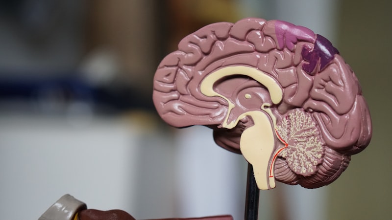
- The Core Definition: Pathways of Motor and Autonomic Control
- Historical Perspectives on Motor Pathway Discovery
- Medial Descending Tracts: Posture and Proximal Control
- Lateral Descending Tracts: Fine Motor and Distal Control
- Autonomic Descending Pathways: Visceral Regulation
- A Practical Example: Reaching for a Coffee Cup
- Significance and Clinical Impact
- Connections to Other Psychological Concepts
Introduction to Descending Tracts
The intricate orchestration of movement, posture, and vital internal functions within the human body relies fundamentally on a complex network of neural pathways. Among these, the descending tracts represent a critical component, serving as the primary communication channels through which the brain transmits commands and regulatory signals to the spinal cord. These pathways are essentially bundles of nerve fibers, or axons, originating from various cortical and subcortical regions of the brain, extending downwards to synapse with motor neurons and interneurons located within the spinal cord’s gray matter. Their function is paramount to nearly all aspects of our physical interaction with the world, from the most precise finger movements to the unconscious maintenance of balance and the regulation of internal organ systems.
The significance of these tracts lies in their direct role in mediating voluntary and involuntary actions. Without them, the sophisticated processing that occurs in the brain, whether it’s the conscious decision to lift an arm or the automatic adjustment of blood pressure, would remain isolated and unable to translate into physical or physiological responses. They act as the efferent arm of the central nervous system’s motor and autonomic control systems, ensuring that intentions are executed and internal homeostasis is maintained. Understanding the structure and function of descending tracts is therefore indispensable for comprehending the neural basis of movement, sensation, and autonomic regulation, offering insights into both normal physiological processes and the debilitating effects of neurological disorders.
The Core Definition: Pathways of Motor and Autonomic Control
At its core, a descending tract is a bundle of axons that conveys efferent signals from higher brain centers to lower motor neurons and interneurons within the spinal cord, ultimately influencing muscle activity or autonomic functions. These pathways are organized into distinct categories based on their anatomical location within the spinal cord’s white matter and their functional roles. The primary division classifies them into three major groups: medial, lateral, and those primarily dedicated to autonomic control. Each group comprises several specific tracts, each originating from particular brain regions and targeting specific sets of spinal cord neurons, thereby enabling a wide range of motor and physiological responses crucial for survival and interaction with the environment.
The fundamental mechanism behind descending tract function involves a hierarchical organization of neural control. Upper motor neurons, located in the cerebral cortex or brainstem, project their axons down through these tracts. These axons then synapse directly or indirectly (via interneurons) with lower motor neurons situated in the ventral horn of the spinal cord. These lower motor neurons, in turn, send their axons out to the skeletal muscles, causing contraction and movement. For autonomic functions, descending pathways from regions like the hypothalamus and brainstem modulate preganglionic autonomic neurons located in the intermediolateral cell column of the spinal cord, influencing organs and glands throughout the body. This intricate relay system ensures that high-level commands from the brain are translated into precise and coordinated peripheral actions.
The distinct categorization into medial, lateral, and autonomic pathways reflects a functional specialization. Medial tracts primarily govern gross movements, posture, and balance, often affecting the musculature of the trunk and proximal limbs. Lateral tracts are predominantly responsible for fine, skilled movements, particularly those involving the distal limbs, such as the hands and fingers. The autonomic pathways, while perhaps less visibly dramatic, are ceaselessly at work maintaining the body’s internal environment, regulating heart rate, digestion, respiration, and myriad other unconscious physiological processes. This division of labor allows for both robust, foundational control and agile, nuanced manipulation, demonstrating the remarkable complexity and efficiency of the central nervous system’s command architecture.
Historical Perspectives on Motor Pathway Discovery
The understanding of descending motor pathways has evolved significantly over centuries, beginning with early anatomical observations and progressively advancing with more sophisticated experimental and imaging techniques. Pioneers in neuroanatomy, such as Andreas Vesalius in the 16th century, laid foundational groundwork by meticulously dissecting and illustrating the gross structures of the brain and spinal cord. However, the functional segregation and specific pathways remained largely unknown until the 19th century, when advancements in microscopy and lesion studies began to unravel the intricate connections. Early hypotheses about nervous system function were often based on philosophical reasoning, but the advent of empirical methods marked a turning point in accurately mapping the brain’s control over the body.
Key breakthroughs occurred in the 19th century with researchers like Charles Bell and François Magendie, who independently demonstrated the functional distinction between dorsal (sensory) and ventral (motor) roots of the spinal nerves, a fundamental principle known as the Bell-Magendie Law. This provided crucial insight into the directionality of neural information flow. Later, precise lesion studies and stimulation experiments, particularly by scientists such as Eduard Hitzig and Gustav Fritsch in the late 1860s, localized the primary motor cortex in the frontal lobe, demonstrating that electrical stimulation of specific cortical areas could elicit movement in corresponding parts of the body. These findings directly implicated the cerebral cortex as a primary origin for descending motor commands, setting the stage for the detailed study of tracts like the corticospinal pathway.
The 20th century brought about an even finer resolution of these pathways, with techniques like degeneration studies (e.g., Wallerian degeneration) tracing the paths of severed axons, and later, electrophysiological methods allowing for the direct recording of neural activity. Santiago Ramón y Cajal’s detailed histological work, using techniques like the Golgi stain, provided unparalleled visualizations of individual neurons and their connections, solidifying the neuron doctrine and offering a morphological basis for understanding neural circuits. In contemporary neuroscience, advanced neuroimaging techniques such as diffusion tensor imaging (DTI) and functional magnetic resonance imaging (fMRI) continue to refine our understanding of these tracts in living humans, revealing their precise trajectories and functional engagement during various motor tasks, thereby continually enhancing our knowledge of the dynamic interplay between the brain and the spinal cord.
Medial Descending Tracts: Posture and Proximal Control
The medial descending tracts are a collection of pathways primarily responsible for controlling gross motor movements, maintaining posture, and regulating the musculature of the trunk and proximal limbs. These tracts generally terminate on interneurons and motor neurons located in the medial aspect of the spinal cord’s ventral horn, which innervate axial and proximal musculature. Their coordinated action is essential for activities that require stability and balance, forming the foundational support upon which finer movements can be executed. Unlike the lateral tracts which emphasize precision, the medial tracts focus on the broader strokes of movement and the constant adjustments necessary to counteract gravity and maintain an upright stance.
One of the most significant medial tracts is the **vestibulospinal tract**, which originates from the vestibular nuclei in the brainstem. These nuclei receive input from the inner ear’s vestibular apparatus, which detects head movements and changes in gravitational pull. The vestibulospinal tract then projects ipsilaterally and bilaterally down the spinal cord, primarily influencing extensor muscles to maintain balance and adjust posture in response to head movements. It plays a crucial role in reflexively stabilizing the body during locomotion and preventing falls, ensuring that our orientation in space is constantly updated and corrected. Without its continuous activity, even simple acts like standing or walking would be profoundly challenging, leading to severe instability and disequilibrium.
Another important medial pathway is the **reticulospinal tract**, which arises from the reticular formation in the brainstem. This tract is broadly categorized into pontine (medial) and medullary (lateral) components, both contributing to posture and gait. The pontine reticulospinal tract primarily facilitates extensor motor neurons, helping to maintain an upright posture and resist gravity, while the medullary reticulospinal tract tends to inhibit these extensor muscles, allowing for more flexible postural adjustments. Together, these tracts regulate muscle tone, especially in the trunk and proximal limbs, and coordinate rhythmic activities such as breathing and walking. They also play a role in integrating sensory information with motor commands, ensuring that postural adjustments are appropriate for the environmental context and the intended movement. Additionally, the **tectospinal tract**, originating from the superior colliculus, is involved in orienting head and eye movements in response to visual and auditory stimuli, further contributing to the body’s overall spatial awareness and reactive postural adjustments.
Lateral Descending Tracts: Fine Motor and Distal Control
In contrast to the medial system, the lateral descending tracts are predominantly responsible for the execution of precise, voluntary movements, particularly those involving the distal musculature of the limbs, such as the hands and fingers. These pathways project to motor neurons in the lateral aspect of the spinal cord’s ventral horn, which innervate the muscles responsible for fine manipulation and skilled actions. The development and functional dominance of these tracts, especially the corticospinal tract, are highly evolved in primates, reflecting their capacity for intricate motor control essential for tool use, communication, and complex interactions with the environment.
The most prominent and clinically significant lateral tract is the **corticospinal tract**, often referred to as the “pyramidal tract” due to its passage through the medullary pyramids where the majority of its fibers decussate (cross) to the contralateral side. Originating primarily from the motor cortex (precentral gyrus) and other cortical areas, this tract is crucial for initiating and controlling voluntary, skilled movements. The lateral corticospinal tract, which comprises about 90% of the fibers and decussates, controls the muscles of the contralateral limbs, enabling movements like writing, grasping, and playing musical instruments. Its direct projections to lower motor neurons allow for highly individualized control over single muscles or small groups of muscles, providing the dexterity that defines human motor capabilities.
Another component, the **corticobulbar tract**, shares a similar cortical origin but projects to cranial nerve nuclei in the brainstem rather than the spinal cord. It controls the voluntary movements of the face, head, and neck, including muscles involved in facial expression, mastication (chewing), deglutition (swallowing), and speech. While not extending to the spinal cord, its close functional relationship with the corticospinal tract underscores the brain’s unified control over motor actions across the entire body. The **rubrospinal tract**, originating from the red nucleus in the midbrain, is another lateral pathway, though its significance in humans is less pronounced than in other mammals. It primarily contributes to the control of upper limb flexion, working in conjunction with the corticospinal tract to modulate movements, particularly in the recovery phase after damage to the corticospinal system. Together, these lateral pathways represent the pinnacle of precise neural command, allowing for the sophisticated and adaptable motor behaviors characteristic of humans.
Autonomic Descending Pathways: Visceral Regulation
Beyond the control of skeletal muscle, the brain also exerts profound influence over the body’s internal environment through autonomic descending pathways. These pathways are responsible for regulating involuntary functions such as heart rate, blood pressure, respiration, digestion, glandular secretions, and pupillary responses, all critical for maintaining homeostasis. Unlike the clearly defined medial and lateral motor tracts, autonomic descending fibers are often more diffuse and integrated into broader brainstem and hypothalamic systems, projecting to preganglionic autonomic neurons located within the intermediolateral cell columns of the thoracic, lumbar, and sacral spinal cord segments.
A key player in autonomic control is the **hypothalamospinal tract**, which originates from various nuclei within the hypothalamus. The hypothalamus is a vital brain region involved in regulating fundamental physiological processes, including temperature, hunger, thirst, and circadian rhythms. Its descending projections to the spinal cord allow it to influence sympathetic and parasympathetic outflow, thereby modulating cardiovascular function, gastrointestinal motility, and sudomotor (sweating) activity. For example, in response to stress, hypothalamic signals descending via this pathway can increase heart rate and blood pressure by activating sympathetic neurons in the spinal cord, preparing the body for a “fight or flight” response.
Furthermore, various nuclei within the brainstem’s reticular formation also contribute significantly to autonomic modulation via descending pathways, often intermingling with the reticulospinal tracts that control somatic motor functions. These brainstem centers integrate sensory information and higher cortical commands to fine-tune autonomic responses. For instance, the medullary reticular formation contains centers that regulate cardiovascular and respiratory rhythms, sending descending commands to the spinal cord to adjust breathing rate and depth or to modify blood vessel constriction. This integrated control ensures that the body’s internal functions are precisely adapted to both internal needs and external demands, maintaining a delicate balance essential for health and survival.
A Practical Example: Reaching for a Coffee Cup
To illustrate the coordinated function of descending tracts, consider the seemingly simple act of reaching out and grasping a coffee cup. This everyday action involves a complex interplay of voluntary motor commands, postural adjustments, and sensory feedback, orchestrated seamlessly by various descending pathways. From the initial decision to the final execution, a cascade of neural events takes place, demonstrating the integrated roles of both lateral and medial systems.
- Initiation and Planning: The desire to drink coffee originates in higher cognitive centers. Once the decision is made, the posterior parietal cortex processes the cup’s location in space, while the premotor and supplementary motor areas in the cerebral cortex formulate a motor plan. This plan, detailing the sequence and trajectory of movements, is then relayed to the primary motor cortex.
- Voluntary Movement via Lateral Tracts: Neurons in the primary motor cortex generate the command to reach. Axons from these neurons form the corticospinal tract, specifically the lateral corticospinal tract, which descends through the brainstem, crosses in the medulla (decussation of the pyramids), and continues down the contralateral side of the spinal cord. These fibers synapse directly or indirectly with motor neurons in the ventral horn that innervate the muscles of the arm and hand. For the precise grasp, the corticobulbar tract also becomes active, coordinating facial and oral movements, such as slightly opening the mouth in anticipation or adjusting head position to better view the cup.
- Postural Support via Medial Tracts: As the arm extends, the body’s center of gravity shifts. To prevent a fall and maintain stability, the medial descending tracts spring into action. The vestibulospinal tract, receiving input from the inner ear, sends signals to the extensor muscles of the trunk and legs, reflexively adjusting muscle tone to counteract the shift and keep the body balanced. Simultaneously, the reticulospinal tract modulates the activity of axial and proximal limb muscles, providing a stable base for the arm’s movement. These involuntary postural adjustments occur concurrently with the voluntary reach, ensuring that the body remains upright and stable throughout the action.
- Grasping and Fine-Tuning: As the hand approaches the cup, visual and proprioceptive feedback (information about limb position and movement) is relayed back to the brain. The cerebellum and basal ganglia continuously modify the ongoing motor commands, refining the reach and grasp. The lateral corticospinal tract’s fine control allows the fingers to precisely wrap around the cup, adjusting grip strength based on the cup’s weight and texture. This intricate coordination of large-scale stability and small-scale dexterity is a testament to the sophisticated design of the descending motor system.
Significance and Clinical Impact
The profound importance of descending tracts extends far beyond their role in everyday movement, permeating various subfields of psychology and medicine. In psychology, understanding these pathways is fundamental to the study of motor control, learning, and skill acquisition. The development of motor skills from infancy through adulthood, and the refinement of complex actions like playing a musical instrument or performing surgery, are directly linked to the maturation and plasticity of these descending neural circuits. Moreover, breakdowns in these pathways can illuminate the neural basis of various psychological conditions where motor symptoms are prominent, such as Parkinson’s disease, where issues in the basal ganglia indirectly affect descending tract function, or in certain psychiatric disorders with motoric components.
Clinically, the integrity of descending tracts is paramount. Damage to these pathways, whether from trauma, stroke, or neurodegenerative diseases, often results in severe and debilitating motor deficits. For instance, a stroke affecting the motor cortex or the corticospinal tract in the brain can lead to hemiparesis (weakness) or hemiplegia (paralysis) on the contralateral side of the body, profoundly impacting a patient’s independence and quality of life. Similarly, a spinal cord injury can transect these tracts, leading to paralysis below the level of the lesion, necessitating intensive rehabilitation and often lifelong assistive devices. The specific symptoms observed (e.g., spasticity, flaccidity, loss of fine motor control) often depend on which particular tracts are affected and at what level.
Knowledge of descending tracts is also critical for developing targeted therapeutic interventions. In physical therapy and rehabilitation, understanding which pathways are compromised allows clinicians to design specific exercises and strategies aimed at promoting neural plasticity and functional recovery. For example, constraint-induced movement therapy for stroke patients capitalizes on the brain’s ability to reorganize, encouraging the use of affected limbs to strengthen remaining or compensatory descending pathways. Furthermore, advances in neuroprosthetics and brain-computer interfaces (BCIs) often rely on decoding signals intended for descending tracts, offering hope for individuals with severe motor impairments to regain some level of control over external devices. Thus, the study of descending tracts continues to be a vibrant area of research, continually yielding insights that improve both our theoretical understanding of the brain and our practical approaches to neurological care.
Connections to Other Psychological Concepts
The concept of descending tracts is deeply intertwined with numerous other key psychological and neurological concepts, serving as a fundamental anatomical and functional bridge across various domains. It forms the efferent arm of the broader field of motor control, which integrates sensory input with motor output to produce coordinated actions. The effectiveness of a descending command is heavily dependent on the context provided by ascending sensory pathways, which inform the brain about the body’s current state and the external environment. This interplay highlights the sensorimotor loop, where sensory feedback is continuously used to refine and adjust motor commands traveling down the descending tracts.
Furthermore, descending tracts are closely related to the functions of other major brain regions involved in movement modulation. The basal ganglia, for instance, play a crucial role in initiating and selecting appropriate movements, while inhibiting unwanted ones. Although not directly part of the descending tracts themselves, the basal ganglia heavily influence the activity of the motor cortex and brainstem nuclei from which many descending tracts originate, thereby indirectly shaping the commands sent down the spinal cord. Similarly, the cerebellum acts as a sophisticated error-correction mechanism, comparing intended movements with actual movements and sending corrective signals back to the motor cortex and brainstem, which then modify the outgoing commands via the descending tracts to ensure accuracy and coordination.
The study of descending tracts falls primarily under the umbrella of neuroanatomy and physiological psychology, but its implications extend into cognitive psychology (e.g., motor planning, executive function in action selection), developmental psychology (e.g., maturation of motor skills), and clinical psychology (e.g., understanding motor symptoms in mental health conditions). Understanding these pathways is also essential for comprehending reflexes, as many descending tracts modulate spinal reflex circuits, influencing their sensitivity and response thresholds. Ultimately, the descending tracts are not isolated entities but integral components of a vast, interconnected neural network that underpins all aspects of physical and much of our psychological experience, demonstrating the holistic nature of brain function.