Flight of Colors: Seeing the Afterimage of Light
- Defining the Phenomenon of Flight of Colors
- Physiological Basis: Retinal Adaptation and Photochemistry
- The Subjective Experience: Succession and Duration
- Historical Context and Early Scientific Inquiry
- Distinction from Other Types of Afterimages
- Experimental Methodologies and Key Observations
- The Role of Intensity and Stimulus Characteristics
- Significance in Modern Visual Perception Theory
Defining the Phenomenon of Flight of Colors
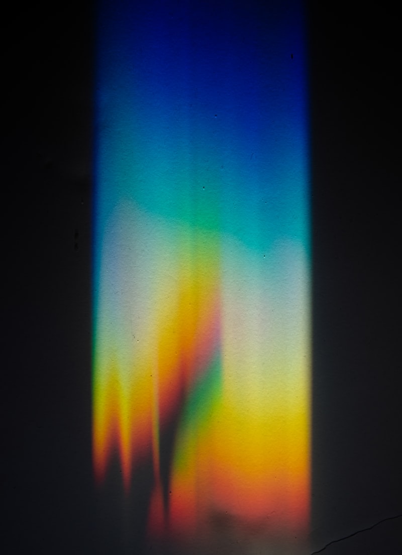
The concept known as the Flight of Colors refers to a specific type of visual afterimage characterized by a dynamic succession of chromatic perceptions that occur immediately following the termination of an intense, brief visual stimulus, typically an extremely bright flash of white light. This phenomenon is categorized as a type of positive afterimage because the initial visual experience shares similarities in brightness and, momentarily, in color distribution with the original stimulus, before devolving into a complex, evolving sequence of hues. Unlike simple, static afterimages, the Flight of Colors is intrinsically kinetic, involving a rapid transition through various saturated colors—including reds, greens, blues, and yellows—which appear to sweep across the visual field before fading into the standard, neutral gray background of stabilized vision. This reaction is a powerful demonstration of the inherent instability and adaptive complexity of the human visual system when exposed to sudden, overwhelming stimulation, revealing the temporal dynamics of the retinal photochemical processes as they attempt to regain equilibrium. Understanding this transitory visual reaction is crucial for grasping the mechanisms underlying color processing and temporal resolution within the optic nerve pathways, particularly concerning the interplay between rods and cones under extreme stimulation conditions.
Psychologically, the Flight of Colors is often described as a spectacular, albeit momentary, visual fireworks display occurring internally within the observer’s perception, providing compelling empirical evidence that visual processing does not cease instantaneously upon removal of the stimulus but rather continues as a cascade of neural and chemical events. The precise sequence of colors experienced by individuals can show some variability, influenced by factors such as the intensity and duration of the initial flash, the ambient lighting conditions prior to exposure, and the individual observer’s unique retinal sensitivity and adaptation state. However, certain patterns tend to predominate, often starting with the complementary color of the adapting light if the stimulus was colored, or initiating a rapid sweep through the primary spectral colors if the stimulus was broadband white light, emphasizing the role of opponent processing mechanisms even in these rapid adaptation phases. This immediate post-stimulus activity underscores the fact that visual sensation is not a static registration of external reality but a continuously reconstructed neural interpretation, highly sensitive to transient changes in energy input and exhibiting predictable oscillatory behavior during recovery.
The core definition emphasizes that the Flight of Colors is fundamentally an after-image that consists of a succession of colors. This definition distinguishes it from simpler afterimages where only one or two colors dominate the recovery phase. The intensity of the initiating stimulus is a necessary precondition; ordinary levels of illumination, even if sustained, typically produce simpler, negative afterimages based on localized photochemical bleaching, whereas the Flight of Colors requires a sudden, massive input of photons to overwhelm the photopigments across large areas of the retina simultaneously. Consequently, this phenomenon is frequently observed in contexts such as sudden exposure to camera flashes, brief electrical sparks, or high-intensity stroboscopic lights, making it a common, yet scientifically profound, visual reaction experienced in daily life. Its study allows researchers to effectively slow down and observe the normally instantaneous recovery process of the photoreceptors, providing invaluable temporal data about the kinetics of rhodopsin regeneration and cone pigment recovery, which are essential for maintaining continuous visual function.
Physiological Basis: Retinal Adaptation and Photochemistry
The underlying physiological mechanism responsible for the Flight of Colors resides primarily within the retinal photoreceptors—the rods and cones—and their subsequent signaling pathways to the bipolar and ganglion cells. When the retina is exposed to an intense flash of light, a massive, near-simultaneous photopigment bleaching event occurs, effectively exhausting the readily available photopigments required for normal transduction. The subsequent perception of a succession of colors is a direct consequence of the differential recovery rates of the three types of cone photopigments (L-cones sensitive to long wavelengths, M-cones sensitive to medium wavelengths, and S-cones sensitive to short wavelengths) and, to a lesser extent, the recovery of the rhodopsin in the rods. Since these pigments operate on distinct photochemical kinetics—recovering and regenerating at slightly different speeds—the retina’s sensitivity to various wavelengths of light shifts moment by moment during the recovery phase, leading to the subjective experience of colors rapidly changing and succeeding one another in the visual field. This temporal desynchronization in receptor recovery generates the dynamic chromaticity that defines the phenomenon.
Furthermore, the mechanism involves the complex interplay of opponent process theory at the post-receptoral level, specifically within the horizontal and bipolar cells. Hering’s theory posits that color information is processed via three antagonistic channels: red/green, blue/yellow, and black/white. When a strong stimulus is suddenly removed, the initial massive neural signal ceases, but the previously hyperpolarized cells that were inhibited by the stimulus rebound and fire spontaneously, generating an excitatory signal in the absence of external light. Because the recovery dynamics of the L, M, and S cones are asynchronous, the balance within the opponent channels is constantly disrupted and re-established. For instance, if the L-cones recover slightly faster than the M-cones, the red/green channel experiences a momentary imbalance favoring the red signal, followed by a shift as the M-cones catch up, causing the perceived color to transition rapidly. The dynamic shifting of these antagonistic balances generates the observable sequence of color changes, confirming that the Flight of Colors is a phenomenon rooted in both photochemical recovery and subsequent neural coding.
The role of the intensity of the flash cannot be overstated, as only a brief, intense stimulus can trigger the requisite level of simultaneous saturation and subsequent synchronized recovery. If the light were sustained, the retina would enter a state of adaptation, and the resulting afterimage would be a negative afterimage dominated by the complementary color due to sustained bleaching and inhibition. The transient nature of the stimulus ensures that the entire recovery process unfolds rapidly and dramatically in the subsequent darkness or against a neutral background. Studies suggest that the initial phase of the Flight of Colors often involves activity dominated by the S-cones (blue perception) due to their relatively lower sensitivity threshold, followed by the activation and subsequent recovery of the M and L cones, creating the characteristic progression from cooler colors (blues, greens) towards warmer colors (yellows, reds) or vice versa, depending on the exact parameters of the flash. This physiological sequence provides a robust explanation for the observed visual instability and the rapid chromatic shifts observed by the subject.
The Subjective Experience: Succession and Duration
The subjective experience of the Flight of Colors is marked by its profound temporal dimension, typically lasting only a few seconds, though the duration is highly variable depending on the intensity of the initial light source. Immediately following the flash, the observer often perceives a bright, localized image—the positive afterimage—which quickly begins to dissolve into the successive color sequence. This succession is not random; observers frequently report an organized progression, although the specific order can be influenced by individual differences in spectral sensitivity and the exact nature of the photopigment recovery curves. Common sequences often include a movement from the primary color of the stimulus to its complementary color, followed by a series of spectral shifts that cycle through the opponent channels, providing a compelling internal demonstration of how the visual system attempts to stabilize its processing capacity. The perceived colors are often described as highly saturated and luminous, standing out vividly against the dark background upon which they are projected.
A key characteristic of this experience is the spatial instability of the image. Unlike a simple negative afterimage which remains fixed in the location where the stimulus was viewed, the Flight of Colors often appears to move, shimmer, or expand, a phenomenon known as Eidinger’s Phenomenon when associated with certain retinal responses. This apparent movement is likely due to the highly dynamic and differential rates of recovery across the retina, where areas exposed to slightly different light levels (due to the non-uniform distribution of the intense flash) recover at slightly different times, creating waves of color change that sweep across the visual field. The observer perceives not a static picture, but a flowing, changing pattern, reinforcing the terminology of a “flight” or sequence of colors. The subjective intensity of the colors decreases exponentially over time until the visual field returns to its baseline state, demonstrating the successful completion of photochemical regeneration.
Psychophysical studies attempting to quantify the subjective duration and perceived color sequence rely heavily on observer reports, which must be carefully analyzed given the rapid nature of the event. Researchers utilize controlled exposure environments to standardize the stimulus and then ask subjects to verbally report the color progression they perceive, often using rapid response mechanisms. These reports consistently confirm that the visual system rapidly cycles through the three opponent color pairs, implying that the chemical recovery kinetics are highly structured. For example, a common progression might start with yellow, immediately transition to blue, move through a phase of green, and conclude with a reddish hue before dissipation. The duration of each distinct color phase is typically measured in milliseconds, highlighting the extraordinary speed at which the visual system processes transient stimuli and recovers from saturation.
Historical Context and Early Scientific Inquiry
The phenomenon of afterimages, including the Flight of Colors, has been a subject of fascination since the earliest days of vision science, predating modern understanding of retinal physiology. Philosophers and early experimentalists recognized that visual sensation persisted beyond the cessation of the stimulus, leading to observations of these fleeting color sequences. Early descriptions often lacked the precise terminology we use today but accurately captured the essence of the phenomenon: the rapid succession of hues following intense exposure. Figures such as Purkinje, who detailed various aspects of visual phenomena in the 19th century, made significant contributions by systematically cataloging the temporal qualities of afterimages, laying the groundwork for later investigations into photochemical dynamics. These early inquiries relied entirely on descriptive phenomenology, using the observer’s detailed report as the primary data point to classify and understand these transient visual reactions.
It was during the 19th and early 20th centuries, coinciding with advances in photochemical and physiological knowledge, that the Flight of Colors began to be rigorously linked to specific biological mechanisms. Researchers like Helmholtz, while focusing heavily on trichromatic theory, also acknowledged the complexities of post-stimulus visual recovery. The definitive connection between the succession of colors and the differential recovery rates of the three hypothesized color receptors (cones) became a central focus, challenging purely psychological interpretations and moving the explanation firmly into the realm of physiological optics. The historical importance of studying this phenomenon lies in its ability to provide tangible, real-time evidence of the photochemical processes occurring within the living eye, processes that are normally too fast to observe under standard viewing conditions.
The formal naming and specific study of the “Flight of Colors” became prominent as experimental psychology matured, particularly when researchers developed methods to produce intense, reproducible light flashes in laboratory settings. These controlled environments allowed for standardization of the stimulus duration and intensity, leading to more consistent and replicable observations of the color sequence. This historical trajectory showcases the evolution of vision science from descriptive observation to quantitative physiological modeling. The phenomenon served as a critical piece of evidence supporting theories of color vision that involved temporal mechanisms, eventually integrating into the comprehensive models that combine trichromacy (at the receptor level) with opponent processing (at the neural level), solidifying its place as a classic demonstration in visual perception studies.
Distinction from Other Types of Afterimages
It is crucial to differentiate the Flight of Colors from other common types of afterimages, particularly the standard negative afterimage. A negative afterimage is typically generated after prolonged exposure to a moderately bright, colored stimulus and appears in the complementary color of the original stimulus. For example, staring at a red square for thirty seconds and then shifting gaze to a white wall results in a green square afterimage. This is caused by localized fatigue or bleaching of the specific cone types sensitive to the original color, resulting in an imbalance where the opponent system overcompensates. In contrast, the Flight of Colors is initiated by a brief, overwhelming flash of light and involves a rapid sequence of colors, not just a single complementary hue. While negative afterimages represent a stable state of localized adaptation, the Flight of Colors represents a dynamic state of global retinal recovery from saturation.
The distinction also extends to positive afterimages. While the Flight of Colors is technically a positive afterimage (meaning the initial phase retains the brightness and rough color characteristics of the stimulus), standard positive afterimages, such as those seen immediately after a flash, are generally short-lived and static, fading quickly without the pronounced, extended chromatic progression. The uniqueness of the Flight of Colors lies specifically in the temporally extended and highly organized succession of hues. If the light flash is not sufficiently intense or brief, the resulting visual artifact might be a simple flash blur or a non-chromatic positive afterimage, failing to trigger the asynchronous recovery necessary for the full “flight” sequence. Therefore, the defining features—intensity of stimulus, brevity, and chromatic succession—must all be present to qualify the event as the Flight of Colors phenomenon.
Furthermore, phenomena related to visual persistence, such as the lingering image seen when rapidly moving a bright object (e.g., a sparkler), are related to the temporal integration capacity of the visual system but are distinct from afterimages. These are primarily related to the integration time of the retina and cortex, merging discrete stimuli into a continuous perception, rather than the photochemical recovery process that characterizes the Flight of Colors. The Flight of Colors stands out as a unique physiological signature of the visual system’s recovery kinetics following massive, instantaneous depolarization, providing a window into the speed and order of receptor regeneration that simple adaptation phenomena cannot offer. Recognizing these distinctions is vital for accurate physiological modeling of human vision and for designing experiments that isolate specific aspects of visual processing.
Experimental Methodologies and Key Observations
Studying the Flight of Colors requires precise control over the visual stimulus, demanding specialized experimental methodologies. Typically, researchers employ a Ganzfeld (uniform visual field) or a dark adaptation room, followed by the delivery of an extremely brief, high-intensity light flash, often generated by a photographic flash unit or a precisely controlled electronic shutter delivering light from a bright source. Crucially, the intensity and duration of the flash must be carefully calibrated to ensure maximal photopigment bleaching without causing retinal damage, usually lasting only a few milliseconds (e.g., 5 to 10 ms). The subsequent observation period is conducted in total darkness or against a neutral gray screen to eliminate external visual input that might interfere with the internally generated afterimage sequence.
One key observation derived from these experiments relates to the influence of the adapting state. If the observer is fully dark-adapted prior to the flash, the resulting Flight of Colors is typically more intense and prolonged, demonstrating the maximum available pool of photopigments ready for bleaching and subsequent regeneration. Conversely, if the observer is light-adapted, the effect is attenuated because a portion of the pigments are already bleached or inhibited. Furthermore, experiments focusing on specific regions of the retina—using small, focused light spots—have confirmed that the Flight of Colors is highly dependent on the local concentration and activity of cones, being most pronounced in the foveal and parafoveal regions where cone density is highest. These localized studies help confirm that the phenomenon is indeed rooted in the differential recovery kinetics of the specific cone types.
A significant methodological advance involves using electrophysiological techniques, such as the electroretinogram (ERG), in conjunction with subjective reports. While the ERG measures the mass electrical response of the retina, it can provide objective temporal data corresponding to the perceived subjective color changes. For instance, researchers have correlated specific oscillatory potentials recorded in the ERG during the post-flash recovery phase with the reported transitions between color phases in the Flight of Colors. These objective measurements provide validation for the subjective reports, linking the perceived sequence of red, green, blue, and yellow phases directly to the asynchronous neural activity generated by the recovering photoreceptors and subsequent retinal neurons, thereby moving the study beyond mere phenomenology into quantitative biological analysis.
The Role of Intensity and Stimulus Characteristics
The precise characteristics of the inducing light stimulus—specifically its intensity, duration, and spectral composition—are the primary determinants governing the nature and quality of the Flight of Colors experienced. As previously noted, intensity is paramount; the stimulus must be powerful enough to saturate the photopigments, ensuring that the recovery process starts from a state of near-total depletion. If the intensity is below this saturation threshold, the recovery will be gradual and uniform across receptor types, resulting in a standard, brief positive afterimage without the characteristic sequential chromatic shifts. High-intensity stimuli guarantee the mass bleaching required for the differential recovery rates to become physiologically significant and perceptually distinct to the observer.
Equally critical is the duration of the flash. The stimulus must be exceedingly brief—typically less than 50 milliseconds—to prevent the visual system from entering a sustained state of adaptation. If the duration is prolonged, the neural feedback mechanisms begin to stabilize the response, leading quickly to the formation of a stable negative afterimage based on the complementary color, thus suppressing the dynamic color sequence that defines the Flight of Colors. The brevity of the flash ensures that the recovery process starts abruptly and globally, maximizing the temporal desynchronization of the cone responses before neural inhibition can normalize the activity patterns. This interaction between duration and intensity defines a narrow operational window in which the phenomenon can be reliably induced and studied.
The spectral composition (color) of the flash also profoundly influences the initial phase and subsequent sequence of the perceived colors. A white light flash (broadband spectrum) stimulates all cone types maximally, leading to a complex recovery sequence where all three cone types compete asynchronously. However, if the flash is monochromatic—for instance, a highly saturated red flash—it predominantly bleaches the L-cones. The resulting Flight of Colors will often initiate with the complementary color (cyan/green) as the less-bleached M and S cones dominate the initial recovery signal, followed by a sequence dictated by the subsequent, slower regeneration of the L-cones. Analyzing these color-specific responses allows researchers to isolate the contribution of each cone type to the overall phenomenon, reinforcing the model that the Flight of Colors is a direct visual manifestation of the kinetics of individual photopigment regeneration.
Significance in Modern Visual Perception Theory
The Flight of Colors remains a phenomenon of high significance in modern visual perception theory because it provides a critical, observable link between the kinetics of molecular biochemistry (photopigment regeneration) and the subjective experience of vision. It offers empirical validation for the temporal aspects of the trichromatic theory, demonstrating that the three cone systems operate independently in their recovery rates, leading directly to the observed chromatic succession. Without this temporal asymmetry, the rapid cycling of colors would not occur; the visual field would simply fade uniformly to gray, validating the necessity of differential kinetics in explaining visual recovery. Therefore, the phenomenon serves as a powerful natural experiment confirming the physiological basis of color coding.
Furthermore, the study of the Flight of Colors strongly supports the opponent process theory by showing how the asynchronous recovery of the receptors feeds into the antagonistic neural channels (red/green, blue/yellow). The perceived transitions between complementary colors during the flight sequence are direct evidence of the neural rebound effect occurring within these opponent channels. As one cone system recovers faster, it temporarily dominates its corresponding opponent channel, only to be supplanted by the recovering antagonistic system moments later, resulting in the rapid, organized shift in perceived hue. This dynamic interaction between the receptor layer and the post-receptoral neural processing is perhaps the most critical insight derived from the analysis of this complex afterimage sequence.
In contemporary vision science, understanding the temporal characteristics of visual persistence and recovery is paramount for applications ranging from display technology (minimizing motion blur and flicker) to clinical ophthalmology (assessing retinal health and adaptation capacity). The Flight of Colors serves as a fundamental benchmark for measuring the absolute time scale required for retinal recovery from saturation. It highlights the profound dependency of our conscious visual experience on underlying biological timing mechanisms. By continuing to study the precise sequence and duration of the Flight of Colors under varied conditions, researchers gain deeper insights into the resilience and temporal limits of the human visual system, contributing to a more complete and temporally accurate model of how we perceive the world.