Ocular Inflammation: The Psychological Impact of Uveitis
The Core Definition of Iridocyclitis
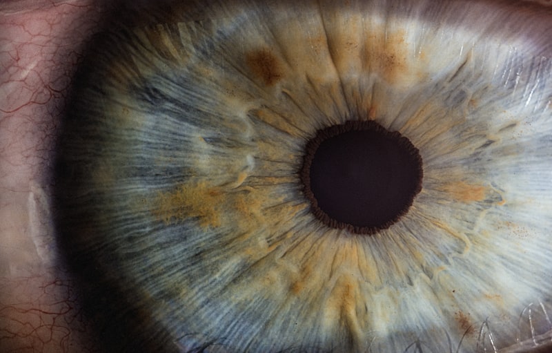
Iridocyclitis is fundamentally defined as the inflammation of the anterior segment of the eye, specifically encompassing the iris and the adjacent ciliary body. This condition represents a localized, acute, or chronic immune response within the delicate structures responsible for controlling light entry and producing the aqueous humor. The term itself is derived from the anatomical structures involved: “irido” referring to the iris, the colored part of the eye that regulates pupil size, and “cyclitis” referring to the ciliary body, a ring of muscle tissue located behind the iris that controls lens accommodation and secretes aqueous humor. Because both structures form the anterior portion of the uveal tract, iridocyclitis is often categorized clinically as a form of anterior uveitis.
The core mechanism behind this inflammatory process involves a breach in the normal immunological privilege of the eye. The eye typically maintains a unique immune environment known as ocular immune privilege, designed to protect the highly sensitive neural tissues from destructive inflammatory responses. When this privilege is compromised, often due to systemic autoimmune conditions, infection, or trauma, inflammatory cells and mediators infiltrate the anterior chamber. This influx of immune components leads to the characteristic symptoms of redness, pain, and photophobia. The severity and persistence of iridocyclitis are directly correlated with the degree of cellular infiltration and the resulting disturbance in the production and flow of the aqueous humor, which fills the anterior chamber and provides nutrients to the cornea and lens.
Although often acute and self-limiting when treated promptly, chronic or recurrent iridocyclitis poses a significant threat to vision. The persistent presence of inflammatory cells and proteins—known clinically as “flare” and “cells”—can damage adjacent structures, leading to secondary complications such as posterior synechiae (adhesions between the iris and the lens), elevated intraocular pressure (secondary glaucoma), and cataract formation. Therefore, understanding the precise location and extent of the inflammation, spanning both the iris stroma and the ciliary processes, is crucial for effective diagnostic categorization and subsequent therapeutic planning.
Anatomical and Pathophysiological Mechanisms
The fundamental principle driving the pathology of iridocyclitis is the disruption of the blood-aqueous barrier (BAB). Under normal conditions, the endothelial cells lining the blood vessels of the iris and the non-pigmented epithelial cells of the ciliary body form tight junctions that regulate the passage of substances from the bloodstream into the anterior chamber fluid, the aqueous humor. This barrier is essential for maintaining the clarity and precise chemical composition of the fluid. In iridocyclitis, whether triggered by infectious agents (e.g., herpes viruses) or non-infectious autoimmune processes (e.g., HLA-B27 associated spondyloarthropathies), these tight junctions are damaged.
Once the blood-aqueous barrier is compromised, plasma proteins, including fibrin, and various inflammatory cells—primarily lymphocytes and macrophages—leak into the anterior chamber. The accumulation of these proteins creates the phenomenon known as aqueous flare, which is visible upon slit-lamp examination as a hazy path of light, similar to the light seen when a dusty room is illuminated. Simultaneously, the inflammatory cells themselves are visible floating in the aqueous humor, referred to as aqueous cells. These cells often aggregate and precipitate onto the inner surface of the cornea, forming characteristic white deposits called keratic precipitates (KPs). The location and appearance of these KPs are vital diagnostic clues, indicating whether the inflammation is acute, chronic, granulomatous, or non-granulomatous.
Inflammation of the ciliary body, the “cyclitis” component, is particularly problematic because this structure is responsible for the production of aqueous humor. Severe inflammation can temporarily suppress the secretory function of the ciliary epithelium, leading paradoxically to an initial drop in intraocular pressure (IOP). However, as the inflammation progresses, the cellular debris and protein exudate can clog the trabecular meshwork—the drainage system of the eye—leading to impaired outflow and a subsequent, often dangerous, rise in IOP, necessitating urgent glaucoma management. Furthermore, the accompanying ciliary spasm contributes significantly to the deep, boring eye pain often reported by patients suffering from acute iridocyclitis.
Historical Understanding and Nomenclature
The recognition of inflammation within the anterior eye structures dates back centuries, though the precise understanding of the iris and ciliary body involvement evolved significantly with the advent of specialized optical tools. Early descriptions often grouped all forms of anterior eye inflammation under generic terms like “iritis” or “ophthalmia,” lacking the precision to distinguish between involvement of the iris alone versus combined involvement of the iris and the ciliary body. The term “iritis” (inflammation of the iris) was widely used, but clinicians gradually recognized that inflammation rarely isolates itself strictly to the iris, given the anatomical contiguity and shared vascular supply with the ciliary body.
The move toward the more comprehensive term iridocyclitis reflects a refined anatomical understanding, particularly supported by microscopic pathology and improved clinical observation methods, notably the introduction of the slit lamp in the early 20th century. The slit lamp allowed for the visualization of cells and flare in the anterior chamber, confirming the presence of exudate originating from the ciliary processes. Key figures in ophthalmology, particularly those studying the uveal tract, like Ernst Fuchs, contributed immensely to classifying these diseases, recognizing distinct clinical entities such as Fuchs’ Heterochromic Iridocyclitis (FHI) in the early 1900s.
Modern nomenclature often prefers the term anterior uveitis as the umbrella diagnosis, with iridocyclitis being the most common clinical presentation under this category. This shift emphasizes that the uvea—the middle vascular layer of the eye comprising the iris, ciliary body, and choroid—is treated as a continuous, functional unit. While the terms iritis and iridocyclitis remain valid and are often used interchangeably in clinical practice when the inflammation is confined to the anterior structure, the use of iridocyclitis is scientifically more accurate as it acknowledges the near-certain involvement of the ciliary processes, regardless of whether symptoms directly related to cyclitis (like ciliary muscle spasm) are dominant.
Clinical Presentation and Diagnostic Markers
Diagnosing iridocyclitis relies heavily on a detailed clinical examination using a slit lamp microscope, serving as the essential “how-to” step in identifying the condition. The typical presentation of acute iridocyclitis includes severe ocular pain, often described as deep and throbbing, associated with intense photophobia (extreme light sensitivity), and significant circumcorneal redness, known as ciliary flush. The patient may also notice a reduction in visual acuity due to corneal edema or inflammatory cells obscuring the visual axis. The presence of these symptoms alerts the clinician to the possibility of serious intraocular inflammation requiring immediate intervention.
The step-by-step diagnostic process begins with assessing visual acuity and then proceeds to the detailed slit-lamp examination. The key diagnostic findings that confirm iridocyclitis are observed in the anterior chamber:
-
Aqueous Cells and Flare: The hallmark sign is the visualization of cells suspended in the aqueous humor, indicating active inflammation. Flare (protein leakage) is graded on a scale from 0 (none) to 4+ (intense).
-
Keratic Precipitates (KPs): Deposits of inflammatory cells adhering to the posterior surface of the cornea. Their size, shape, and distribution (e.g., small, diffuse; or large, “mutton-fat” granulomatous KPs) guide the physician toward the etiology (infectious vs. autoimmune).
-
Pupillary Involvement: The iris often becomes sluggish in response to light, and inflammation can lead to posterior synechiae, where the iris adheres to the anterior capsule of the lens.
-
Intraocular Pressure (IOP): While IOP may initially be low due to ciliary body suppression, it can rapidly become elevated if the trabecular meshwork is blocked by inflammatory debris.
A comprehensive evaluation must also include a search for underlying systemic conditions. Because many cases of iridocyclitis are linked to autoimmune diseases like ankylosing spondylitis or juvenile idiopathic arthritis, diagnostic testing may involve blood work (e.g., HLA-B27 typing, complete blood count, erythrocyte sedimentation rate) and referral to a rheumatologist. The classification of iridocyclitis is often based on its cause (infectious or non-infectious), duration (acute or chronic), and pattern (granulomatous or non-granulomatous), ensuring that treatment targets the specific underlying pathology.
Therapeutic Approaches and Management
The significance of correctly identifying and treating iridocyclitis lies in the need to quickly quell the inflammation to prevent permanent structural damage and maintain vision. The cornerstone of therapy for non-infectious iridocyclitis is the use of corticosteroids, powerful anti-inflammatory agents. These are typically administered topically via eye drops in high frequency (e.g., every hour initially) to achieve high concentrations in the anterior chamber with minimal systemic side effects. In severe cases or when the inflammation is unresponsive, periocular steroid injections or oral corticosteroids may be necessary.
A second critical component of management involves cycloplegic and mydriatic agents, such as atropine or cyclopentolate. These drugs serve two primary purposes: first, they paralyze the ciliary muscle, relieving the painful ciliary spasm associated with cyclitis; and second, they dilate the pupil, which helps prevent the formation of posterior synechiae (adhesions between the iris and the lens capsule). Preventing synechiae is crucial because they can disrupt the flow of aqueous humor, potentially leading to a severe form of secondary glaucoma known as pupillary block glaucoma.
For cases where a specific infectious etiology is identified (e.g., viral iridocyclitis caused by Herpes Simplex or Varicella Zoster), the treatment regimen must be adapted to include appropriate antiviral medications in addition to the anti-inflammatory drugs. Furthermore, chronic or refractory cases linked to systemic autoimmune diseases often require immunosuppressive therapy managed in collaboration with a rheumatologist. These may include systemic agents like methotrexate or biologics, used to modulate the underlying immune dysregulation and prevent recurrent episodes of uveitis, thereby protecting long-term visual function. The long-term prognosis is dependent on the patient’s adherence to the medication schedule and consistent monitoring for complications like glaucoma and cataracts.
The Impact of Heterochromic Iridocyclitis
One distinct and historically important subtype of this condition is Fuchs’ Heterochromic Iridocyclitis (FHI), a chronic, low-grade, non-granulomatous anterior uveitis that presents unique challenges. FHI is characterized by its insidious onset, often lacking the acute pain, redness, and photophobia typical of other forms of iridocyclitis. This subtle presentation means that FHI often goes undiagnosed until significant vision loss or complications arise. The most striking clinical feature, which is encapsulated in the historical source content, is the resulting heterochromia—a difference in the color of the two eyes—due to the progressive loss of pigment in the affected iris.
In FHI, the affected iris typically appears lighter or slightly faded compared to the healthy eye. This loss of pigment is accompanied by fine, stellate (star-shaped) keratic precipitates that are widely distributed across the entire corneal endothelium. Unlike acute iridocyclitis, FHI is less responsive to traditional topical corticosteroids, and its management focuses primarily on monitoring and treating the two most common complications associated with its chronic nature: cataracts and secondary glaucoma. Cataract surgery in FHI patients is often successful, but the underlying, persistent inflammation requires careful management during and after the procedure to prevent complications.
The mechanism of pigment loss in FHI is complex but involves atrophy of the iris pigment epithelium. Although the exact etiology remains debated—with theories ranging from infectious causes (e.g., rubella virus) to localized immunological deficiencies—its clinical course is predictably chronic and generally unilateral. FHI illustrates the critical link between inflammation and long-term structural change, confirming that even low-grade, simmering inflammation can drastically alter the anatomy of the eye, leading to irreversible loss of pigment in the iris and profound functional impairment if the subsequent complications are not managed aggressively.
Connections and Relations in Ophthalmology
Iridocyclitis belongs to the broader field of Ophthalmology, specifically within the subspecialty focusing on ocular inflammatory diseases, or uveitis. It is intimately connected to several other key concepts based on the anatomical distribution of the inflammation. As mentioned, iridocyclitis is the common term for anterior uveitis, which distinguishes it from intermediate uveitis (pars planitis, involving the vitreous and pars plana), posterior uveitis (involving the choroid and retina), and panuveitis (involving all layers of the uvea). The classification system ensures that diagnosis and treatment are tailored to the specific structures involved, as inflammation in the posterior segment carries a much higher risk of immediate, irreversible vision loss.
Furthermore, iridocyclitis is often studied alongside systemic autoimmune disorders. The relationship between acute anterior uveitis and HLA-B27 positivity is particularly strong, meaning patients with this genetic marker are highly susceptible to recurrent episodes. This connection places iridocyclitis firmly within the domain of Immunology, highlighting the need for a multidisciplinary approach involving both ophthalmologists and rheumatologists. Understanding these systemic links is essential, as the ocular manifestation may be the first clinical sign of a broader, undiagnosed condition such as ankylosing spondylitis or inflammatory bowel disease.
Finally, the chronic complications of iridocyclitis, namely secondary glaucoma and cataracts, connect it directly to the fields of cataract and glaucoma surgery. The management of secondary glaucoma in uveitic eyes is notoriously difficult, requiring specialized surgical techniques and careful control of the underlying inflammation. Thus, iridocyclitis serves as a prime example of how localized ocular pathology can quickly cascade into major surgical and chronic disease management challenges within the broader scope of clinical medicine.