Pupillary Light Reflex: How Your Eyes Reveal Inner States
Introduction and Nomenclature
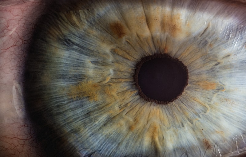
The Iritic Reflex, though a less commonly used descriptor, is scientifically synonymous with the Pupillary Light Reflex (PLR). This involuntary neurological mechanism governs the constriction and dilation of the pupil in response to changes in light intensity. The term “iritic” derives directly from the iris, the pigmented structure of the eye responsible for controlling the diameter and size of the pupil and thus regulating the amount of light reaching the retina. Functionally, the reflex serves as a critical homeostatic mechanism, ensuring that the visual system is protected from damaging high luminance while simultaneously optimizing visual acuity across various lighting conditions, acting effectively as the eye’s automatic aperture control system.
Understanding the Iritic Reflex is fundamental to both ophthalmology and clinical neurology, as its integrity reflects the health of specific cranial nerves and brainstem structures. Unlike voluntary muscle movements, this reflex operates entirely within the autonomic nervous system, specifically the parasympathetic and sympathetic branches, which exert opposing effects on the intrinsic muscles of the iris. The speed and symmetry of this response are key indicators of neurological function, prompting clinicians to assess the PLR immediately during routine examinations and emergency trauma evaluations.
While the primary stimulus is light, the pupil size is also modulated by other factors, including arousal, emotional state, and the act of focusing on near objects (the accommodation reflex). However, when discussing the Iritic Reflex specifically, the focus rests overwhelmingly on the light-induced response. The reflex involves a sophisticated arc spanning from the retina, through the midbrain, and back to the effector muscles within the iris, a pathway so precise that localized damage at any point can be identified by characteristic pupillary abnormalities.
Anatomical Pathways of the Iritic Reflex Arc
The Iritic Reflex operates via a complex three-neuron pathway, traditionally divided into the afferent (sensory) limb and the efferent (motor) limb. The afferent pathway begins when light strikes specialized photoreceptors in the retina, primarily the intrinsically photosensitive retinal ganglion cells (ipRGCs), which contain the photopigment melanopsin. These cells are particularly crucial for mediating the light reflex, complementing the input from traditional rods and cones. Signals generated here travel along the optic nerve (Cranial Nerve II) and continue past the optic chiasm and optic tract.
Crucially, the fibers carrying pupillary information diverge from the main visual pathway before reaching the lateral geniculate nucleus (LGN), which processes conscious sight. Instead, these fibers enter the pretectal nucleus, located in the dorsal midbrain. This anatomical separation highlights the reflexive, non-conscious nature of pupillary control. The pretectal nucleus acts as the coordinating center, relaying signals bilaterally to the paired parasympathetic nuclei responsible for constriction, ensuring that light entering one eye results in the constriction of both pupils simultaneously.
The efferent pathway, responsible for pupil constriction (miosis), is mediated by the parasympathetic division of the Oculomotor Nerve (Cranial Nerve III). The pathway originates in the Edinger-Westphal nucleus (EWN), which receives input from the pretectal nucleus. From the EWN, preganglionic parasympathetic fibers travel along the third cranial nerve until they synapse in the ciliary ganglion, situated within the orbit. Postganglionic fibers then emerge from the ciliary ganglion as the short ciliary nerves, innervating the effector muscle, the sphincter pupillae.
Physiology of Miosis and Mydriasis
The mechanical action of the Iritic Reflex relies on the antagonistic functions of two smooth muscles located within the iris stroma: the sphincter pupillae and the dilator pupillae. Miosis, or pupil constriction, is the primary response to increased light, driven entirely by the parasympathetic efferent limb. When the short ciliary nerves release the neurotransmitter acetylcholine (ACh), it binds to muscarinic M3 receptors on the sphincter pupillae muscle fibers. These circular fibers contract concentrically, causing the pupil aperture to rapidly decrease, thus limiting light entry and protecting the delicate photoreceptors of the retina from phototoxicity.
Conversely, mydriasis, or pupil dilation, is largely mediated by the sympathetic nervous system, often in response to low light levels, pain, or emotional stress. Although the sympathetic system is not strictly part of the classic light reflex arc, it constantly modulates pupillary size. The sympathetic pathway involves a three-neuron chain originating in the hypothalamus. The final neuron releases norepinephrine, which acts on alpha-1 adrenergic receptors located on the dilator pupillae muscle. These muscle fibers are arranged radially, and their contraction pulls the iris periphery outward, increasing the pupil diameter.
The balance between these two systems dictates the resting pupil size, which is known as the pupillary tone. In normal ambient lighting, the parasympathetic tone slightly dominates, maintaining a moderate pupil size. Dysfunction in either the parasympathetic (constricting) or sympathetic (dilating) input disrupts this delicate balance, leading to observable pathological asymmetry (anisocoria). The speed of the Iritic Reflex is highly impressive; the pupil can constrict in mere milliseconds to adapt to sudden changes in luminance, reflecting the rapid synaptic transmission and muscle response enabled by the parasympathetic system.
Consensual and Direct Responses
A defining characteristic of the Iritic Reflex is its bilateral nature, resulting in both a direct and a consensual response. The Direct Response refers to the constriction of the pupil in the eye that is directly stimulated by light. The Consensual Response refers to the simultaneous and equal constriction of the pupil in the contralateral (unstimulated) eye. This synchronized action is a hallmark of a healthy reflex arc and is essential for maintaining symmetrical vision.
The anatomical mechanism underlying the consensual response is the partial decussation, or crossing, of nerve fibers within the pretectal nucleus in the midbrain. When light stimulates the afferent fibers, the pretectal nucleus ensures that the signal is split and sent equally to the Edinger-Westphal nucleus (EWN) on the ipsilateral side (for the direct response) and the EWN on the contralateral side (for the consensual response). Because the motor output (Efferent limb, CN III) is entirely unilateral to its corresponding eye, the bilateral input at the midbrain level guarantees synchronized efferent activity.
The assessment of both the direct and consensual responses is invaluable in clinical localization of neurological lesions. If light shone into the right eye causes both the right and left pupils to constrict (direct and consensual responses intact), the afferent pathway (CN II) and the efferent pathways (CN III) are functioning normally. However, if light shone into the right eye yields no response in either eye, but light shone into the left eye causes both pupils to constrict, it strongly indicates a lesion of the right optic nerve (the afferent pathway). Conversely, if the direct response is present but the consensual response is absent, the issue lies in the efferent pathway (CN III) or the sphincter muscle of the non-responsive eye.
Clinical Significance and Assessment
The assessment of the Iritic Reflex is one of the most fundamental components of a neurological examination, often summarized by the acronym PERRLA (Pupils Equal, Round, Reactive to Light and Accommodation). The clinical utility of this reflex is profound because the reflex arc traverses critical structures in the brainstem, making it a reliable, non-invasive indicator of central nervous system compression or damage, particularly in emergency settings such as head trauma or suspected stroke.
Assessment typically involves the use of a focused light source, such as a penlight, shone obliquely across the pupil while the patient fixates on a distant object to minimize interference from the accommodation reflex. The clinician observes several key characteristics: the size of the pupils in darkness and light, the speed of constriction (reactivity), and the symmetry between the two eyes (equality). A fixed, dilated pupil that is unresponsive to light, often described as a “blown pupil,” is a dire sign, frequently indicating compression of the third cranial nerve due to rising intracranial pressure.
For detecting subtle afferent defects, the specialized Swinging Flashlight Test is employed. In this test, the light is rapidly moved from one eye to the other, observing the subsequent reactions. If the affected eye has a damaged afferent pathway (e.g., optic neuritis or severe retinal disease), the signal sent to the midbrain is weaker. When the light is swung to the affected eye, both pupils may paradoxically appear to dilate, despite being illuminated. This phenomenon is known as a Relative Afferent Pupillary Defect (RAPD), or a Marcus Gunn pupil, and is a crucial sign of unilateral optic nerve dysfunction.
Pharmacological Modulation
The Iritic Reflex is highly susceptible to manipulation by pharmacological agents, a capability routinely utilized in clinical medicine for diagnostic and therapeutic purposes. These drugs fall broadly into two categories based on their effect on pupil diameter: miotics (causing constriction) and mydriatics (causing dilation). Understanding their mechanism of action is essential for interpreting pupillary responses in patients receiving systemic medications or topical ophthalmic drops.
Miotics are agents that enhance the parasympathetic tone, thus promoting pupil constriction. They include direct-acting cholinergic agonists, such as pilocarpine, which directly stimulate the muscarinic receptors on the sphincter pupillae muscle. Another class includes acetylcholinesterase inhibitors, which prevent the breakdown of endogenous acetylcholine, prolonging its effect. Miotics are medically significant in the treatment of glaucoma, as pupillary constriction slightly alters the angle of the anterior chamber, often improving the outflow of aqueous humor and reducing intraocular pressure.
Mydriatics are drugs used to dilate the pupil, facilitating the examination of the retina and lens. They achieve dilation through two primary mechanisms. The first involves parasympathetic antagonists (anticholinergics), such as atropine or tropicamide, which block the action of acetylcholine at the sphincter muscle, paralyzing the constriction mechanism. The second involves sympathetic agonists, such as phenylephrine, which stimulate the alpha-adrenergic receptors on the dilator pupillae muscle, actively pulling the pupil open. Mydriatics are essential for retinal photocoagulation and other procedures requiring maximal visual access to the posterior segment of the eye.
Pathologies Associated with Iritic Dysfunction
Dysfunction of the Iritic Reflex often heralds significant underlying neurological or ocular pathology. Pathological changes in pupil size, symmetry (anisocoria), or reactivity provide vital clues to the location of the lesion within the reflex arc. Conditions affecting the efferent parasympathetic pathway often result in a dilated, poorly reactive pupil, whereas conditions affecting the sympathetic pathway result in a constricted pupil.
One of the most clinically urgent pathologies is compression of the Oculomotor Nerve (CN III), frequently caused by cerebral aneurysms or uncal herniation secondary to mass lesions. Because the parasympathetic fibers responsible for the Iritic Reflex run superficially along the nerve, they are the first to be damaged by external pressure, leading to a fixed, dilated pupil on the side of the compression—a key indicator of rising intracranial pressure and neurological emergency.
Conversely, damage to the sympathetic pathway results in Horner’s Syndrome, characterized by the triad of unilateral miosis (constriction), ptosis (droopy eyelid), and often anhidrosis (lack of sweating) on the affected side of the face. This condition indicates damage to the sympathetic chain anywhere from the hypothalamus down to the superior cervical ganglion or the plexus surrounding the carotid artery. Other notable pathological conditions affecting the reflex include:
- Argyll Robertson Pupil: Associated historically with neurosyphilis, where the pupils constrict poorly or not at all to light, but react briskly during accommodation (near effort).
- Adie’s Tonic Pupil: A benign disorder resulting from damage to the ciliary ganglion, causing unilateral, sluggish, and delayed constriction and redilation, often accompanied by absent deep tendon reflexes.
- Optic Neuritis: Inflammation of the optic nerve often results in a severe Relative Afferent Pupillary Defect (RAPD), reflecting the reduced conduction of sensory information along the affected afferent pathway.