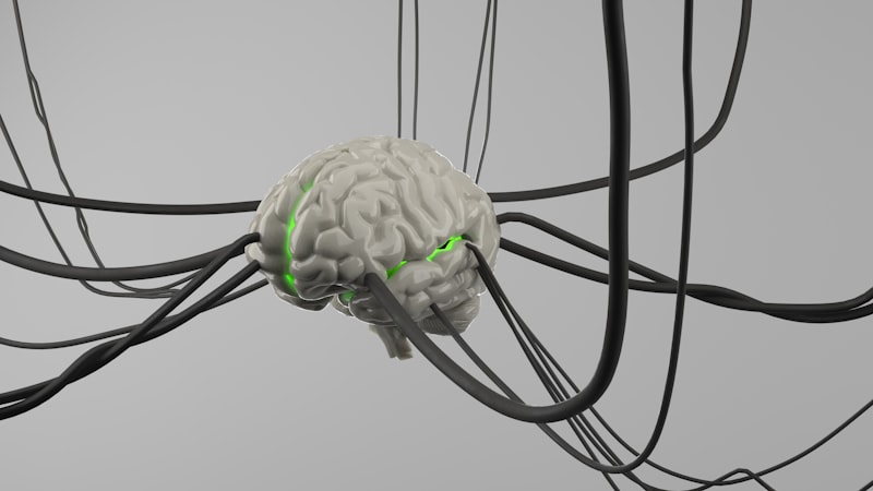
Neural Foundations: Mapping the Mind’s Hidden Architecture
- The Core Definition: Unveiling the Invisible World Relevant to Psychology
- Historical Context: The Dawn of Neuropsychological Observation
- Techniques for Microscopic Exploration in Psychological Science
- Applications: Unraveling the Biological Basis of Mind and Behavior
- A Practical Example: Observing Neuronal Communication
- Significance and Impact: Bridging Biology and Psychology
- Connections and Relations: The Interdisciplinary Landscape
The Core Definition: Unveiling the Invisible World Relevant to Psychology
The microscopic level refers to the scale of observation encompassing entities too minute to be discerned by the unaided human eye, requiring specialized magnification tools for their visualization and study. This realm exists between the macroscopic level, which is readily visible to the naked eye, and the subatomic level, where individual atoms and their constituent particles reside, often requiring even more advanced instrumentation or theoretical frameworks for comprehension. In the context of psychology and its foundational discipline, neuroscience, understanding the microscopic level is paramount, as it provides the fundamental biological framework upon which all mental processes, behaviors, and experiences are built. It is within this intricate, unseen world that the neural machinery underlying cognition, emotion, and perception operates, making its exploration indispensable for a holistic understanding of the human psyche.
At its core, the microscopic level in psychology involves the examination of the biological components of the nervous system, particularly the neurons and glial cells that constitute the brain and spinal cord. These cellular structures, along with their intricate connections known as synapses, are the basic units of information processing in the brain. The ability to observe these elements, their internal organelles, and the molecular machinery that governs their function, allows researchers to bridge the gap between biological events and psychological phenomena. Without access to this microscopic perspective, our understanding of how thoughts form, memories are stored, or emotions are experienced would remain largely speculative and incomplete, lacking empirical grounding in the physical reality of the brain.
The fundamental principle driving the study of the microscopic level in psychology is the belief that complex psychological states and behaviors emerge from the coordinated activity of vast networks of individual cells. By dissecting these networks at an increasingly finer resolution, from the overall architecture of a neuron down to the molecular interactions at a synapse, scientists can identify the specific biological mechanisms that contribute to various psychological functions and dysfunctions. This reductionist approach, while acknowledging the emergent properties of complex systems, provides critical insights into the underlying causes of conditions such as depression, anxiety, learning disabilities, and neurodegenerative disorders, fostering the development of targeted interventions.
Historical Context: The Dawn of Neuropsychological Observation
The journey into the microscopic world began long before its direct application to psychology, with early pioneers like Anton van Leeuwenhoek and Robert Hooke in the 17th century utilizing primitive light microscopy to observe “animalcules” and cellular structures. However, the specific relevance to psychology truly began to unfold in the late 19th and early 20th centuries, as advancements in microscopy and staining techniques allowed for the detailed visualization of the nervous system. Key figures in this era were Santiago Ramón y Cajal and Camillo Golgi, whose groundbreaking work laid the foundation for modern neuroscience and, by extension, biopsychology.
Camillo Golgi, an Italian physician and scientist, developed a revolutionary silver staining method in 1873, known as the “black reaction,” which allowed for the complete visualization of individual neurons, including their cell bodies, axons, and dendrites, against a clear background. This technique was crucial because, unlike previous methods, it only stained a small percentage of cells, making it possible to trace their intricate shapes and connections. While Golgi believed the nervous system was a continuous network (the reticular theory), his method provided the essential tool for others to challenge this view and ultimately establish the discrete nature of neurons.
It was Santiago Ramón y Cajal, a Spanish neuroscientist, who meticulously applied Golgi’s staining method to various parts of the nervous system across different species and developmental stages. Through his exquisite drawings and detailed observations, Cajal provided compelling evidence that neurons are indeed distinct, individual cells that communicate at specialized junctions, rather than forming a continuous reticulum. This concept, known as the neuron doctrine, became a cornerstone of modern neuroscience. Cajal’s work, which earned him a shared Nobel Prize with Golgi in 1906, fundamentally shifted the understanding of brain organization, moving the field towards a cellular and microscopic perspective that is indispensable for understanding the biological basis of psychological functions.
Techniques for Microscopic Exploration in Psychological Science
The ability to observe the microscopic level in psychology relies heavily on a sophisticated array of imaging techniques, each offering unique insights into the structure and function of the nervous system. Light microscopy remains a fundamental tool, employing visible light and a series of lenses to magnify specimens. Modern iterations, such as fluorescence microscopy and confocal microscopy, have dramatically enhanced its capabilities. Fluorescence microscopy uses fluorescent dyes or proteins to label specific cellular components or molecules, allowing researchers to visualize particular neurons, proteins, or even the activity of specific genes within brain tissue or living cells. Confocal microscopy further refines this by using a pinhole to eliminate out-of-focus light, producing sharp, three-dimensional images of thick specimens, which is invaluable for studying complex neuronal networks and their intricate structures like dendritic spines, crucial for synaptic plasticity and learning.
For an even higher resolution, researchers turn to electron microscopy, which uses a beam of electrons instead of light to illuminate and magnify specimens. Scanning electron microscopy (SEM) provides detailed three-dimensional surface images, allowing for the visualization of the intricate external morphology of cells and tissues, such as the architecture of neuronal circuits or the surfaces of cellular organelles. In contrast, Transmission electron microscopy (TEM) passes electrons through ultrathin sections of tissue, revealing the internal ultrastructure of cells with unparalleled detail. TEM is critical for examining the precise morphology of synapses, identifying the vesicles containing neurotransmitters, and observing the fine details of organelles within neurons, providing insights into the physical substrates of neural communication and cellular health, which are directly relevant to understanding the biological underpinnings of psychological states.
Beyond traditional microscopy, advanced techniques continue to push the boundaries of microscopic observation in psychological and neuroscientific research. Super-resolution microscopy techniques, for instance, overcome the diffraction limit of light, allowing scientists to visualize structures and molecular interactions within cells at a resolution previously only achievable with electron microscopy. Furthermore, techniques like optogenetics combine genetic engineering with light to control the activity of specific neurons, often observed at the microscopic level, enabling researchers to investigate the causal roles of particular neural circuits in behavior and psychological processes. These cutting-edge tools collectively empower researchers to explore the dynamic interplay of structure and function at the microscopic scale, revealing the intricate mechanisms that give rise to the complexities of the human mind.
Applications: Unraveling the Biological Basis of Mind and Behavior
The applications of studying the microscopic level within psychology are vast and transformative, providing a foundational understanding of how biological processes give rise to psychological phenomena. One primary application is the detailed study of neurons and glial cells, which are the fundamental building blocks of the nervous system. By observing their morphology, density, and connectivity under the microscope, researchers can discern differences in brain structure associated with various cognitive abilities, developmental stages, or neurological conditions. For example, microscopic analyses can reveal changes in dendritic arborization or synaptic density in response to learning experiences, illustrating the brain’s remarkable plasticity at the cellular level, which underpins the psychological concept of adaptation and skill acquisition.
Another crucial application lies in understanding the mechanisms of synaptic plasticity and memory formation. Memories are not stored as single entities but rather as patterns of altered synaptic strengths and connections between neurons. Microscopic techniques allow scientists to visualize these changes directly, such as the growth of new dendritic spines or the strengthening of existing synapses following learning. This direct observation provides empirical evidence for long-term potentiation and depression, which are cellular mechanisms thought to underlie learning and memory. This microscopic understanding is critical for developing interventions for memory disorders and for enhancing cognitive function, directly impacting fields like educational psychology and cognitive rehabilitation.
Furthermore, the microscopic level is indispensable for investigating the biological basis of neurological and psychological disorders. Many conditions, such as Alzheimer’s disease, Parkinson’s disease, and even certain psychiatric disorders like schizophrenia, are characterized by specific cellular or molecular pathologies. Microscopic examination of brain tissue from affected individuals can reveal hallmarks like amyloid plaques and neurofibrillary tangles in Alzheimer’s, or Lewy bodies in Parkinson’s. Identifying these microscopic changes helps in diagnosis, understanding disease progression, and developing targeted therapies. Similarly, microscopic studies of receptor distributions and neurotransmitter pathways inform the field of psychopharmacology, revealing how psychiatric medications exert their effects at the cellular and molecular levels to alleviate symptoms of mood disorders or anxiety.
A Practical Example: Observing Neuronal Communication
To illustrate the profound utility of the microscopic level in psychology, consider the seemingly simple act of deciding to lift your arm. While this is a macroscopic behavioral output, its initiation and execution are orchestrated by an intricate dance of microscopic events within your brain and nervous system. The process begins with electrical signals, known as action potentials, propagating along the axons of motor neurons. These action potentials are microscopic electrical impulses, fundamentally changes in ion concentrations across the neuronal membrane, occurring on the scale of nanometers and milliseconds, entirely invisible without specialized tools.
As an action potential reaches the end of a motor neuron’s axon terminal, it triggers the release of neurotransmitters into the synaptic cleft, a microscopic gap between neurons. These neurotransmitters, which are tiny molecular messengers, then diffuse across this cleft and bind to specific receptor proteins on the dendrite of the next neuron or muscle cell. This binding event initiates a new electrical signal in the postsynaptic cell, continuing the chain of communication. All these events—the release of vesicles, the diffusion of molecules, the binding to receptors—occur at an incredibly small scale, measured in nanometers, making them prime targets for microscopic investigation.
Researchers employ advanced microscopy techniques to visualize and measure these microscopic events. For instance, electrophysiology, often combined with microscopy, allows scientists to record the minute electrical currents (action potentials) flowing through individual neurons. Fluorescence microscopy can be used to label and track neurotransmitter vesicles as they move towards the synaptic membrane, or to observe the real-time binding of neurotransmitters to their receptors using fluorescent indicators. By combining these microscopic observations with behavioral studies, researchers can establish direct links between cellular-level activities and macroscopic psychological outputs, such as movement, perception, or decision-making. This “how-to” of observing neuronal communication at the microscopic level provides concrete evidence for the biological underpinnings of all psychological functions.
Significance and Impact: Bridging Biology and Psychology
The study of the microscopic level holds profound significance for the field of psychology, serving as a critical bridge between the biological sciences and the understanding of mind and behavior. It moves beyond purely theoretical or observational psychological models by providing empirical evidence for the physical realities that underpin mental processes. By revealing the intricate cellular and molecular mechanisms of the brain, microscopic research has transformed psychology from a purely behavioral or cognitive discipline into one deeply integrated with neuroscience and biology, fostering a more holistic and comprehensive biopsychosocial perspective. This integrated approach acknowledges that psychological phenomena are not merely abstract concepts but are rooted in the complex interactions occurring at the cellular and subcellular levels within the nervous system.
The impact of microscopic investigation is evident in its wide-ranging applications across various domains. In clinical psychology, it has revolutionized our understanding and treatment of mental health disorders by identifying specific cellular pathologies or molecular imbalances that contribute to conditions like depression, anxiety, and schizophrenia. This knowledge directly informs the development of more effective psychopharmacological treatments, as drugs are designed to interact with specific receptors or enzymes at the microscopic level. In developmental psychology, microscopic studies illuminate the processes of brain development, from neurogenesis and neuronal migration to synaptic pruning, helping to explain critical periods of learning and vulnerability to developmental disorders. Understanding these microscopic changes is crucial for early intervention strategies and educational approaches.
Furthermore, microscopic research has significantly advanced cognitive psychology by providing insights into the neural correlates of cognitive functions such as attention, perception, and memory. For instance, visualizing the activity of specific neurons or synapses during learning tasks helps to unravel the neural circuits involved in memory consolidation. In social psychology, while direct microscopic applications are less common for macroscopic social behaviors, understanding the microscopic basis of emotion, empathy, and decision-making provides a foundational layer for explaining social interactions. Ultimately, the microscopic level is indispensable for a complete, evidence-based understanding of the human mind, pushing the boundaries of what is known about consciousness, free will, and the biological underpinnings of all psychological experience.
Connections and Relations: The Interdisciplinary Landscape
The concept of the microscopic level in psychology is not an isolated domain but is intricately connected to numerous other key psychological terms and theories, primarily through the lens of neuroscience. Its most direct relationship is with Biopsychology (also known as Physiological Psychology or Behavioral Neuroscience), which is the subfield explicitly dedicated to studying the biological bases of behavior and mental processes. Biopsychologists routinely employ microscopic techniques to examine brain structure, neuronal activity, and genetic expression to understand how these biological factors influence psychological phenomena such as emotion, motivation, learning, and perception. Without microscopic analysis, much of biopsychology’s empirical foundation would be inaccessible.
The microscopic level also forms a crucial foundation for Cognitive Neuroscience, a field that seeks to understand the neural mechanisms underlying cognitive processes like memory, attention, language, and problem-solving. While cognitive neuroscience often utilizes macroscopic imaging techniques like fMRI, the interpretation of these findings is deeply informed by microscopic knowledge of neuronal networks, synaptic plasticity, and cellular functions. For example, understanding how individual neurons process information and form synapses is essential for building comprehensive models of how brain regions contribute to complex cognitive tasks. Similarly, Developmental Neuroscience heavily relies on microscopic observation to track changes in brain structure and connectivity from conception through aging, explaining how microscopic processes like neurogenesis, neuronal migration, and synaptic pruning contribute to cognitive and behavioral development across the lifespan.
Furthermore, the microscopic level is intimately linked with Psychopharmacology, the study of drug effects on psychological states and behavior. All psychoactive drugs exert their effects by interacting with specific molecules, receptors, or enzymes at the cellular and subcellular levels within the nervous system. Microscopic analysis is essential for understanding drug-receptor binding, changes in neurotransmitter release and reuptake, and the resulting cascades of intracellular signaling that ultimately alter neuronal function and behavior. Finally, while less directly, fields like Clinical Psychology and Educational Psychology draw upon the findings from microscopic research to inform therapeutic interventions and learning strategies, particularly when addressing disorders with known biological underpinnings. The broader category to which the microscopic level most directly belongs within psychology is Biopsychology and the overarching discipline of Neuroscience, which serves as an interdisciplinary foundation for understanding the biological machinery of the mind.