Embryonic Development: The Blueprint of Human Cognition
- The Core Definition and Structure
- The Process of Cleavage and Formation
- Historical Understanding and Early Discoveries
- Transition to the Blastocyst: Differentiation
- Practical Relevance: Assisted Reproductive Technologies (ART)
- Maternal Factors and Environmental Influence
- Connections and Relations in Developmental Biology
The Core Definition and Structure
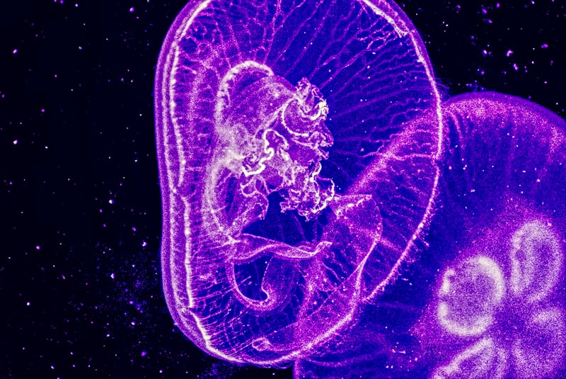
The Morula is a vital, transient stage in the process of early embryonic development, occurring shortly after the successful fertilization of the ovum. Defined structurally, the morula is a solid ball of cells, typically comprising between 16 and 32 blastomeres, which resembles a small mulberry (the literal translation of the Latin term morula). This stage marks the culmination of the initial rapid mitotic divisions, known as cleavage, but precedes the formation of the fluid-filled cavity characteristic of the subsequent blastocyst stage. The fundamental mechanism underlying the morula’s formation is the maintenance of cell mass within the confines of the original zona pellucida, meaning that while the number of cells increases exponentially, the overall size of the embryo remains relatively constant, distinguishing this stage from later growth phases.
During the morula stage, the cells are tightly packed together, a crucial process achieved through cellular interactions that maximize surface contact. While the individual cells, or blastomeres, are initially loosely aggregated, they undergo a phenomenon called compaction, which transforms the loosely organized cluster into a tight, cohesive sphere. This structural integrity is paramount, as it is the prerequisite for the first major cellular differentiation event that will follow. Before compaction, all blastomeres are considered totipotent, meaning they could theoretically form an entire organism; however, the physical forces and molecular signaling established during the morula stage begin to restrict the developmental potential of the outer cells versus the inner cells, setting the stage for the establishment of the embryonic axis.
The morula typically forms between 72 and 96 hours (Day 3 to Day 4) post-fertilization, while the embryo is still navigating the journey down the fallopian tube toward the uterus. The cells at this point are still nourished primarily by cytoplasmic resources inherited from the original oocyte, though some early metabolic demands are met by the surrounding maternal environment. The internal structure, though seemingly uniform, contains the seeds of future complexity, with cells already beginning to align themselves into groups that will eventually give rise to the embryo itself and the necessary supportive structures required for successful implantation and gestation.
The Process of Cleavage and Formation
The formation of the morula is the direct consequence of cleavage, a unique sequence of mitotic divisions that lack intervening growth phases (G1 and G2). This process begins immediately after the fusion of the sperm and egg, resulting in the zygote. The zygote first divides into two blastomeres, then four, and subsequently eight, with each division occurring approximately every 12 to 24 hours. These initial divisions are synchronous; however, as the cell count surpasses eight, the divisions often become asynchronous, leading to transitional stages with odd numbers of cells. It is generally accepted that once the embryo reaches the 16-cell stage, it is designated as a morula, though the functional definition relies more heavily on the occurrence of compaction.
Compaction represents a critical developmental milestone. Before this process, the blastomeres are spherical and easily distinguishable. During compaction, the outer blastomeres flatten against one another, forming tight junctions and gap junctions. These specialized cellular connections mediate communication and adhesion, effectively sealing the outer layer. This action creates a distinction between the external cells, which will interact with the environment, and the internal cells, which are protected from the exterior and are destined to form the organism. This mechanical rearrangement is driven by the expression and localization of cell adhesion molecules, most notably E-cadherin, which ensures the stability and cohesion of the developing structure as it prepares for the next phase of differentiation.
The timing and success of morula formation are highly dependent on the quality of the initial gametes and the integrity of the zygotic genome. Any significant chromosomal anomalies or severe limitations in inherited cytoplasmic components can lead to developmental arrest prior to or during the morula stage. The process is remarkably efficient in healthy embryos, ensuring that the necessary number of cells is generated quickly while the embryo remains mobile within the fallopian tube. The completion of the morula stage signifies that the embryo is ready to enter the uterine cavity, where it will begin the preparations for fluid accumulation and the transformation into the blastocyst, which requires a new set of metabolic adaptations.
Historical Understanding and Early Discoveries
The historical identification and description of the morula stage are closely tied to the advent of microscopy and the systematic study of comparative embryology in the 18th and 19th centuries. Pioneer developmental biologists, such as Caspar Friedrich Wolff and later Karl Ernst von Baer, were instrumental in laying the groundwork for understanding mammalian development. Von Baer, often credited as the “Father of Embryology,” made fundamental observations regarding the initial stages of development in various species, including the identification of the mammalian ovum. While early observations were limited by technology, the concept of rapid cell division post-fertilization leading to a tightly packed cellular structure was recognized as a universal feature of early life.
The specific term “morula” derived its descriptive power from its visual resemblance to a mulberry fruit, which provided a clear morphological marker for researchers attempting to chart the sequence of embryonic development. The ability to precisely stage embryos became paramount for comparative studies across different taxa. Detailed understanding of the internal cellular dynamics, such as the mechanism of compaction and the functional distinction between inner and outer cells, required more advanced techniques, including histochemistry and electron microscopy, which became widely available only in the mid-20th century. These later findings confirmed that the morula is not merely a collection of cells but a highly organized, polarized structure.
Early embryological studies often focused on easily accessible non-mammalian models, such as sea urchins and frogs, where the cleavage stages were readily observable. Applying these principles to mammalian systems, which exhibit internal development, presented significant challenges. It was the careful observation of tubal flushing and subsequent culturing techniques that allowed researchers to isolate and study the morula stage in mammals, confirming that the fundamental process of cleavage, compaction, and subsequent differentiation into the blastocyst was conserved across diverse vertebrate species, validating the initial morphological descriptions established centuries prior.
Transition to the Blastocyst: Differentiation
The most significant role of the morula stage is serving as the bridge between undifferentiated cleavage divisions and the first major morphological and functional differentiation event in the embryo’s life cycle: the formation of the blastocyst. This transition, which occurs around Day 4 or 5 post-fertilization, involves a process called cavitation. As the morula enters the uterine environment, the external cells (now tightly bound by compaction) begin to actively pump fluid from the outside environment into the center of the cellular mass. This influx of fluid creates a central cavity, known as the blastocoel.
As the blastocoel expands, the solid morula is transformed into a hollow sphere—the blastocyst. This physical transformation solidifies the distinction between the two primary cell lineages established during compaction. The outer layer of cells surrounding the blastocoel becomes the trophoblast, which is responsible for forming the fetal portion of the placenta and mediating implantation into the uterine wall. Conversely, the cluster of cells sequestered internally, known as the Inner Cell Mass (ICM), are those cells that will ultimately give rise to the actual embryo and all its tissues.
The successful transition from morula to blastocyst is a critical checkpoint for embryonic viability. If the morula fails to compact correctly or if the necessary molecular pumps fail to initiate cavitation, the embryo will not be able to hatch from the zona pellucida or implant into the endometrium, leading to developmental failure. This transition represents the point where cells transition from being totipotent (capable of forming the entire organism plus supportive structures) to pluripotent (capable of forming only the embryo proper), highlighting the morula as the final stage of truly unrestricted cellular potential.
Practical Relevance: Assisted Reproductive Technologies (ART)
The morula stage holds immense practical significance within the field of Assisted Reproductive Technologies (ART), particularly in In Vitro Fertilization (IVF). Embryologists closely monitor the developmental progression of embryos in culture dishes to assess their viability before uterine transfer. The health and timing of morula formation serve as a robust indicator of the embryo’s developmental competency. A high-quality embryo should achieve the morula stage—demonstrating effective cleavage and compaction—by the end of Day 3 or the beginning of Day 4 in the laboratory setting.
The “how-to” of clinical assessment involves meticulous microscopic observation. Embryos that fail to compact or show excessive fragmentation during the morula stage are often deemed less viable for transfer. In modern IVF protocols, clinics frequently prefer to culture embryos to the blastocyst stage (Day 5 or 6) because this allows for a more rigorous natural selection process, ensuring that only the most robust embryos are transferred. However, some clinical situations necessitate a Day 4 transfer, in which case the morphology and timing of the morula are the primary criteria for selection. The appearance of a tightly compacted, symmetrical morula is highly correlated with the successful development into a healthy blastocyst and subsequent implantation.
Furthermore, the morula stage is sometimes used as a crucial time point for biopsy in Preimplantation Genetic Diagnosis (PGD). Although blastocyst biopsy (sampling trophoblast cells) is now more common, historical and specialized PGD techniques sometimes involved removing one or two blastomeres at the 8-cell stage or the early morula stage for genetic testing. The precise knowledge of the cellular state—whether the cells are still loosely aggregated or fully compacted—is essential for minimizing damage to the embryo while ensuring an adequate sample for detecting chromosomal abnormalities or single-gene disorders before transfer.
Maternal Factors and Environmental Influence
The successful progression through the morula stage is intrinsically linked to the optimal maternal environment, even though the embryo is still contained within the zona pellucida. While the initial cleavage is supported by stored maternal messenger RNA and proteins, the transition into the morula stage often coincides with the critical event of genomic activation, where the embryo’s own DNA takes over control of development. The surrounding environment in the fallopian tube, and later the uterus, provides essential metabolic substrates and signaling molecules necessary for this genetic transition.
Maternal lifestyle factors, including nutrition, age, and exposure to harmful substances (teratogens), significantly impact the quality of the ovum and the environment encountered by the developing morula. For instance, deficiencies in crucial micronutrients, or exposure to toxins like alcohol or certain medications during this sensitive developmental window, can disrupt the complex molecular signaling pathways required for proper compaction and cavitation, potentially halting development before implantation can occur. Therefore, maintaining a healthy physiological state is critical for ensuring the proper transition from the morula to the implanting blastocyst.
The subtle interactions between the morula and the tubal epithelium are also necessary for regulating the embryo’s transit time. Hormonal balances in the mother, particularly levels of progesterone and estrogen, influence the ciliary action and muscular contractions of the fallopian tube, ensuring the morula arrives in the uterus at the optimal time for implantation (around Day 5 or 6). A premature or delayed arrival can lead to developmental difficulties or ectopic implantation. The morula stage thus serves as a period of intense developmental vulnerability where the embryo relies on the precision of both its internal genetic programming and the supportive consistency of the maternal environment.
Connections and Relations in Developmental Biology
The morula is not an isolated event but a critical node within the continuous sequence of human development, firmly situated within the broader subfield of Developmental Biology and Embryology. It is directly preceded by the zygote and the initial cleavage stages (2-, 4-, and 8-cell stages) and is immediately followed by the blastocyst stage. Understanding the morula requires appreciating its position as the last solid, pre-cavitation structure.
Its relationship to the blastocyst is perhaps the most crucial: the morula’s successful compaction dictates the formation of the outer trophoblast layer and the inner cell mass, effectively separating the future embryonic tissues from the future extra-embryonic tissues. This initial cell fate decision is foundational to all subsequent development, including the formation of the germ layers during gastrulation. Failure at the morula stage means a failure to establish the necessary cellular polarity required for successful implantation and the initiation of organogenesis.
Furthermore, the morula stage is intimately related to the concept of cellular potency. While the cells leading up to and including the early morula are often considered totipotent, capable of forming all cells of the body and placenta, the moment compaction establishes the outer and inner cell populations, true totipotency is lost. The inner cell mass cells of the subsequent blastocyst are pluripotent, capable of forming the three germ layers but not the necessary extra-embryonic membranes, thus marking the morula as the final transitional stage where cell fate has not yet been irrevocably restricted. This concept is central to modern stem cell research and regenerative medicine.