Oculovestibular Response: Why Your Eyes Track the World
- The Core Definition of the Oculovestibular Response
- Neural Pathways and Mechanism of Action
- Historical Perspectives and Early Investigations
- A Practical Illustration: The Sudden Noise Scenario
- The Broader Significance in Neurophysiology and Clinical Practice
- Interconnections with Related Reflexes and Sensory Systems
- Classification within Psychology and Neuroscience
The Core Definition of the Oculovestibular Response
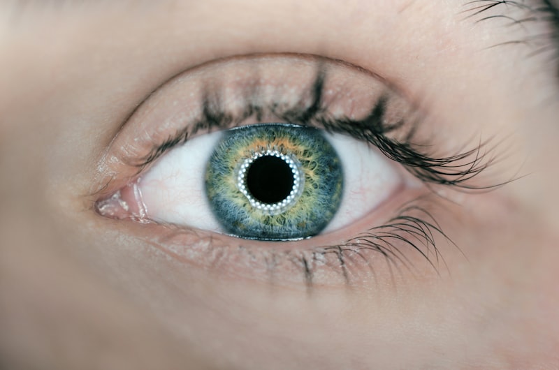
The Oculovestibular Response (OVR) is a fundamental, involuntary physiological reflex that integrates sensory information from the inner ear’s vestibular system with the motor control of eye movements, managed by the ocular system. At its most basic level, the OVR ensures the coordination between head position and eye movements, playing a crucial role in maintaining visual stability and spatial orientation, particularly during unexpected or sudden changes in auditory or vibratory stimuli. This intricate reflex is essential for an organism’s ability to react swiftly to its environment, safeguarding balance and facilitating clear vision even when confronted with abrupt sensory inputs.
More specifically, the OVR is activated by various stimuli directed at the ear, such as an intense sound, a distinct vibration, or an electrical stimulus. Upon detection of such a stimulus, the reflex elicits a characteristic set of responses: the eyes execute rapid, involuntary, and often jerky movements, while simultaneously, the body might experience a compensatory tilt or rotation in the direction opposite to the perceived stimulus. This complex interplay of eye movements and postural adjustments is not merely a localized reaction but a coordinated effort designed to counteract potential disequilibrium, thereby preserving an individual’s balance and enabling the precise synchronization of head and eye motions under dynamic conditions.
The key idea behind the Oculovestibular Response lies in its capacity to process sudden sensory information and translate it into immediate motor actions that protect stability and visual focus. Unlike some other reflexes that respond to head motion directly, the OVR’s unique trigger mechanism – external stimuli impacting the ear – highlights its role as an alarm system. It serves as a rapid defense mechanism, allowing an individual to quickly reorient themselves and stabilize their visual field, preventing disorientation or falls that might otherwise occur due to abrupt environmental disturbances. This reflex underscores the profound interconnectedness of our sensory and motor systems in maintaining our interaction with the world.
Neural Pathways and Mechanism of Action
The sophisticated coordination observed in the Oculovestibular Response is orchestrated by a precise neural circuit involving key structures within the brainstem. Central to this pathway are the vestibular nuclei, a collection of four distinct nuclei situated in the upper medulla and lower pons of the brainstem. These nuclei serve as the primary relay centers for sensory input originating from the inner ear’s vestibular organs, which detect head movements and gravitational forces. When an auditory or vibratory stimulus impacts the ear, specialized sensory cells within the inner ear transmit signals to the vestibular nuclei, signaling an unexpected environmental event that could potentially disrupt balance or visual stability.
From the vestibular nuclei, neural signals are then rapidly transmitted to the oculomotor nuclei, which are located in the midbrain. The oculomotor nuclei are critically responsible for controlling the extrinsic muscles of the eye, which dictate all eye movements. Upon receiving excitatory signals from the vestibular nuclei, the oculomotor nuclei activate specific eye muscles, prompting the characteristic jerky or compensatory eye movements that define a part of the OVR. This direct neuronal link ensures an almost instantaneous coordination between the incoming sensory information about a potential perturbation and the motor response required to stabilize the visual field.
Beyond the direct eye movements, the OVR also involves efferent pathways that modulate postural muscles, leading to the observed body tilt or rotation. This involves further projections from the vestibular nuclei down the spinal cord to influence motor neurons controlling trunk and limb musculature. The entire process, from stimulus reception to coordinated eye and body movements, occurs with remarkable speed, reflecting the evolutionary importance of this reflex in maintaining an organism’s orientation and balance in a dynamic environment. Understanding this intricate neural mechanism is vital for diagnosing and treating conditions that impair vestibular and oculomotor function.
Historical Perspectives and Early Investigations
The study of reflexes involving the vestibular system and eye movements has a rich history within neuroscience and physiology, dating back to the late 19th and early 20th centuries. While the term “Oculovestibular Response” itself gained prominence as research became more specialized, the underlying principles of how the inner ear influences eye motion and posture have been subjects of intensive investigation for decades. Early researchers, often pioneers in neurophysiology, meticulously documented involuntary eye movements, such as nystagmus, in response to various forms of vestibular stimulation, laying the groundwork for understanding reflexes like the OVR.
Throughout the 20th century, the OVR and related reflexes, like the Vestibulo-Ocular Reflex (VOR), were extensively studied in a variety of animal models, including cats, dogs, and monkeys. These animal studies were instrumental in mapping the neural pathways, identifying the specific brainstem nuclei involved – such as the vestibular nuclei and oculomotor nuclei – and elucidating the synaptic connections that govern these rapid, involuntary responses. The controlled experimental conditions offered by animal research allowed for precise manipulations of stimuli and detailed observations of neural and behavioral outcomes, providing foundational insights into the reflex’s mechanics.
In parallel with animal research, the Oculovestibular Response has also been a significant focus of study in humans, both in healthy individuals and those presenting with neurological disorders. Clinical investigations have sought to characterize the normal range of OVR responses and to identify deviations indicative of pathology. The references cited in the original text, dating from the late 1990s and early 2000s, underscore a more recent era of research that has built upon earlier foundations. These studies have utilized advanced techniques to explore the OVR’s nuances in conditions such as multiple sclerosis, stroke, and traumatic brain injury, further solidifying its importance in clinical diagnosis and our understanding of human neurophysiology.
A Practical Illustration: The Sudden Noise Scenario
To fully grasp the essence of the Oculovestibular Response, consider a common, relatable scenario from everyday life: walking down a quiet street when suddenly, an unexpected and extremely loud car horn blares from directly behind you. This abrupt and intense auditory stimulus serves as an ideal trigger for the OVR, demonstrating its protective and orienting functions in a real-world context. The reflex is not a conscious decision but an automatic, hardwired response designed to help you quickly assess and react to a potentially threatening or startling event, ensuring your immediate safety and awareness.
Here’s a step-by-step breakdown of how the OVR manifests in this scenario: First, the sudden loud noise is detected by the sensory receptors in your inner ear. This auditory information, particularly its intensity and suddenness, is quickly relayed through the auditory pathways, with critical cross-talk occurring with the vestibular system‘s sensory processing centers in the brainstem. The vestibular nuclei interpret this abrupt input as a significant environmental perturbation that requires immediate compensatory action, even if your head itself has not yet moved.
Next, in response to this interpreted threat or disorientation signal, the vestibular nuclei rapidly send signals to the oculomotor nuclei. This neural communication triggers a reflexive, often jerky movement of your eyes, which may dart quickly in a specific direction, typically towards the perceived source of the sound or in a compensatory manner to stabilize the visual field against a perceived shift. Simultaneously, signals are also sent to your postural muscles, causing your body to involuntarily stiffen or subtly shift, perhaps tilting slightly away from the sound or rotating to better orient your body. This coordinated reaction – rapid eye movement and subtle body adjustment – is the OVR in action, helping you to quickly locate the source of the startling noise and stabilize your body, preparing you to either avoid danger or continue your activity without losing balance.
The Broader Significance in Neurophysiology and Clinical Practice
The Oculovestibular Response holds profound significance for both theoretical neurophysiology and practical clinical applications. From a fundamental scientific perspective, studying the OVR provides invaluable insights into the intricate mechanisms of sensory integration, motor control, and rapid reflexive behaviors. It allows researchers to unravel how the brainstem acts as a vital hub for processing diverse sensory inputs – auditory, vibratory, and vestibular – and translating them into coordinated motor outputs that ensure stability and effective interaction with a dynamic environment. Understanding the OVR contributes to a broader comprehension of how the central nervous system maintains balance, stabilizes gaze, and orchestrates rapid responses to unexpected stimuli.
Clinically, the integrity of the OVR serves as an important diagnostic marker for various neurological disorders, particularly those affecting the brainstem, vestibular system, or oculomotor pathways. Abnormal or absent OVR can indicate damage or dysfunction within these critical neural structures. For instance, in comatose patients, testing the oculovestibular reflex (often via caloric testing, which induces specific ear stimuli) is a standard procedure to assess brainstem function. The presence or absence of a normal OVR can provide crucial information regarding the level and extent of brain injury, guiding prognosis and treatment decisions in critical care settings.
Furthermore, research into the OVR has direct implications for rehabilitation and therapeutic interventions. For patients recovering from conditions like stroke, multiple sclerosis, or traumatic brain injury, who often experience impairments in balance, gaze stability, and spatial orientation, understanding the OVR can inform targeted therapies. Vestibular rehabilitation exercises, for example, often aim to recalibrate and strengthen these fundamental reflexes. By leveraging knowledge of the OVR’s pathways and functions, clinicians can develop more effective strategies to help patients regain lost neurological function, improve their quality of life, and enhance their ability to navigate their surroundings safely and confidently.
Interconnections with Related Reflexes and Sensory Systems
The Oculovestibular Response does not operate in isolation but is intimately connected with a network of other reflexes and sensory systems that collectively maintain our spatial orientation and visual stability. One of the most closely related and frequently discussed reflexes is the Vestibulo-Ocular Reflex (VOR). While both reflexes involve the vestibular system and eye movements, the VOR is primarily triggered by head movements (e.g., turning your head), whereas the OVR responds to non-head-motion stimuli delivered to the ear (e.g., loud sounds, vibrations). Both reflexes serve the overarching goal of gaze stabilization, ensuring that our eyes remain fixed on a target despite movements of the head or body, thereby preventing blurry vision.
Another related phenomenon frequently observed in the context of vestibular and oculomotor dysfunction is nystagmus. Nystagmus refers to involuntary, rhythmic eye movements characterized by a slow phase in one direction and a rapid corrective phase in the opposite direction. It can be physiological (e.g., optokinetic nystagmus) or pathological, often indicating issues with the vestibular system or its central connections. The jerky eye movements characteristic of the OVR can sometimes resemble or contribute to a form of nystagmoid activity, especially during diagnostic tests like caloric stimulation, which specifically excites the vestibular apparatus to evoke such eye responses.
Beyond these direct reflex connections, the OVR also interacts with broader sensory systems crucial for balance and spatial awareness. The proprioceptive system, which provides sensory information about the position and movement of our limbs and body in space, works in conjunction with the vestibular and visual systems to create a comprehensive internal map of our body’s orientation. The postural adjustments that accompany the OVR are influenced by and, in turn, influence, these proprioceptive inputs, contributing to the overall sense of bodily equilibrium. Therefore, understanding the OVR requires appreciating its integration within this complex sensory web, where signals from multiple modalities converge to create a coherent perception of self-motion and environmental interaction.
Classification within Psychology and Neuroscience
The Oculovestibular Response falls primarily within the domains of Neurophysiology and Sensory Neuroscience. As a reflexive behavior mediated by specific neural pathways in the brainstem, its study delves into the fundamental electrical and chemical processes that govern how the nervous system functions. Neurophysiologists investigate the cellular and synaptic mechanisms underlying the OVR, analyzing nerve impulse transmission, neurotransmitter actions, and the precise timing of neural circuits. This detailed exploration contributes to our understanding of basic brain function and how specific sensory inputs are transduced into motor commands.
Within the broader field of psychology, the OVR is often considered part of Perceptual Psychology or Cognitive Psychology, particularly when examining its role in spatial orientation, attention, and awareness. While the reflex itself is involuntary, its outcome — improved visual stability and bodily reorientation — directly impacts how an individual perceives their environment and allocates attentional resources. For instance, the OVR’s ability to stabilize vision after a sudden startling sound allows for a more rapid and accurate perceptual assessment of the source of the sound, which is a cognitive function critical for survival and interaction with the world.
Furthermore, due to its clinical significance, the OVR is a crucial topic within Clinical Neurology and Neurorehabilitation. In these applied fields, the OVR is not just a theoretical construct but a practical tool for diagnosing neurological disorders and designing therapeutic interventions. Its study helps clinicians understand the pathological processes affecting balance and eye movements in various patient populations and develop strategies to restore function. Thus, the Oculovestibular Response bridges basic scientific inquiry into brain function with applied clinical practice, offering insights into both the healthy and disordered nervous system.