ON Cells: How Light Shapes Your Visual Perception
The Core Definition
ON cells, a specialized type of neuron, are fundamental components of the visual system, particularly within the retina and subsequent visual pathways. They are characterized by their excitatory response to an increase in light intensity within a specific area of their receptive field, known as the “ON-center.” This selective sensitivity to incremental light stimuli is crucial for detecting changes in illumination, identifying bright objects, and perceiving contrast, laying a foundational layer for complex visual processing. Fundamentally, these cells operate like other neurons, comprising a variety of molecular components such as DNA, proteins, lipids, and carbohydrates, which collectively enable their specialized function.
The operational mechanism of an ON cell involves intricate signal transduction pathways. When light hits the photoreceptors (rods and cones) within the retina, it causes a hyperpolarization. This hyperpolarization then affects bipolar cells. In the case of ON bipolar cells, this leads to depolarization, which in turn excites the ON ganglion cells. This process signifies a complex interplay of neurotransmitters and ion channels, orchestrated by the cell’s internal machinery. Like all cells, ON cells possess a nucleus, which serves as the control center, housing the genetic material that dictates the cell’s structure and function. The surrounding cytoplasm contains numerous organelles, each performing specialized tasks essential for the cell’s vitality and responsiveness.
These internal organelles are integral to an ON cell’s ability to process and transmit visual information. Mitochondria, often referred to as the cell’s powerhouses, generate the necessary energy (ATP) to sustain the high metabolic demands of continuous neuronal firing and signal processing. The endoplasmic reticulum is crucial for synthesizing and modifying proteins, including those vital for neurotransmitter production and receptor function. Subsequently, the Golgi apparatus packages and transports these molecules to their appropriate destinations, ensuring that the cell can maintain its complex structure and execute its specialized functions effectively. This intricate cellular machinery allows ON cells to detect subtle changes in light and relay this critical information further along the visual pathway to the brain.
Historical Context
The understanding of specialized retinal cells, including the concept of ON and OFF responses, emerged primarily in the mid-20th century, building upon earlier electrophysiological studies of the visual system. Key insights were provided by researchers like Stephen W. Kuffler in the 1950s. Kuffler, working at the Johns Hopkins University, conducted pioneering experiments on the cat retina, using microelectrodes to record the activity of individual retinal ganglion cells. His groundbreaking work revealed that these cells did not simply respond to light uniformly across the retina but rather to specific patterns of light and dark within distinct areas, which he termed receptive fields.
Kuffler’s experiments demonstrated that some ganglion cells exhibited an excitatory response when light was shone in the center of their receptive field (the “ON-center”), while others responded excitedly when light was turned off in the center (the “OFF-center”). This discovery was revolutionary, fundamentally changing the understanding of how the visual system encodes information. It moved beyond the idea of simple point-to-point transmission of light information and introduced the concept of feature detection at the earliest stages of visual processing. These findings laid the groundwork for subsequent detailed studies by Nobel laureates David Hubel and Torsten Wiesel, who further elucidated the hierarchical processing of visual information in the visual cortex, building directly on the principles of receptive fields established by Kuffler.
The historical development of understanding ON cells is intrinsically linked to the advancement of electrophysiology and neuroscience. Early researchers faced significant technical challenges in isolating and recording from single neurons within complex neural networks. The refinement of techniques for recording neural activity in living organisms allowed for the precise mapping of receptive fields and the characterization of different cell types. This meticulous empirical work provided the initial evidence for specialized cellular responses to specific visual stimuli, transforming theoretical models of vision into empirically supported frameworks. The insights gained from these studies have profoundly influenced our understanding of sensory perception and continue to be foundational in modern visual neuroscience research.
A Practical Example
To illustrate the function of ON cells in a relatable, real-world scenario, consider the act of reading text on a digital screen, such as a white background with black letters. When your eyes scan a page, whether on a computer monitor or a smartphone, your visual system constantly processes changes in light intensity to delineate shapes, letters, and words. An ON cell plays a crucial role in detecting the brighter areas, such as the white background, and the transitions from dark to light, which help define the edges of the black characters against the white.
Specifically, imagine your gaze resting on a segment of a white page where a black letter is displayed. As your eye moves from a dark part of a letter to the bright white background, the ON cells with receptive fields centered on the transition point become highly active. The “how-to” of this process involves the ON cell’s receptive field, which is structured with an excitatory center and an inhibitory surround. When light increases in the center of an ON cell’s receptive field—as it would when your vision shifts from a dark letter to the surrounding bright white space—the cell fires a rapid burst of action potentials. This burst of activity signals to higher visual centers in the brain that a significant increase in light intensity has occurred at that specific location.
Conversely, if the eye were to move from a bright area to a darker area, the ON cells would reduce their firing rate, or be inhibited by the dark stimulus in their center, allowing other cells, such as OFF cells (which respond to decreases in light), to become active. This complementary action of ON and OFF cells creates a robust system for detecting both increases and decreases in light, thereby enhancing contrast perception and enabling the precise discrimination of edges and boundaries. Without the specialized response of ON cells to increments in light, our ability to perceive bright objects, distinguish figures from grounds, and ultimately read text would be significantly impaired, making them essential for a clear and detailed visual experience.
Significance and Impact
The discovery and characterization of ON cells represent a cornerstone in our understanding of visual perception and the broader field of sensory neuroscience. Their specialized response to light increments is not merely a biological curiosity but a fundamental mechanism that underlies our ability to navigate and interpret the visual world. By specifically signaling increases in luminance, ON cells contribute to the initial encoding of visual information, forming a crucial first step in the complex cascade of neural processing that eventually leads to conscious perception. This selective sensitivity allows the visual system to efficiently detect bright objects, discern positive contrast (e.g., a white object on a grey background), and perceive the edges of illuminated forms, which are all vital for tasks ranging from object recognition to spatial orientation.
The impact of understanding ON cells extends deeply into various applications within psychology, medicine, and technology. In psychology, the knowledge of these cells helps explain phenomena related to visual adaptation, contrast sensitivity, and certain visual illusions. For instance, understanding how ON cells contribute to edge detection provides insights into theories of contour perception. Clinically, insights into ON cell function are invaluable in diagnosing and understanding conditions affecting the retina, such as retinitis pigmentosa or glaucoma, where the integrity and function of these cells can be compromised, leading to significant visual impairment. Researchers can use electroretinography (ERG) to assess the health and activity of these cells, providing diagnostic markers for retinal diseases.
Furthermore, the principles derived from ON cell function have inspired technological advancements in artificial vision systems and computer vision. Algorithms designed for edge detection and feature extraction in digital image processing often mimic the center-surround receptive field organization found in these biological neurons. This bio-inspired approach enhances the efficiency and accuracy of machine perception, allowing for better object recognition in robotics, autonomous vehicles, and security systems. The study of ON cells also continues to drive basic research into neural plasticity, how visual circuits develop, and the mechanisms of neural coding, continually pushing the boundaries of our comprehension of the brain’s remarkable ability to construct a coherent visual reality from incoming light signals.
Connections and Relations
ON cells are inextricably linked to several other fundamental concepts and structures within neuroscience and sensory psychology, primarily within the domain of the visual system. Their most direct counterparts are OFF cells. While ON cells are excited by light increments in their receptive field center, OFF cells exhibit the opposite response: they are excited by light decrements (i.e., when light is turned off in their center). This complementary pairing of ON and OFF pathways is crucial for encoding both increases and decreases in light intensity, providing a robust mechanism for detecting contrast and defining edges, which are essential for forming a detailed visual image. The balance and interaction between these two cell types are fundamental for accurate visual processing.
Beyond their direct counterparts, ON cells are integrated into broader visual pathways. Information processed by ON retinal ganglion cells is transmitted via the optic nerve to the lateral geniculate nucleus (LGN) in the thalamus, and then onwards to the visual cortex. Along this pathway, the information is further refined and integrated, contributing to more complex visual features. The concept of the receptive field, first elucidated for retinal ganglion cells (including ON cells), is a foundational principle that extends throughout the visual system, with cortical neurons having more complex and sophisticated receptive fields that integrate inputs from many ON and OFF cells. This hierarchical processing allows for the construction of increasingly complex representations of the visual world, from simple light changes to intricate patterns and objects.
The study of ON cells falls primarily under the broader category of sensory psychology and physiological psychology (or neuroscience, specifically visual neuroscience). These fields explore how sensory information is transduced, processed, and ultimately leads to perception and behavior. Understanding ON cells is also critical to cognitive psychology, especially concerning topics like attention, object recognition, and visual memory, as the initial encoding by ON cells provides the raw data for these higher-level cognitive processes. The general cellular mechanisms described earlier (energy production, communication, molecular components) are universal to all neurons, including ON cells, emphasizing that specialized psychological functions are built upon fundamental biological principles, highlighting the interdisciplinary nature of psychological science.
Impact of Modern Technology on the Study of ON Cells
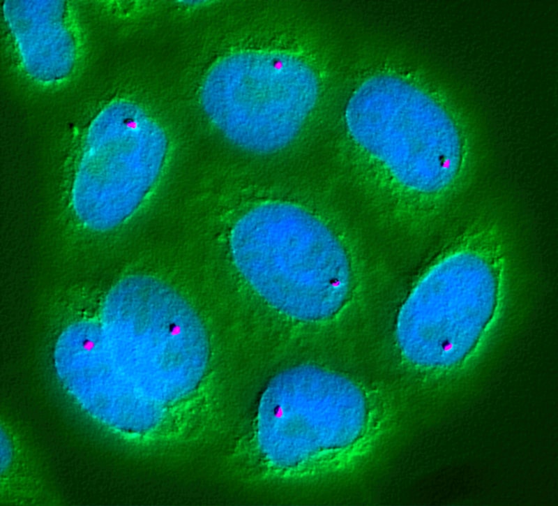
Modern technology has revolutionized the study of ON cells, allowing researchers to delve into their intricate functions with unprecedented detail and precision. Advances in microscopy, particularly confocal microscopy and two-photon microscopy, enable scientists to visualize the morphology of individual ON cells, their dendritic arbors, and synaptic connections in living tissue. This capability has been crucial for understanding the structural basis of their receptive fields and how they integrate inputs from bipolar cells. Furthermore, super-resolution microscopy techniques push the boundaries even further, revealing subcellular structures and the precise localization of proteins involved in signal transduction within these specialized neurons.
Beyond imaging, genetic engineering techniques have opened new avenues for manipulating and studying ON cells. Techniques such as optogenetics allow researchers to selectively activate or inhibit ON cells using light, providing causal links between their activity and visual perception or behavior. Similarly, chemogenetics offers chemical control over ON cell activity. These tools, coupled with gene knockout or knockdown strategies, enable the investigation of specific genes or proteins involved in ON cell development, function, and their role in visual disorders. Such precise control over neural activity was unimaginable just a few decades ago and has significantly accelerated our understanding of complex visual circuits.
Moreover, advanced electrophysiological methods, including patch-clamp recording and multi-electrode arrays, provide high-resolution data on the electrical activity of ON cells. Flow cytometry, initially used for analyzing general cell populations, can be adapted to isolate specific types of retinal cells, including ON cells, for molecular analysis, though its direct application to live functional ON cell activity is less common than in other cell biology contexts. However, the broader principle of measuring cellular activity, often through calcium imaging in live cells, allows researchers to map the responses of large populations of ON cells simultaneously. These technological advancements collectively contribute to a more holistic understanding of ON cells, from their molecular machinery to their network-level contributions to visual processing, and their implications for human perception and disease.
Future Directions and Research
The ongoing study of ON cells continues to be a vibrant area of research, with future directions focusing on several key frontiers. One major area involves a deeper exploration of the neural circuits in which ON cells are embedded. Researchers are employing advanced connectomics techniques, such as electron microscopy reconstruction, to map the precise synaptic connections of ON cells within the retina and their projections to higher brain areas. Understanding these intricate wiring diagrams is essential for fully comprehending how visual information is transformed and integrated across different layers of the visual system, ultimately leading to coherent perception.
Another critical direction involves investigating the role of ON cells in various neurodevelopmental disorders and neurodegenerative diseases affecting vision. For instance, understanding how ON cell function is altered in conditions like amblyopia, diabetic retinopathy, or age-related macular degeneration could pave the way for novel therapeutic interventions. Research is exploring targeted gene therapies or pharmacological approaches to restore or enhance ON cell function in diseased retinas, aiming to preserve or improve visual acuity and quality of life for affected individuals. The potential for retinal prostheses to selectively stimulate ON cells to restore vision in blind patients also represents a significant and promising area of ongoing development.
Furthermore, research is delving into the plasticity of ON cell responses. How do these cells adapt to different lighting conditions, and what mechanisms underlie their ability to maintain functionality across a wide range of luminances? Investigations into the molecular and cellular mechanisms of adaptation, long-term potentiation, and depression in ON cells are critical for understanding the dynamic nature of visual processing. Such studies not only deepen our understanding of fundamental visual physiology but also contribute to broader principles of neuronal adaptation and learning, reinforcing the central role of ON cells as a model system for exploring the complexities of the brain’s interaction with the sensory world.