Partial Lipodystrophy: The Psychology of Body Image Loss
- Introduction and Definition of Partial Lipodystrophy (PLD)
- Classification and Genetic Basis
- Clinical Presentation and Characteristic Adipose Tissue Redistribution
- Associated Metabolic Complications
- Etiology and Pathogenesis
- Diagnostic Criteria and Assessment
- Management and Therapeutic Approaches
- Psychological and Quality of Life Implications
Introduction and Definition of Partial Lipodystrophy (PLD)
Partial Lipodystrophy (PLD) refers to a heterogeneous group of rare, chronic disorders characterized primarily by selective, abnormal redistribution of adipose tissue. This condition is fundamentally a severe lipid-metabolism disorder, resulting in the inability of the body to store fat safely within appropriate subcutaneous depots. Instead, patients exhibit localized, often severe, loss of subcutaneous fat (lipoatrophy) in specific anatomical regions, concomitant with paradoxical accumulation of fat (lipoaccumulation) in others, particularly the lower abdomen, buttocks, and thighs. The clinical recognition of PLD often occurs during early childhood or adolescence, although the underlying metabolic derangements may manifest later in life, contributing significantly to morbidity.
The core defining feature of PLD, distinguishing it from generalized lipodystrophy, is the regional specificity of the fat loss. Historically, and as highlighted in clinical vignettes, the condition is often noted when parents or clinicians observe a significant and symmetric lack of adipose tissue in the facial area, leading to a gaunt appearance. This facial wasting is coupled with a prominent muscular and venous structure beneath the skin. While the upper body—including the face, neck, and upper limbs—typically displays severe lipoatrophy, the regions below the waist might or might not be lacking in fat, often presenting a stark contrast of upper-body wasting and lower-body adiposity.
The critical physiological consequence of this dysfunctional fat distribution is not merely cosmetic; functional adipose tissue is vital for buffering excess lipids and maintaining metabolic homeostasis. When fat storage capacity is compromised or exhausted in the safe subcutaneous depots, lipids spill over and are deposited ectopically in organs such as the liver, muscle, and pancreas. This ectopic fat deposition is the primary driver of severe metabolic complications that define the pathology of PLD, leading to conditions far more debilitating than the initial aesthetic changes.
Classification and Genetic Basis
Partial Lipodystrophy is generally categorized into two main forms: Familial Partial Lipodystrophy (FPLD), which is genetically inherited, and Acquired Partial Lipodystrophy (APL), also known as Barraquer-Simons syndrome, which is believed to have an autoimmune etiology. The delineation between these forms is crucial for diagnosis and guiding therapeutic intervention, as their underlying pathogenic mechanisms differ significantly despite overlapping clinical presentations related to metabolic dysfunction.
Familial Partial Lipodystrophy is the most commonly recognized form, with several subtypes designated based on the specific gene mutation involved. The most prevalent and well-studied subtype is FPLD Type 2, or the Dunnigan variety, which is caused by dominant mutations in the LMNA gene. This gene encodes A-type lamins, crucial structural proteins of the nuclear envelope. Mutations in LMNA disrupt the stability of the nuclear structure in pre-adipocytes, leading to premature cell death and differentiation arrest, thereby severely impairing the development and function of mature fat cells in the affected regions. Other, less common genetic forms involve mutations in genes such as PPARG (Peroxisome Proliferator-Activated Receptor Gamma) and AKT2, each contributing to unique variations in the extent and distribution of lipoatrophy.
Acquired Partial Lipodystrophy (APL) presents a distinct clinical challenge. Its pathogenesis often involves an autoimmune process, typically initiated after an infection or during early childhood, frequently noted between the ages of five and fifteen. A hallmark of APL is the progressive, cephalocaudal loss of subcutaneous fat—starting in the face and neck and descending gradually to the trunk. Unlike FPLD, APL is associated with immunological markers, specifically the presence of C3 nephritic factor (C3NeF) and low levels of the complement component C3, suggesting that complement system dysregulation contributes to the destruction of adipocytes. This progressive nature underscores the need for continuous monitoring and management, as the associated metabolic abnormalities may develop years after the initial aesthetic changes are observed.
Clinical Presentation and Characteristic Adipose Tissue Redistribution
The clinical presentation of PLD is highly distinctive and often serves as the initial pointer toward diagnosis. The classic feature involves the symmetric, pronounced wasting of fat in the face, leading to prominent cheekbones, sunken eyes, and a taut appearance of the skin over the musculature. This atrophy extends to the neck, shoulders, and sometimes the arms, creating a marked contrast between the upper and lower halves of the body. For instance, in FPLD Type 2, the onset often correlates with puberty, where the normal expected accumulation of fat in the face and upper body fails to occur or regresses dramatically, leading to significant body image distress in affected individuals.
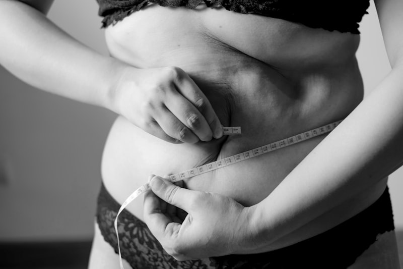
Crucially, the fat depletion in the upper body is often accompanied by a compensatory, paradoxical increase in adipose tissue in the lower body, particularly the hips, thighs, and gluteal region, and often severe intra-abdominal (visceral) fat accumulation. This redistribution is highly detrimental, as visceral fat is metabolically active and proinflammatory. Furthermore, females with PLD commonly exhibit fat accumulation in the neck and supraclavicular regions, sometimes referred to as a “buffalo hump,” despite the overall generalized inability to store lipids safely. The specific pattern of fat loss—facial and upper-body wasting combined with lower-body or truncal lipoaccumulation—is pathognomonic for PLD.
The progression of the disorder can vary significantly depending on the subtype. In acquired forms, the loss may be slow and gradual, spanning years, whereas in genetic forms, the changes are often more abrupt during periods of hormonal flux, such as early adolescence. Regardless of the speed, the selective destruction or failure of adipocytes profoundly impacts the patient’s health and psychological well-being. This highly visible, unusual distribution of fat often leads to significant misunderstanding and misdiagnosis, underscoring the need for specialized clinical evaluation when this specific pattern of adipose deficiency is noted.
Associated Metabolic Complications
The most severe consequences of Partial Lipodystrophy stem from the metabolic syndrome that universally accompanies the condition. Because functional subcutaneous adipose tissue is depleted, the body lacks sufficient capacity to store dietary triglycerides, leading to a flood of lipids into the bloodstream and ectopic storage in vital organs. This state results in profound and often refractory metabolic abnormalities that require aggressive pharmacological management.
A primary and highly dangerous complication is severe insulin resistance. Adipose tissue normally secretes adipokines (such as leptin and adiponectin) that regulate insulin sensitivity. In PLD, the dysfunctional fat cells fail to release sufficient protective adipokines, and the ectopic lipid deposition in muscle and liver tissues interferes directly with insulin signaling pathways. This resistance is often severe enough to require extremely high doses of insulin, yet glycemic control remains challenging, often leading to early onset Type 2 Diabetes Mellitus (T2DM). The severity of T2DM in PLD patients is often greater than in typical T2DM patients due to the underlying severe lack of fat storage capacity.
Furthermore, PLD is characterized by severe dyslipidemia, specifically marked hypertriglyceridemia and very low levels of High-Density Lipoprotein (HDL) cholesterol. High circulating triglycerides are a direct result of the inability to clear chylomicrons and VLDL particles effectively, posing an immediate risk of acute pancreatitis, a life-threatening inflammatory condition. The chronic dyslipidemia significantly accelerates the progression of atherosclerosis, placing patients at a heightened risk for premature cardiovascular disease, including coronary artery disease and stroke, often decades earlier than the general population.
The ectopic lipid deposition also manifests as significant organ damage, most notably hepatic steatosis (fatty liver disease), which can progress to Non-Alcoholic Steatohepatitis (NASH) and cirrhosis. The severity of these metabolic complications necessitates a comprehensive, multidisciplinary approach to care. Key metabolic sequelae include:
- Refractory Hyperglycemia: Requiring intensive insulin therapy.
- Severe Hypertriglyceridemia: Leading to risk of acute pancreatitis.
- Premature Atherosclerosis: Increasing cardiovascular morbidity.
- Hepatic Steatosis: Risk of progression to end-stage liver disease.
Etiology and Pathogenesis
The underlying mechanisms driving PLD involve distinct pathways depending on whether the condition is familial or acquired, but both result in the failure of adipocyte function. In FPLD, the pathogenesis is rooted in genetic defects that compromise the structural integrity or regulatory functions of adipocytes. For instance, the LMNA gene mutations lead to the production of mutated lamin proteins that structurally destabilize the nuclear membrane, rendering the pre-adipocytes highly sensitive to mechanical stress and leading to their premature apoptosis, particularly in areas of high mechanical strain, such as the face.
The specific anatomical distribution of fat loss in FPLD is thought to be related to the unique developmental timing and metabolic activity of different fat depots. Adipose tissue in the face and upper extremities tends to be more metabolically active and susceptible to the structural defects caused by lamins, leading to early and severe atrophy in these regions. Conversely, the fat that accumulates in the lower body, particularly in the gluteal and femoral regions, may represent a metabolically less-active depot that is resistant to the effects of the genetic mutation, or perhaps a compensatory mechanism attempting to sequester excess lipids.
Conversely, the pathogenesis of Acquired Partial Lipodystrophy (APL) is strongly linked to immune system dysregulation. The gradual destruction of adipocytes in a cephalocaudal pattern suggests an ongoing inflammatory or immune assault. The strong association with C3 nephritic factor (C3NeF), an autoantibody that stabilizes the C3 convertase, leading to excessive consumption of C3, indicates a connection to the alternative pathway of the complement cascade. This sustained activation of complement components is hypothesized to target and destroy adipocytes selectively. Furthermore, APL is often associated with immune-mediated renal diseases, such as membranoproliferative glomerulonephritis, reinforcing the underlying autoimmune etiology that drives the localized destruction of fat cells and subsequent metabolic chaos.
Diagnostic Criteria and Assessment
Diagnosis of Partial Lipodystrophy requires a high index of clinical suspicion combined with detailed biochemical and, often, genetic confirmation. Given the rarity of the condition, it is frequently missed or misdiagnosed as Cushing’s syndrome, anorexia nervosa, or simple obesity, especially in patients presenting primarily with severe metabolic syndrome.
The initial clinical assessment must focus on documenting the specific pattern of adipose tissue loss and accumulation. This involves a thorough physical examination to confirm the symmetric facial wasting and the contrast between upper-body lipoatrophy and lower-body lipoaccumulation. Once PLD is suspected, laboratory testing is crucial to quantify the severity of the associated metabolic dysregulation. Key diagnostic markers include fasting plasma glucose and insulin levels (to assess insulin resistance), lipid panels showing profound hypertriglyceridemia, and liver function tests to check for hepatic steatosis. Genetic testing is essential for confirming FPLD subtypes, particularly screening for LMNA mutations, which establishes a definitive diagnosis and assists in prognosis.
Imaging techniques provide objective confirmation of fat redistribution. Dual-energy X-ray Absorptiometry (DEXA) scans and whole-body Magnetic Resonance Imaging (MRI) are used to accurately map and quantify subcutaneous and visceral fat depots. These imaging studies can precisely measure the percentage of fat in specific body segments, providing concrete evidence of the specific regional atrophy and paradoxical accumulation. For APL, immunological testing, including complement factor assays (C3, C4) and screening for C3NeF, is vital to support the acquired, autoimmune etiology. The integration of clinical presentation, metabolic profile, and specialized testing is mandatory for accurate diagnosis and the initiation of appropriate, highly specialized treatment protocols.
Management and Therapeutic Approaches
The management of Partial Lipodystrophy is complex and focuses on mitigating the severe metabolic consequences stemming from ectopic lipid deposition, alongside addressing the physical symptoms. Standard treatments for T2DM and dyslipidemia are often insufficient due to the profound nature of the underlying metabolic defect.
Pharmacological management involves aggressive control of hyperglycemia and dyslipidemia. Due to severe insulin resistance, patients often require high doses of insulin, sometimes combined with insulin sensitizers like metformin and thiazolidinediones (although the latter must be used cautiously). For hypertriglyceridemia, high-dose fibrates and omega-3 fatty acids are typically employed. However, the cornerstone of advanced metabolic therapy for lipodystrophy patients who are leptin deficient is the use of leptin replacement therapy.
Metreleptin, a synthetic analog of the hormone leptin, represents a breakthrough in treating the severe metabolic aspects of PLD, particularly in patients with low endogenous leptin levels resulting from inadequate fat mass. Leptin replacement significantly improves insulin sensitivity, reduces hypertriglyceridemia, and often leads to the resolution of hepatic steatosis by redirecting lipids away from ectopic sites and enhancing their safe metabolism. The introduction of metreleptin has dramatically altered the prognosis for many patients with severe PLD. Furthermore, addressing the physical manifestations of lipoatrophy, particularly the facial wasting, often involves plastic surgery techniques such as autologous fat grafting or dermal fillers, although these procedures can be challenging as the underlying metabolic environment may impair the long-term viability of the grafted fat.
Psychological and Quality of Life Implications
The visible and often disfiguring nature of Partial Lipodystrophy carries profound psychological and social consequences, significantly impacting the patient’s quality of life. The onset of symptoms, especially during infancy or early adolescence, coincides with crucial periods of self-identity formation, leading to severe body image distress and psychological morbidity.
The stark contrast between the emaciated appearance of the face and upper body and the accumulation of fat in other regions often leads to social stigma, bullying, and feelings of isolation. Patients frequently report anxiety, depression, and avoidance of social situations due to self-consciousness about their appearance. This psychological burden is compounded by the chronic nature of the metabolic complications, which require continuous self-monitoring, dietary restriction, and frequent medical appointments, leading to significant emotional fatigue.
Therefore, comprehensive care for PLD must extend beyond endocrinology and metabolism to include robust psychological and psychiatric support. A multidisciplinary team approach is essential, involving dietitians for specialized nutritional counseling, social workers, and mental health professionals to address body image issues and coping strategies. Recognizing that the psychosocial sequelae are integral to the disease presentation ensures that patients receive holistic treatment aimed not only at prolonging life but also at enhancing their overall well-being and successful integration into social and professional life.