The Phrenic Nerve: Breathing Life into Mind-Body Connection
- Anatomical Origin and Course of the Phrenic Nerve
- Physiological Function: The Engine of Respiration
- Sensory Innervation and Referred Pain Phenomena
- Etiology of Phrenic Nerve Injury and Dysfunction
- Clinical Presentation and Diagnostic Assessment
- Surgical and Therapeutic Management Strategies
- Relationship to the Autonomic Nervous System
Anatomical Origin and Course of the Phrenic Nerve
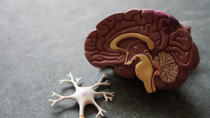
The Phrenic Nerve (from the Greek word phren, meaning diaphragm) represents a critical component of the somatic nervous system, tasked primarily with controlling respiration. Its initialization occurs within the **cervical plexus** of the neck, derived predominantly from the anterior rami of the third, fourth, and fifth cervical spinal nerves (C3, C4, and C5). This specific root contribution is often summarized clinically by the mnemonic: “C3, 4, 5 keeps the diaphragm alive,” highlighting the absolute necessity of these segments for maintaining autonomous breathing. Immediately upon formation, the nerve descends almost vertically through the neck, traversing the anterior surface of the **anterior scalene muscle**, a key anatomical landmark used by clinicians and surgeons to locate and protect the nerve during procedures involving the cervical region. This long, uninterrupted course demonstrates the evolutionary importance of securing the neural pathway responsible for the body’s most fundamental rhythmic process.
After navigating the neck, the Phrenic Nerve enters the thorax, passing between the subclavian artery and vein, and continues its descent within the **mediastinum**. The trajectory differs slightly between the right and left nerves due to the asymmetry of the thoracic cavity and the presence of major vascular structures. The right phrenic nerve travels alongside the right brachiocephalic vein and the superior vena cava, often passing through the caval opening to reach the inferior surface of the diaphragm. Conversely, the left phrenic nerve follows a more tortuous path, crossing anteriorly over the arch of the aorta and the root of the left lung, before descending alongside the pericardium of the heart to reach the left hemidiaphragm. This close anatomical relationship with the heart and major blood vessels explains why the nerve, in addition to its motor function, transmits sensory branches to the pericardium and pleura, integrating it into the sensory feedback loop governing thoracic and abdominal comfort.
The nerve’s terminal destination is the diaphragm, the primary muscle of inspiration, which it reaches by penetrating the muscle fibers from below. This precise anatomical arrangement ensures that the phrenic nerve is the **sole motor supply** to the diaphragm, granting it complete control over the movement necessary for pulmonary ventilation. The substantial length and complex path through the neck and mediastinum render the phrenic nerve vulnerable to various forms of injury, including mechanical trauma, compression from tumors, and iatrogenic damage during surgical interventions, particularly those involving the neck, thoracic cavity, or cardiac structures. Understanding the minute details of its course is paramount for accurate diagnosis of respiratory distress stemming from neurological causes and for meticulous surgical planning aimed at preserving its integrity.
Physiological Function: The Engine of Respiration
The fundamental physiological role of the Phrenic Nerve is the motor control of the diaphragm, making it the indispensable engine driving pulmonary ventilation. Through the transmission of action potentials, the nerve instructs the large, dome-shaped sheet of muscle fibers that forms the floor of the thoracic cavity to contract. During the contraction phase, the central tendon of the diaphragm is pulled inferiorly, causing the dome to flatten significantly. This flattening action dramatically increases the vertical dimension and overall volume of the thoracic cavity. According to Boyle’s law, this increase in volume leads to a corresponding decrease in intra-thoracic pressure relative to atmospheric pressure, creating a vacuum effect that draws air into the lungs—the process known as **inhalation** or inspiration.
While the act of quiet expiration is largely passive, relying on the elastic recoil of the lungs and chest wall, the initial, necessary expansion is entirely dependent on the intact function of the phrenic nerves. A fully functional phrenic nerve allows for efficient, deep, and rhythmic breathing, which is essential for adequate gas exchange (oxygenation and carbon dioxide removal). The rhythmic firing of the phrenic nerve is controlled centrally by the respiratory centers located in the brainstem, specifically the medulla oblongata and the pons, which adjust the rate and depth of breathing based on metabolic demands signaled by chemoreceptors monitoring blood pH and gas concentrations. This complex reflex loop ensures that breathing occurs automatically and subconsciously, unless voluntarily overridden.
Disruption to the Phrenic Nerve’s function, even unilaterally, severely compromises respiratory mechanics. If the nerve is damaged or severed, the corresponding hemidiaphragm becomes paralyzed. Instead of contracting downward during inspiration, the paralyzed side moves paradoxically upward, pushed by the negative pressure generated by the contracting, healthy hemidiaphragm and the abdominal contents. This phenomenon, known as **paradoxical breathing**, significantly reduces tidal volume and vital capacity, leading to symptoms of **dyspnea** (shortness of breath), particularly when lying flat (orthopnea) or during periods of increased physical exertion. Bilateral phrenic nerve paralysis is a life-threatening emergency, often requiring immediate mechanical ventilatory support, underscoring the vital, non-redundant nature of its motor supply.
Sensory Innervation and Referred Pain Phenomena
Although overwhelmingly recognized for its motor role, the Phrenic Nerve also contains crucial afferent (sensory) fibers. These sensory components originate primarily from the central tendon of the diaphragm, the parietal pleura (the membrane lining the thoracic cavity), and the parietal pericardium (the sac surrounding the heart). These sensory inputs provide feedback regarding the stretching, irritation, or inflammation of these internal structures. This dual functionality means that the nerve acts not only as a conduit for movement commands but also as a monitor for the internal environment of the chest and upper abdomen, alerting the central nervous system to potential distress or pathological changes in these areas.
Perhaps the most clinically significant aspect of the phrenic nerve’s sensory contribution is the phenomenon of **referred pain**. Because the nerve roots (C3, C4, C5) that contribute to the phrenic nerve also contribute to the supraclavicular nerves and other sensory nerves supplying the shoulder and neck region, irritation of the diaphragm or adjacent structures innervated by the phrenic nerve can be perceived by the brain as pain originating in the ipsilateral shoulder tip or lateral neck. For instance, irritation of the diaphragm caused by conditions like peritonitis (inflammation of the peritoneal lining), subphrenic abscesses, or even effusions of blood or fluid following abdominal trauma, frequently manifests as discomfort reported specifically in the shoulder area.
This referral pattern is a critical diagnostic clue. A patient presenting with unexplained shoulder pain, particularly if it is sharp, constant, and not easily explained by musculoskeletal injury, may be experiencing diaphragmatic or visceral pathology. Physicians are trained to recognize that pain referred via the phrenic nerve requires investigation of internal organs, including the liver, spleen, and gallbladder, whose capsules or surrounding fluid might impinge upon the diaphragm. The sensory fibers thus serve as an early warning system, utilizing a shared neural pathway to project deep visceral pain to a more superficial, recognizable dermatomal area.
Etiology of Phrenic Nerve Injury and Dysfunction
Dysfunction of the Phrenic Nerve (PNI) can arise from a wide array of pathological processes, which are broadly categorized into traumatic, compressive, infectious, and neuropathic causes. **Traumatic injury** is common, particularly in cases involving severe neck or chest trauma, such as motor vehicle accidents, where sharp bone fragments or excessive stretching of the cervical spine can directly damage the C3-C5 roots or the nerve trunk itself. Furthermore, **iatrogenic injury**, or damage caused unintentionally during medical procedures, is a frequent cause; procedures involving the neck (e.g., radical neck dissections), thoracic surgery, or cardiac procedures (especially cryoablation for atrial fibrillation) carry a risk of mechanical or thermal damage to the nerve due to its close proximity to the operative field.
**Compressive lesions** represent another major category of PNI. Since the phrenic nerve has such a long, defined course, it is highly susceptible to compression by expanding masses. Malignant tumors, particularly **Pancoast tumors** located in the apex of the lung, are notorious for involving the lower cervical and upper thoracic nerve roots, often leading to combined phrenic nerve and brachial plexus symptoms. Non-malignant masses, such as large mediastinal lymphadenopathy, aortic aneurysms, or even enlarged pericardial sacs, can also exert sufficient pressure to induce demyelination and functional paresis of the nerve. The slow onset of symptoms associated with compression often allows the body to partially adapt, making the subtle signs of diaphragmatic weakness easier to miss in early stages.
Finally, **neuropathic and infectious etiologies** contribute significantly to phrenic nerve impairment. Systemic diseases such as **Guillain-Barré Syndrome (GBS)**, an acute autoimmune polyneuropathy, frequently affects the phrenic nerve, sometimes leading to rapidly progressive, life-threatening respiratory failure requiring intubation. Viral infections, including Herpes Zoster or certain post-viral syndromes, can specifically target the nerve roots. Chronic conditions like **motor neuron disease** (e.g., Amyotrophic Lateral Sclerosis or ALS) or muscular dystrophies also eventually compromise phrenic nerve function, leading to progressive respiratory decline that is typically resistant to direct nerve intervention.
Clinical Presentation and Diagnostic Assessment
The clinical presentation of Phrenic Nerve Injury (PNI) is dependent on whether the damage is unilateral or bilateral, and the extent of the paresis or paralysis. Unilateral paralysis may be asymptomatic at rest, especially in young, otherwise healthy individuals, as the remaining hemidiaphragm and accessory breathing muscles can compensate. However, patients typically report **dyspnea upon exertion** and often experience **orthopnea**, a difficulty breathing when lying flat, due to the upward pressure exerted by abdominal contents on the already weak diaphragm. Bilateral paralysis, conversely, presents as severe respiratory failure, profound dyspnea, and often necessitates immediate ventilatory support due to the complete loss of the primary inspiratory muscle.
Diagnosis begins with a thorough clinical examination, observing for signs of paradoxical breathing. The definitive diagnostic tool for assessing diaphragmatic function is typically **fluoroscopy** (a dynamic X-ray examination), often performed during a procedure called the “sniff test.” During the sniff test, the patient is asked to inhale sharply. In a healthy individual, both sides of the diaphragm move sharply downward. If PNI is present, the paralyzed hemidiaphragm will move paradoxically upward into the chest cavity, pushed by the rapid drop in intrathoracic pressure and the simultaneous downward movement of the intact side. Chest X-rays can also reveal a permanently elevated hemidiaphragm, suggesting chronic paralysis.
To determine the extent and location of the nerve damage, electrophysiological studies are essential. **Electromyography (EMG)** of the diaphragm, performed by inserting fine needle electrodes into the muscle, can confirm denervation (lack of electrical activity) or reinnervation patterns. **Nerve Conduction Studies (NCS)** can assess the latency and amplitude of the action potential traveling along the phrenic nerve, often stimulated non-invasively in the neck. These tests help differentiate between damage to the nerve itself (neuropathy) versus primary muscle failure (myopathy) and are crucial for prognosis, as the presence of even small electrical signals suggests potential for recovery.
Surgical and Therapeutic Management Strategies
The therapeutic management of Phrenic Nerve Injury is highly individualized and depends heavily on the underlying cause and the prognosis for spontaneous recovery. If the injury is acute, such as following surgical trauma, management often involves a period of conservative observation, as nerve function may return within 6 to 12 months, particularly in cases of neurapraxia (temporary conduction block). During this observational phase, **respiratory muscle training** and physical therapy are employed to maximize the efficiency of accessory breathing muscles and prevent lung collapse.
For cases of permanent, non-recovering paralysis, particularly bilateral paralysis leading to ventilator dependence, advanced surgical techniques are considered. One of the most significant advances is **diaphragmatic pacing**. This technique involves surgically implanting electrodes directly onto the phrenic nerve or into the diaphragm muscle, which are connected to an external or implanted stimulator. The pacemaker delivers rhythmic electrical impulses to the nerve, mimicking the natural signals from the brainstem and causing the diaphragm to contract rhythmically, thereby restoring functional respiration and potentially liberating the patient from mechanical ventilation. Diaphragmatic pacing has proven transformative for patients with high spinal cord injury or central hypoventilation syndromes.
In situations where the nerve has been anatomically severed or severely damaged, requiring reconstruction, **nerve transfer procedures** may be performed. This involves harvesting a less critical motor nerve (such as a branch of the intercostal nerves or a motor fascicle from the brachial plexus) and surgically connecting it to the distal stump of the damaged phrenic nerve. Although the recovery of functional strength is slow and often incomplete, nerve transfer offers a chance for reinnervation and improvement in diaphragmatic function, leading to greater respiratory independence and a significantly improved quality of life. The decision to proceed with surgery is always balanced against the risks and the patient’s overall respiratory reserve.
Relationship to the Autonomic Nervous System
While the Phrenic Nerve is classically categorized as a somatic nerve—meaning it controls voluntary, striated muscle (the diaphragm)—its functions are deeply integrated with the autonomic nervous system (ANS), particularly in the regulation of visceral reflexes and cardiac function. The nerve sheath contains autonomic fibers, and its close physical proximity to the vagus nerve (Cranial Nerve X) and sympathetic chains within the mediastinum ensures a complex interplay of signals. For instance, strong stimulation or irritation of the phrenic nerve roots can trigger sympathetic responses, affecting heart rate and blood pressure, demonstrating that its role extends beyond simple motor command.
The sensory feedback the phrenic nerve provides to the central nervous system also influences autonomic responses. Irritation of the pericardium, conveyed via phrenic afferents, can trigger reflexes that alter heart rate or vascular tone. Furthermore, the ability of the phrenic nerve to drive respiration is fundamentally regulated by the autonomic control centers in the brainstem that monitor blood gases. When blood carbon dioxide levels rise (hypercapnia), the respiratory centers increase the firing rate of the phrenic nerve, causing deeper and faster contractions of the diaphragm, a purely autonomic, life-sustaining response that overrides the somatic control pathways during periods of high metabolic stress.
Understanding this interrelation is crucial in clinical settings. Conditions like hiccups (singultus), which are involuntary spasms of the diaphragm, represent a temporary, often benign, disruption to the phrenic nerve’s rhythmic function, possibly initiated by irritation of the phrenic afferents in the abdomen or chest, or sometimes originating from the central respiratory pacemaker itself. Conversely, high spinal cord injuries that spare the C3-C5 segments can leave the diaphragm intact, even as the patient loses control over lower body movement, highlighting the phrenic nerve’s unique and isolated position as the critical conduit between the brainstem’s autonomic respiratory drive and the mechanical process of breathing.