The Pinna: Decoding How Our Ears Shape Sensory Perception
Definition and Nomenclature
The structure known anatomically as the pinna, or more commonly as the auricle, represents the visible, projecting component of the external ear apparatus. This cartilaginous flap is affixed to the side of the head, typically adjacent to the temporal bone, and serves as the initial collector and funnel for airborne sound waves before they enter the external acoustic meatus. The pinna is fundamentally distinct from the middle and inner ear structures, which are embedded within the cranial vault. While the term auricle is often used synonymously in clinical settings, pinna is preferred in precise anatomical description, originating from the Latin word for feather or wing, reflective of its shape. Understanding the nomenclature is crucial because defects or variations in the pinna are often termed auricular anomalies, highlighting the interchangeability of these specific terms within the sphere of otolaryngology. Its primary superficial role is aesthetic, yet its complex, convoluted shape is integral to the sophisticated process of directional hearing and sound localization within the environment.
The human pinna is composed predominantly of elastic cartilage, providing both flexibility and structural integrity, allowing it to withstand minor physical stresses without permanent deformation. The sole exception to this cartilaginous framework is the inferior-most portion, termed the lobule, which consists primarily of dense connective tissue and adipose material, a feature that holds significant cultural and clinical importance, particularly related to practices like ear piercing. The orientation of the pinna is subtle but crucial; it is positioned at an angle of approximately 15 to 30 degrees relative to the mastoid process, maximizing its efficiency in gathering sounds that originate from the frontal and lateral fields. This complex three-dimensional curvature ensures that sound entering the ear is subtly modified—a process essential for the central nervous system to accurately interpret the source of acoustic stimuli.
From a psychological perspective, the pinna provides immediate external identity for an individual, and deviations from typical size or shape can lead to significant psychological distress, particularly during formative years. The structure acts as the sentinel of the auditory system, directing the vibratory energy down the external acoustic canal toward the tympanic membrane. Therefore, while often viewed merely as an appendage, the pinna is the necessary gateway for translating environmental pressure waves into neurological signals. Its specific geometry, involving numerous ridges and depressions, creates minute time differences and spectral cues in the incoming sound, which are decoded by the brain to determine whether a sound originates from above, below, or level with the listener.
Gross Anatomy and Structure
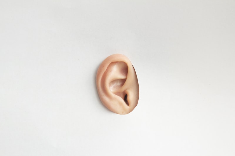
The morphological complexity of the pinna is defined by a series of specific anatomical landmarks, each contributing to its overall function and unique appearance. The prominent outer rim is known as the helix, which curls inward and terminates inferiorly at the lobule. Running parallel to the helix is the antihelix, a curved ridge that divides superiorly into two separate crura: the superior and inferior crura. The depression created between these two crura is termed the triangular fossa. Immediately anterior to the antihelix lies the deepest depression of the external ear, the concha, which funnels directly into the external auditory meatus. The concha itself is divided into the cymba conchae (superior part) and the cavum conchae (inferior part), both acting as resonant chambers.
Protecting the entrance to the external acoustic meatus is the tragus, a small, triangular projection anteriorly situated. Opposite the tragus, and separated by the intertragal notch, is the antitragus, marking the posterior boundary of the conchal bowl. These small cartilaginous structures play a minor role in filtering high-frequency sounds originating from behind the head, contributing subtly to localization. The skin covering the pinna is tightly adherent to the underlying perichondrium, especially over the antihelix and helix, except at the lobule, where the skin is loose and mobile. This tight adherence is clinically relevant because trauma or infection can easily strip the perichondrium, jeopardizing the blood supply to the cartilage and leading to subsequent tissue necrosis or deformity.
Although functionally vestigial in humans, the pinna possesses rudimentary extrinsic and intrinsic musculature. The extrinsic muscles—the anterior, superior, and posterior auricular muscles—connect the pinna to the skull and scalp fascia. While these muscles are highly developed and functional in many mammals, allowing for focused movement (pinniped movement) toward a sound source, in humans they are typically non-functional or capable of only minimal, involuntary twitching, representing an evolutionary remnant. The primary neurovascular supply is derived from branches of the external carotid artery, notably the posterior auricular and superficial temporal arteries, and venous drainage follows corresponding pathways. Sensory innervation is remarkably complex, involving contributions from the greater auricular nerve, the lesser occipital nerve, the auriculotemporal nerve (a branch of the trigeminal nerve), and even the auricular branch of the vagus nerve (Arnold’s nerve), explaining the variability in pain referral patterns experienced during ear manipulation or infection.
Physiological Function and Acoustic Role
The most critical physiological function of the pinna is its role in the capture, amplification, and spatial processing of sound. As an acoustical antenna, the pinna enhances the intensity of sounds within the frequency range most important for human speech (approximately 2 kHz to 5 kHz) by directing the sound waves into the external auditory canal. This natural amplification, often referred to as the ear canal resonance effect, can provide a gain of up to 10 to 12 decibels in this critical range, significantly improving audibility. However, the pinna’s unique anatomical convolutions are what provide the crucial information necessary for sound localization, particularly regarding the vertical plane.
When sound waves strike the various ridges (helix, antihelix) and depressions (concha, fossae) of the pinna, they are reflected and diffracted. These reflections create slightly delayed secondary wave fronts that interfere with the primary wave entering the canal. Crucially, the pattern of interference—and thus the resulting spectral changes in the sound reaching the eardrum—is entirely dependent upon the sound source’s elevation. For instance, sounds originating from above the head will interact with the pinna differently than those coming from below, resulting in subtle spectral notches or peaks at high frequencies (above 5 kHz). The auditory cortex interprets these unique spectral cues, effectively differentiating between vertical elevations, a process that cannot be accomplished solely by comparing the arrival time differences between the two ears (interaural time differences), which only provide horizontal location information.
This complex filtering mechanism is mathematically described through the Head-Related Transfer Function (HRTF), which measures how the head, torso, and pinnae modify a sound signal traveling from a point in space to the eardrum. Because the geometry of every individual’s pinna is unique—like a fingerprint—each person possesses a unique HRTF. The brain learns and adapts to this specific filter during early childhood, establishing the necessary neural map for spatial hearing. If the pinna is significantly altered by injury or surgery, the brain may temporarily struggle to localize sounds until it recalibrates its interpretation of the new spectral cues. Furthermore, the pinna acts as a baffle, partially shielding the contralateral ear from sounds originating directly opposite the head, thereby enhancing the interaural level difference (ILD) cues necessary for precise horizontal localization.
Embryological Development
The formation of the pinna is a critical and highly complex process occurring early in gestation, primarily between the fourth and ninth weeks. The structure originates from six mesenchymal swellings, known as the hillocks of His, which arise from the mesenchyme of the first and second branchial (pharyngeal) arches. Specifically, the first three hillocks originate from the caudal border of the first arch, and the remaining three hillocks derive from the cephalic border of the second arch. The precise arrangement and fusion of these six hillocks dictate the final morphology of the ear.
During development, the six hillocks coalesce and differentiate to form the major structures of the pinna. For instance, the tragus is generally formed from the first hillock, while the helix and antihelix often arise from the fusion of the second and third hillocks (first arch) and the fourth, fifth, and sixth hillocks (second arch). Concurrently with the fusion and proliferation of the hillocks, the developing auricle undergoes a crucial process of migration and rotation. Initially, the developing pinna is situated low in the neck region. As the mandible and associated facial structures grow, the pinna migrates superiorly and posteriorly to its final position adjacent to the temporal bone, achieving the necessary tilt and placement relative to the skull.
Disruptions during this critical developmental window are the etiology of numerous congenital anomalies of the pinna. Failure of the hillocks to fuse properly can result in preauricular sinuses or tags, which are small pockets or skin remnants anterior to the ear. More significant defects, such as Microtia (underdevelopment of the pinna) or Anotia (complete absence of the pinna), occur when the mesenchymal migration or the differentiation of the hillocks of His is severely impaired, often related to defects in the first and second arch development. These congenital conditions are not merely cosmetic; they often indicate coexisting developmental issues in the middle ear structures, which also derive from the pharyngeal arches, potentially leading to conductive hearing loss.
Clinical Significance and Pathologies
The exposed location and delicate cartilaginous framework of the pinna make it susceptible to various forms of trauma, infection, and chronic disease. One of the most common acute injuries is auricular hematoma, often resulting from blunt force trauma typical in contact sports such as wrestling or boxing. This injury causes a separation of the perichondrium from the underlying cartilage, leading to a collection of blood in the potential space. If not promptly drained and managed, the underlying cartilage is deprived of its essential blood supply, leading to necrosis, fibrosis, and the characteristic irreversible deformity known as Cauliflower Ear (or boxer’s ear).
Infections of the pinna, particularly perichondritis, are highly significant due to the limited vascularity of the cartilage. Perichondritis, an infection of the perichondrium, is frequently caused by Pseudomonas aeruginosa following trauma, insect bites, or poorly sterilized ear piercings. If the infection spreads to the cartilage itself (chondritis), the rapid destruction of the structural framework can lead to severe disfigurement. Furthermore, the pinna is a common site for various skin cancers, most notably basal cell carcinoma and squamous cell carcinoma, due to its continuous exposure to ultraviolet radiation, underscoring the necessity of dermatological vigilance regarding chronic lesions or non-healing ulcers on the ear.
Congenital anomalies, while discussed embryologically, present significant clinical challenges. Microtia, ranging from a slightly small ear (Grade I) to the near-complete absence of the external ear structure (Grade III/IV), requires specialized surgical planning. Management often involves collaboration between otolaryngologists, plastic surgeons, and audiologists to address both the aesthetic deficit and the conductive hearing loss that frequently accompanies these deformities. Other minor anomalies, such as prominent ears (often termed “bat ears”), while structurally normal, are commonly corrected surgically via otoplasty due to the potential for severe psychological impact and social ostracization.
Cosmetic and Cultural Significance
Beyond its physiological utility, the pinna holds profound cultural, social, and cosmetic importance across human history. The modification of the pinna, particularly through piercing, is arguably the oldest and most widespread form of body modification globally. As noted in the original entry, the piercing of the pinna, specifically the lobule, is a globally common practice rooted in diverse traditions, ranging from rites of passage to declarations of status or wealth. In various cultures, the size, number, and material of ear ornaments serve as complex visual communication markers.
The practice of extreme modification, such as gauge stretching of the lobule or complex helix piercings, reflects contemporary aesthetic trends but carries inherent clinical risks, including infection, tissue migration, and keloid formation. Historically, modifications have been non-decorative; for example, in some cultures, stretching the earlobes was a sign of nobility or spiritual attainment, reflecting a conscious alteration of natural anatomy to conform to specific societal ideals of beauty or power. Because the pinna is a highly visible, stable structure, its modification is often viewed as a permanent representation of identity or affiliation.
Furthermore, the shape and size of the pinna contribute significantly to perceived facial symmetry and aesthetics. Conditions like prominent ears, even when functionally normal, are frequently targeted for surgical correction (otoplasty). This trend emphasizes the psychological dimension of the pinna; its appearance can heavily influence self-perception and interaction with peers. The development of techniques for ear reconstruction, utilizing materials ranging from autologous rib cartilage to porous polyethylene implants, highlights the societal value placed on achieving an aesthetically typical pinna structure, demonstrating that the external ear is viewed as a critical component of facial harmony.
Comparative Anatomy
A comparative analysis of the pinna across the mammalian kingdom reveals the structure’s evolutionary adaptation to specific auditory and thermoregulatory needs. In many species, particularly those that rely on acute hearing for survival (e.g., rabbits, bats, deer, and many predatory carnivores), the pinna is dramatically larger relative to body size and, crucially, is highly mobile. These animals possess robust and functional extrinsic auricular muscles, allowing them to independently rotate their ears (a process called pinniped movement) to localize sound sources with extreme precision without moving the head. This mobility significantly enhances the acoustic gain and directional sensitivity, offering a clear evolutionary advantage in complex acoustic environments.
Conversely, some aquatic mammals, such as seals (pinnipeds, though the name is coincidental) and whales (cetaceans), have either reduced or completely lost the external pinna. In these environments, a projecting external ear would create significant hydrodynamic drag and offer no acoustic advantage, as sound travels efficiently through water and is primarily received via bone conduction or specialized internal structures. Their auditory systems have evolved to bypass the need for an air-filled funnel. This evolutionary trade-off underscores the fact that the human pinna is optimized for airborne terrestrial sound collection and localization.
A notable example of dual function is observed in animals like the African elephant, whose enormous pinnae serve not only a minor auditory role but, more importantly, a vital thermoregulatory function. Highly vascularized, these large, thin flaps act as radiators, dissipating excess internal heat through convection and cooling the blood, demonstrating that the pinna’s utility can extend far beyond basic acoustics depending on the environmental pressures faced by the species. The human pinna, being relatively small and fixed, retains its primary role in sound processing, relying on the head and body for thermal regulation.
Surgical Reconstruction and Modification
Surgical interventions involving the pinna fall broadly into two categories: cosmetic correction and complex reconstruction. Otoplasty, or ear pinning, is the most frequently performed cosmetic procedure, designed to correct protruding or overly prominent ears (the condition often referred to as ‘lop ear’ or ‘cup ear’). The goal of otoplasty is to reshape the cartilage—usually by creating or enhancing the antihelical fold and reducing the depth of the concha—to bring the ear closer to the head, thereby improving facial symmetry and minimizing the psychological stress often associated with the anomaly. These procedures are typically performed on children around the age of five or six, when the ear cartilage is mature but before the child enters schooling where peer judgment often begins.
The most challenging surgical procedures involve the reconstruction of the pinna following severe congenital defects (Microtia or Anotia) or significant traumatic loss. For Microtia reconstruction, the gold standard technique involves the harvesting of autologous costal cartilage (rib cartilage) from the patient, typically between the ages of 8 and 10. This cartilage is meticulously carved and shaped into a three-dimensional framework resembling a normal pinna, which is then implanted beneath the skin of the mastoid region. This multi-stage procedure requires exceptional surgical skill and patience, often spanning several years to achieve satisfactory projection and detail.
Alternatively, in cases where autologous cartilage is unavailable or unsuitable, alloplastic implants (such as porous polyethylene framework) or osseointegrated prosthetic ears may be utilized. Prosthetic ears, anchored to the skull by small titanium fixtures, offer excellent aesthetic results but require daily maintenance. The decision between autologous reconstruction, synthetic implantation, or prosthetic fitting depends heavily on the extent of the defect, the patient’s age, the presence of associated middle ear anomalies, and the psychological readiness of the patient and family to undergo multiple complex surgeries. The successful reconstruction of the pinna provides not only an anatomical structure but also a vital psychological restoration of self-image and normalcy.