The Vestibular System: Why Your Balance Controls Your Mind
- The Core Definition and Vestibular Function
- Detailed Anatomy of the Three Canals
- Historical Understanding of Balance and Equilibrium
- A Practical Example: Maintaining Gaze During Movement
- Significance: The Vestibulo-Ocular Reflex (VOR)
- Clinical Implications and Pathologies
- Connections to Broader Psychological and Physiological Concepts
The Core Definition and Vestibular Function
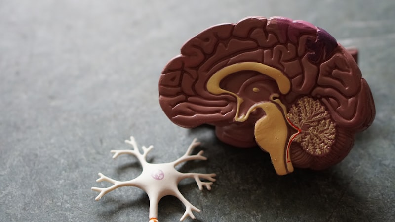
The vestibular system, often termed the sixth sense, is the sensory apparatus responsible for providing the brain with information about motion, head position, and spatial orientation, which is crucial for maintaining posture and balance. At the heart of this system lie the semicircular canals, three delicate, arching structures housed within the bony labyrinth of the inner ear. These canals function specifically as highly sensitive biological gyroscopes, detecting rotational or angular movements of the head. Without their constant, accurate input, the world would appear to spin wildly with every movement, rendering coordinated action and stable vision virtually impossible. Their structure and precise anatomical arrangement allow them to monitor movement along all three dimensions of space, ensuring comprehensive coverage of the head’s rotational dynamics relative to gravity.
The primary function of the semicircular canals is to transduce mechanical energy—the movement of fluid caused by head rotation—into electrical signals that the central nervous system (CNS) can interpret. This process is fundamental to the body’s sense of equilibrium. Each canal is filled with a fluid known as endolymph, and the base of each canal features a swelling called the ampulla, which houses the sensory receptors. When the head moves, the inertia of the endolymph causes it to lag behind the movement of the canal walls, deflecting the specialized hair cells within the ampulla. This mechanical deflection opens ion channels, leading to the generation of action potentials that travel via the vestibular nerve to the brainstem and cerebellum, ultimately informing the brain about the speed and direction of rotation.
It is important to understand that the canals respond not to constant velocity, but exclusively to changes in velocity, or angular acceleration. If a person spins at a constant rate for an extended period, the endolymph eventually catches up to the movement of the canal walls, ceasing the deflection of the hair cells and leading to a temporary sensation that the movement has stopped. This mechanism explains why dizziness or vertigo often occurs when rapid movement ceases; the fluid continues to move due to inertia, signaling rotation even though the body has stopped. This intricate sensory feedback loop is essential for generating compensatory reflexes that stabilize both posture and vision during locomotion.
Detailed Anatomy of the Three Canals
The efficiency of the semicircular canals stems directly from their elegant anatomical organization. There are three canals on each side of the head—the anterior (or superior), the posterior, and the horizontal (or lateral) canal—and each is oriented perpendicular to the other two, forming the basis of a three-dimensional coordinate system. This arrangement ensures that every possible direction of head rotation in space can be precisely monitored. Because the canals on the left side work in complementary pairs with those on the right side, a movement that excites a canal on one side will simultaneously inhibit its functional counterpart on the opposite side, providing the brain with push-pull signals that enhance sensitivity and accuracy regarding the axis of rotation.
The Horizontal Canal, positioned roughly 30 degrees upward from the horizontal plane when the head is upright, is primarily sensitive to rotations in the transverse plane, corresponding to the “no” motion or yaw movement, such as turning the head side-to-side while stationary. The input from this canal is crucial for monitoring movements that occur parallel to the ground. Conversely, the Anterior Canal and the Posterior Canal are oriented vertically. The anterior canal detects rotation in the sagittal plane, corresponding to the “yes” motion or pitch movement (nodding), while the posterior canal responds to rotations in the coronal plane, or roll movement (tilting the head towards the shoulder).
Each canal opens into a central chamber called the utricle and contains its sensory organ, the crista ampullaris, located within the ampulla. The crista is topped by a gelatinous structure known as the cupula, which acts like a swing door. The hair cells—the actual sensory receptors—are embedded within the crista, and their stereocilia project into the cupula. When the head rotates, the endolymph movement pushes the cupula, bending the hair cells. This bending is the critical transduction step; movement in one direction causes depolarization (excitation), while movement in the opposite direction causes hyperpolarization (inhibition). This binary signaling mechanism provides the central nervous system with clear, unambiguous information about the precise rotational vector.
Historical Understanding of Balance and Equilibrium
The understanding of the inner ear’s role in balance evolved slowly, initially being overshadowed by its auditory function. Early anatomical observations identified the structures, but their physiological purpose remained elusive for centuries. The critical shift occurred in the mid-19th century, spearheaded by researchers like Jean Pierre Flourens. In the 1820s, Flourens conducted pioneering experiments involving lesioning the semicircular canals in pigeons. He meticulously documented that surgical disruption of specific canals led to predictable, characteristic disturbances in posture and movement, definitively proving that the canals were organs of balance, not just secondary structures related to hearing.
Further advancements were made by Ernst Mach and Josef Breuer in the 1870s, who formalized the hypothesis regarding the mechanical operation of the canals. They proposed that the flow of endolymph acted upon the sensory receptors, linking fluid dynamics directly to the perception of rotation. This theoretical framework was solidified by Ewald’s Law, derived from the experiments of physiologist Richard Ewald in the late 19th century. Ewald’s Law confirmed that movement of the endolymph toward the ampulla (ampullopetal flow) in the horizontal canal causes excitation, while movement away (ampullofugal flow) causes inhibition. Although the excitation/inhibition pattern is reversed in the vertical canals, Ewald’s work established the fundamental relationship between fluid movement and neural signaling for each pair of canals, paving the way for modern clinical testing methods.
The recognition of the semicircular canals as the primary sensors for angular motion marked a profound conceptual shift in neuroscience and psychology. Prior to this, balance was often attributed vaguely to muscular coordination or visual input alone. The discovery of a dedicated, internal sensory organ for rotational movement provided a physiological basis for understanding symptoms like vertigo and motion sickness, moving the study of equilibrium from speculative philosophy into the realm of experimental psychophysiology. This historical foundation remains critical, as the anatomical and physiological principles established during this period are still used to diagnose and treat vestibular disorders today.
A Practical Example: Maintaining Gaze During Movement
A perfect illustration of the semicircular canals in action is the unconscious, yet vital, process of maintaining a stable visual image while the head is moving—a requirement for activities ranging from driving a car to simply walking down a street. Consider a real-world scenario where a person is rapidly shaking their head “no” while attempting to read a sign across the room. If the visual system alone were responsible for gaze stability, the image on the retina would blur instantly, making the text unreadable. However, thanks to the input from the horizontal semicircular canals, the image remains remarkably clear.
The “How-To” of this stabilization involves a rapid, automatic reflex known as the Vestibulo-ocular reflex (VOR). The process unfolds in a fraction of a second:
- Head Movement Detected: The person initiates a rapid head turn to the left.
- Canal Activation: The endolymph in the left horizontal canal lags, pushing the cupula and exciting the hair cells in that canal. Simultaneously, the corresponding right horizontal canal is inhibited.
- Neural Transmission: The strong excitatory signal is immediately transmitted via the vestibular nerve to the vestibular nuclei in the brainstem.
- Signal Routing: The brainstem processes this signal and sends compensatory motor commands through the cranial nerves that control eye muscles.
- Compensatory Eye Movement: For a head turn to the left, the brain commands the eyes to move an equal and opposite distance to the right. This counter-rotation stabilizes the eyes in space, keeping the image of the sign fixed on the fovea, ensuring continuous, sharp vision despite the head motion.
This example highlights the incredible speed and precision of the canal system. The VOR operates with an exceptionally short latency, often less than 10 milliseconds, which is necessary because visual stabilization must occur faster than any voluntary reaction. The semicircular canals provide the initial, crucial signal for rotational speed, allowing the reflex to calculate the necessary counter-movement instantaneously, thereby bridging the sensory gap between body motion and visual perception.
Significance: The Vestibulo-Ocular Reflex (VOR)
The significance of the semicircular canals to human function is best encapsulated by their role in generating the VOR. The VOR is perhaps the most important reflex tied to the canals, serving as the foundation for spatial awareness during dynamic movement. In essence, the VOR acts as a high-gain, high-fidelity stabilization system, ensuring that the movement of the eyes perfectly compensates for the movement of the head. Without the VOR, activities requiring accurate vision while moving, such as tracking a ball or navigating uneven terrain, would be impossible, leading to chronic oscillopsia (the illusory movement of stationary objects).
In the field of clinical medicine and applied psychology, the VOR is extensively studied and tested. Its integrity is a reliable indicator of vestibular system health. For example, tests like the head impulse test (HIT) or rotational chair testing directly measure the functionality and gain of the VOR. If the canals are damaged, the VOR gain decreases, meaning the eyes do not move far enough to compensate for head movement. This deficit forces the visual system to make corrective movements (saccades) to re-fixate the target, a sign of vestibular failure. Thus, the VOR not only maintains vision but also serves as a crucial diagnostic window into inner ear function.
Beyond simple gaze stabilization, the continuous, accurate data provided by the semicircular canals feeds into deeper cognitive processes related to spatial orientation and navigation. Psychologists recognize that the integration of vestibular input with visual and proprioceptive signals creates our internal map of space. Disturbances in canal function can therefore lead to more than just physical imbalance; they can cause significant psychological distress, including spatial anxiety, disorientation, and difficulty with cognitive tasks requiring spatial memory, underscoring the canals’ deep connection to higher-order brain functions.
Clinical Implications and Pathologies
When the delicate mechanics of the semicircular canals are disrupted, the resulting symptoms, collectively known as vestibular dysfunction, can be debilitating. The most common and distressing symptom is vertigo, which is the hallucination of movement—the feeling that oneself or the surroundings are spinning. This occurs because the damaged canal sends asymmetric signals to the brain, which interprets the discrepancy as constant, rapid rotation, even when the head is still. Causes of damage are varied, ranging from trauma to infection to age-related degeneration.
One prominent pathology directly affecting the canals is Ménière’s disease, an inner ear disorder characterized by episodic vertigo, fluctuating hearing loss, tinnitus, and a feeling of fullness in the ear. While the exact etiology is complex, it is believed to involve an excess of endolymph (hydrops), which distorts the membranes and hair cells within the canals and the cochlea, leading to intermittent signal failure. Another extremely common canal-related disorder is Benign Paroxysmal Positional Vertigo (BPPV). BPPV occurs when tiny calcium carbonate crystals (otoconia) normally located in the utricle dislodge and migrate into one of the semicircular canals, most commonly the posterior canal. When the head moves into a specific position, these stray particles cause abnormal, heavy movement of the endolymph and cupula, leading to brief, intense bursts of vertigo.
The treatment of canal disorders often relies on understanding their precise biomechanics. For BPPV, clinicians use specialized maneuvers, such as the Epley maneuver, which are physical procedures designed to use gravity to guide the misplaced crystals out of the affected canal and back into the utricle where they can be reabsorbed. For chronic balance issues resulting from canal hypofunction (reduced output), rehabilitation techniques known as Vestibular Rehabilitation Therapy (VRT) are utilized. VRT involves specific exercises aimed at retraining the brain to rely more heavily on visual and proprioceptive inputs to compensate for the faulty vestibular information, demonstrating the brain’s remarkable capacity for sensory plasticity following injury to these vital sensors.
Connections to Broader Psychological and Physiological Concepts
The semicircular canals are not isolated structures; their function is intimately linked to several broader psychological and physiological concepts. They belong fundamentally to the field of Sensory Psychology, specifically within the domain of the somatosensory system, even though they are located in the head. Their input must be continuously integrated with information from the visual system (sight) and the proprioceptive system (muscle and joint receptors, which sense limb position). This process of sensory integration, managed largely by the cerebellum and vestibular nuclei, is what allows us to form a coherent, stable perception of our body’s position and movement in the environment.
Furthermore, the canal system plays a critical role in Motor Control. Accurate signaling is prerequisite for the coordinated movements required for complex tasks like walking, running, or catching a ball. For example, the automatic postural adjustments needed to prevent falling when stepping on an unstable surface rely heavily on immediate feedback from the canals regarding unexpected head movement. In a broader psychological context, the canals influence our internal sense of verticality and orientation, contributing to cognitive mapping and spatial reasoning abilities, which are essential for navigation and memory.
Finally, the link between the vestibular system and the autonomic nervous system is profound. The neural pathways connecting the vestibular nuclei to brain regions responsible for nausea, heart rate, and digestion explain why motion sickness—a condition caused by a sensory mismatch between the canals (detecting motion) and the eyes (seeing a static environment, or vice versa)—can trigger severe physiological responses. This connection underscores that the integrity of the semicircular canals is not just a matter of physical balance, but a foundational element influencing both our physical health and our psychological experience of the world.