Sensory Homunculus: Mapping Your Brain’s Hidden Map
- The Core Definition and Fundamental Mechanism
- Historical Foundations of the Homunculus Concept
- Anatomical Location and Specific Mapping
- A Practical Example: Two-Point Discrimination
- Significance, Impact, and Clinical Application
- Limitations and Modern Revisions
- Connections and Relations to Other Psychological Concepts
The Core Definition and Fundamental Mechanism
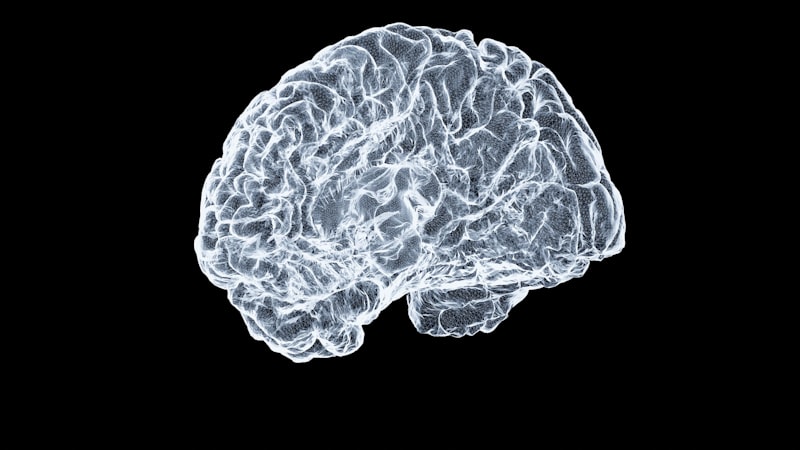
The Sensory Homunculus, translated literally as “little man,” is a foundational conceptual and visual tool in neuroscience and psychology used to illustrate the spatial representation of the human body within the primary somatosensory cortex of the brain. It is not a physical structure itself, but rather a distorted map or topographical organization where specific areas of the body surface—such as the fingers, lips, and torso—correspond directly to dedicated regions of the cerebral cortex. This arrangement ensures that sensory information received from the periphery is processed systematically within the central nervous system, maintaining the relative order of body parts, though drastically altering their proportional size based on sensitivity. The initial, simple definition establishes the homunculus as the neural blueprint of our tactile, proprioceptive, and nociceptive awareness.
The fundamental mechanism driving the homunculus’s unique structure is known as cortical magnification. This principle dictates that the amount of cortical tissue dedicated to processing sensory input is not proportional to the physical size of the body part it represents, but rather to the density of sensory receptors and the behavioral importance of that area. Areas of the body critical for fine motor control, detailed tactile exploration, or high sensitivity—most notably the hands, lips, tongue, and genitalia—occupy vastly disproportionate areas on the sensory map compared to larger but less sensitive regions like the back or the legs. This differential allocation of neural resources highlights the evolutionary prioritization of sensory detail necessary for survival and interaction with the environment.
Understanding the sensory homunculus requires recognizing its location deep within the brain’s processing centers. The map resides primarily in the Somatosensory Cortex, situated immediately posterior to the central sulcus in the Parietal Lobe. When an external stimulus, such as a gentle touch or a change in temperature, activates receptors on the skin, these signals travel via peripheral nerves and the spinal cord before ascending to the thalamus and finally projecting onto the specific, corresponding region of the homunculus. This systematic relay ensures rapid and accurate perception, forming the basis of our conscious experience of touch, pressure, pain, and body position.
Historical Foundations of the Homunculus Concept
While the most recognizable depictions of the homunculus solidified in the mid-20th century, the foundational idea that the brain contains a dedicated, organized representation of the body dates back much further. The earliest philosophical murmurings of such a relationship can be traced to figures like the Czech physiologist and anatomist, Johannes Evangelista Purkinje, who proposed in 1837 that individual sensory organs were represented by specific neuronal groups, laying the groundwork for spatial mapping theories. However, Purkinje’s work was largely theoretical and preceded the advanced electrophysiological techniques required to empirically verify such organization.
The concept gained significant momentum and formal structure in the late 19th and early 20th centuries through the exhaustive work of prominent neurologists and physiologists. Crucially, the Nobel laureate Sir Charles Sherrington, often regarded as the father of modern neurology, significantly expanded on the understanding of reflex arcs and the integrated action of the nervous system. While Sherrington did not create the iconic visual image of the homunculus, his meticulous studies on sensory pathways, nerve integration, and the organization of spinal cord reflexes provided the detailed empirical evidence necessary to support the theory that sensory processing was topographically organized within the cortex. His work provided the anatomical and functional blueprint upon which later researchers would overlay the actual body map.
The definitive and enduring visual representation of the sensory homunculus was ultimately mapped and popularized by Canadian neurosurgeon Wilder Penfield and his colleague, Herbert Jasper, primarily in the 1930s and 1940s. During complex brain surgeries performed under local anesthesia to treat epilepsy, Penfield utilized mild electrical stimulation directly on the exposed cortex of conscious patients. By observing the specific physical sensations (like a tingling finger or a twitching toe) reported by the patients when particular cortical areas were stimulated, Penfield systematically cataloged and drew the precise map of the somatosensory and motor cortices. It was Penfield’s iconic, grotesque, and spatially distorted drawing—with massive hands and lips—that fully captured the concept of the “little man” and cemented the sensory homunculus in both popular science and academic study.
Anatomical Location and Specific Mapping
The primary locus of the sensory homunculus is the Primary Somatosensory Cortex (S1), which is functionally defined by Brodmann area 3, 1, and 2. This narrow strip of cortex runs superiorly from the lateral fissure, spanning the depth of the longitudinal fissure. The organization within S1 is contralateral, meaning the somatosensory cortex in the right hemisphere processes sensory information received from the left side of the body, and vice-versa. This anatomical arrangement is fundamental to all sensory and motor processing pathways in the brain.
The mapping within the somatosensory cortex follows a strict, inverted, and specific spatial sequence. Generally, the lower extremities (feet, legs, and genitals) are mapped onto the medial surface of the hemisphere, buried deep within the longitudinal fissure. As one moves laterally (down the side of the brain), the map progresses through the torso, shoulder, and arm. The areas requiring the highest magnification—the hands, especially the thumb and fingers—are represented laterally, followed by the face, jaw, pharynx, and finally, the tongue, which resides closest to the temporal lobe. This detailed, point-to-point correspondence between the body surface and the cortical area is what allows the brain to accurately localize touch and pain.
The disproportionate scale of the homunculus is critical to grasp. For instance, the lips and hands combined often occupy more cortical space than the entire torso and back combined. This unequal representation reflects the density of sensory receptors (mechanoreceptors, thermoreceptors, etc.) in these areas. The hands require vast cortical space because they are essential for object manipulation, texture discrimination, and tool use, demanding high resolution. Similarly, the lips and tongue, packed with sensory cells, are crucial for speech articulation, eating, and emotional expression, necessitating a massive dedicated processing region. The density of incoming Afferent Neurons dictates the size of the corresponding cortical territory.
A Practical Example: Two-Point Discrimination
To illustrate the practical application and implications of the sensory homunculus’s distorted map, the concept of two-point discrimination is perhaps the most accessible real-world example. Two-point discrimination refers to the minimum distance needed between two simultaneous stimulation points on the skin (like two tips of a caliper or two pen points) before the person perceives them as two distinct stimuli, rather than just one. This measure directly correlates with the amount of cortical space dedicated to that body part.
The step-by-step application in a real-world scenario highlights the homunculus’s topography. If a researcher performs this test, they would find that on the fingertips, two points can be discriminated even when they are separated by only 2 to 3 millimeters. This exceptional sensitivity is possible because the receptive fields in the fingertips are very small and densely packed, and consequently, the corresponding region on the somatosensory cortex is enormous. Each millimeter of fingertip skin projects to a unique, dedicated cluster of neurons.
In contrast, if the same test is performed on the middle of the back or the calf, the two points may need to be separated by 40 to 60 millimeters before they are perceived as distinct. The back has far fewer sensory receptors, and their receptive fields are massive, meaning a large area of skin is monitored by a single sensory neuron. Because the back occupies a relatively small area on the sensory homunculus, sensory input from a large patch of skin converges onto a smaller cortical area, resulting in poor spatial resolution and a high threshold for two-point discrimination. This simple test is a direct functional demonstration of the structural layout depicted by the homunculus.
Significance, Impact, and Clinical Application
The sensory homunculus is not merely a historical curiosity; it remains fundamentally important because it provides a simple, predictive model for understanding the complex organization of sensation and how it can be affected by injury or disease. By detailing the precise anatomical relationship between the brain and the body, the homunculus allows clinicians and researchers to diagnose the location of neurological damage. For example, if a patient experiences numbness only in their right hand, but not the arm or face, it suggests a highly localized lesion or stroke affecting only the corresponding hand region of the left somatosensory cortex, rather than damage lower down in the spinal cord or peripheral nerves.
Furthermore, the concept is central to understanding phenomena related to cortical reorganization, particularly Neuroplasticity. While the homunculus was initially depicted as a static map, modern research confirms that the organization is dynamically modifiable throughout life. If a person loses a digit or limb (amputation), the cortical area previously dedicated to that body part does not simply become silent. Instead, the adjacent cortical territories—often representing the face or the remaining fingers—will expand and take over the now-vacant cortical space. This explains phenomena such as the experience of phantom limb sensations, where stimulating the face might generate a sensation in the missing hand, as the facial representation has encroached upon the hand’s former territory.
In clinical settings, the homunculus informs targeted rehabilitation strategies. Techniques such as constraint-induced movement therapy (CIMT) for stroke patients or sensory retraining after nerve damage aim to exploit this plasticity. By forcing the use of a damaged limb or providing intense, focused sensory input to a specific area, therapists intentionally attempt to induce beneficial cortical reorganization, effectively enlarging the functional representation of that body part on the Somatosensory Cortex and improving sensory and motor function. The recognition of the homunculus’s plasticity has revolutionized physical and occupational therapy.
Limitations and Modern Revisions
While the sensory homunculus is an invaluable pedagogical and diagnostic tool, it is crucial to acknowledge its limitations, particularly when viewed through the lens of modern imaging and electrophysiology. The classic homunculus, as drawn by Penfield, is a two-dimensional, fixed, and discrete representation. Reality, however, is far more complex. First, the brain’s representation of the body is not neatly segmented; there is significant overlap and integration between adjacent body parts, meaning stimulating one small area might activate responses in several neighboring representations.
Second, the homunculus model does not adequately account for the multimodal nature of sensory processing. Sensation is rarely purely tactile; it involves the integration of proprioception (body position), nociception (pain), and temperature, which are processed not just in S1, but also in secondary somatosensory areas (S2) and deeper cortical and subcortical structures. The simple map fails to capture the intricate feedback loops and descending modulation that shape how we perceive sensory input, which is influenced by attention, expectation, and emotional state.
The most significant modern revision addresses the dynamic nature of the map, emphasizing Neuroplasticity. The amount of cortex dedicated to a body part changes based on experience and usage. A musician who practices the violin for hours daily will develop an enlarged representation of their left-hand fingers compared to a sedentary individual. This constant reshaping demonstrates that the homunculus is not a static blueprint but a dynamic, ever-changing portrait reflecting current behavioral relevance and sensory demands, leading to more sophisticated, high-dimensional models that surpass the simple “little man” drawing.
Connections and Relations to Other Psychological Concepts
The sensory homunculus is part of the broader field of Cognitive Neuroscience and specifically falls under the domain of sensory processing and perception. It is intrinsically linked to several other major psychological and neurological concepts. Chief among these is its close neighbor, the Motor Homunculus.
- The Motor Homunculus: Situated anterior to the central sulcus in the primary motor cortex, the motor homunculus mirrors the sensory map almost exactly in its topographical organization and its disproportionate scaling. While the sensory homunculus processes incoming tactile information, the motor homunculus is responsible for initiating and executing voluntary movements. The close anatomical and functional proximity of these two “little men” highlights the critical sensory-motor loop: sensory input informs motor output, and motor actions constantly generate new sensory feedback, ensuring coordinated interaction with the world.
- Body Schema and Body Image: The homunculus provides the neural substrate for the body schema, which is the unconscious, sensorimotor representation of the body used for controlling posture and movement. It is distinct from the body image, which is the conscious, cognitive, and affective perception of one’s own body. While the homunculus is purely anatomical and functional, it contributes the raw data necessary for the brain to construct the body schema used in everyday actions.
- Phantom Limb Phenomena: As mentioned previously, the homunculus is key to explaining phantom sensations. The persistence of neural activity in the cortical area formerly representing the missing limb, combined with the invasion of neighboring cortical maps, directly illustrates the tension between a fixed anatomical organization and the flexible power of Neuroplasticity.
In summary, the sensory homunculus serves as a fundamental pillar in understanding how the brain organizes and processes the continuous stream of sensory data received from the environment. Its study bridges basic neuroscience, clinical neurology, and cognitive psychology, providing a powerful visualization of the highly organized, yet dynamically flexible, relationship between our physical body and our neural architecture.