Skeletal Muscle: The Mind-Body Connection in Movement
- Understanding Skeletal Muscle: A Fundamental Definition
- The Microscopic Architecture: Sarcomeres and Myofilaments
- Pioneering Discoveries in Muscle Physiology
- The Unveiling of the Sliding Filament Theory
- The Act of Lifting: A Step-by-Step Illustration of Muscle Contraction
- From Neural Impulse to Mechanical Force: Excitation-Contraction Coupling in Action
- Beyond Movement: The Broader Importance of Skeletal Muscle
- Clinical and Applied Relevance in Modern Science
- Distinguishing Skeletal Muscle from Other Muscle Types
- Skeletal Muscle in the Context of Psychology and Neuroscience
Understanding Skeletal Muscle: A Fundamental Definition
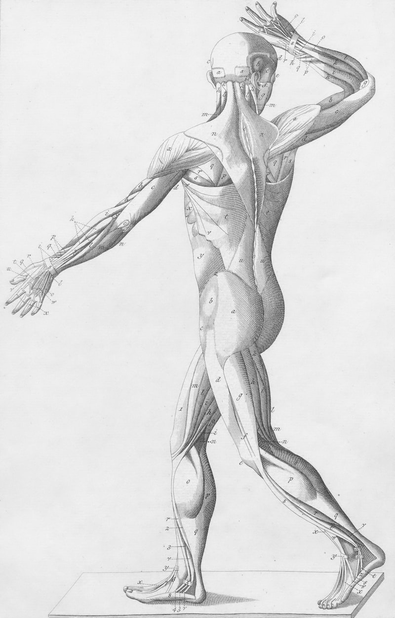
Skeletal muscle represents the most prevalent tissue type within the human body, constituting approximately 40% of body weight and serving as the primary effector for voluntary movement. It is a highly specialized tissue characterized by its striated appearance under a microscope, a result of the orderly arrangement of contractile proteins. Unlike cardiac or smooth muscle, skeletal muscle is under conscious, voluntary control, enabling a vast repertoire of actions from subtle facial expressions to powerful locomotive movements. Its fundamental role extends beyond locomotion, encompassing crucial functions such as maintaining posture, facilitating respiration, and generating heat to regulate body temperature.
The intricate structure of skeletal muscle underpins its remarkable functional capabilities. Each muscle is an organ composed of numerous muscle fibers, which are essentially elongated cells. These fibers are bundled together, surrounded by connective tissue, and supplied by an extensive network of blood vessels and nerves. The defining characteristic of these fibers is their internal organization into repeating contractile units known as sarcomeres. These sarcomeres are the fundamental units of muscle contraction, where the mechanical force necessary for movement is generated through the precise interaction of protein filaments.
At its core, the mechanism of skeletal muscle function revolves around the dynamic interaction between two primary contractile proteins: actin and myosin. These proteins are organized into thin and thick filaments, respectively, which slide past each other during contraction. This intricate process, known as the sliding filament theory, is initiated by a neural signal and involves a cascade of biochemical events that ultimately lead to the shortening of the sarcomere and, consequently, the entire muscle fiber. The coordinated contraction of countless muscle fibers then translates into observable macroscopic movement.
The Microscopic Architecture: Sarcomeres and Myofilaments
The highly organized internal structure of a skeletal muscle fiber is paramount to its function. Within each muscle fiber are numerous cylindrical organelles called myofibrils, which run the entire length of the cell. These myofibrils are composed of repeating units called sarcomeres, which are the fundamental contractile units responsible for the muscle’s striated appearance. Each sarcomere is delimited by two Z-discs, which serve as anchoring points for the thin filaments.
Within the sarcomere, the principal contractile proteins, myofilaments, are arranged in a precise, overlapping pattern. The thin filaments are primarily composed of actin, a globular protein that polymerizes into a double helix. Associated with actin are two regulatory proteins: troponin and tropomyosin. Tropomyosin forms a strand that spirals around the actin filament, covering the myosin-binding sites in a resting muscle. Troponin is a complex of three proteins attached to tropomyosin, playing a crucial role in the calcium-dependent regulation of contraction.
The thick filaments are primarily composed of myosin, a motor protein characterized by its long tail and a globular head. These myosin heads are capable of binding to actin and hydrolyzing ATP, the energy currency of the cell, to power the contraction cycle. The central region of the sarcomere, the A-band, contains the full length of the thick myosin filaments, while the I-band contains only thin actin filaments. During contraction, the thin filaments slide inward, pulling the Z-discs closer together and shortening the sarcomere, a process that collectively shortens the entire muscle.
Pioneering Discoveries in Muscle Physiology
The scientific understanding of skeletal muscle has evolved significantly over centuries, beginning with early anatomical observations and progressing to detailed molecular insights. One of the earliest pivotal discoveries came from Luigi Galvani in the late 18th century, who demonstrated that electricity could stimulate muscle contraction, laying the groundwork for the field of electrophysiology and hinting at the neural control of muscles. His experiments on frog legs revealed the intrinsic electrical excitability of muscle tissue, a concept that was revolutionary for its time and challenged prevailing theories about vital forces.
Further advancements in the 19th century involved microscopic examinations that gradually unveiled the striated nature of skeletal muscle. Researchers began to discern the repeating bands and zones within muscle fibers, leading to initial hypotheses about how these structures might contribute to contraction. However, the precise mechanism remained elusive, largely due to the limitations of microscopy and biochemical techniques available during that era. These early observations were crucial, providing the morphological context upon which later, more detailed physiological explanations would be built.
The Unveiling of the Sliding Filament Theory
A monumental breakthrough in understanding muscle contraction occurred in the mid-20th century with the independent proposals of the sliding filament theory. In 1954, two research teams, one led by Andrew Huxley and Rolf Niedergerke, and another by Hugh Huxley and Jean Hanson, published their findings almost simultaneously. Their work, based on advanced electron microscopy and X-ray diffraction studies, revealed that muscle contraction does not involve the shortening of the actin or myosin filaments themselves, but rather the sliding of these filaments past one another within the sarcomere.
This theory provided a coherent explanation for the observed changes in sarcomere banding patterns during contraction and relaxation, fundamentally shifting the paradigm of muscle physiology. It established that the myosin heads form cross-bridges with actin filaments, pulling them inward towards the center of the sarcomere. The energy for this process was subsequently identified as coming from the hydrolysis of ATP, confirming the active, energy-dependent nature of muscle contraction.
The elucidation of the sliding filament theory paved the way for understanding the molecular machinery of contraction, including the critical roles of calcium ions and regulatory proteins like troponin and tropomyosin. This comprehensive model allowed scientists to delve into the intricate details of excitation-contraction coupling, linking the electrical signal from a nerve to the mechanical event of muscle shortening. The profound impact of this theory continues to influence research in fields ranging from exercise physiology to the study of neuromuscular diseases.
The Act of Lifting: A Step-by-Step Illustration of Muscle Contraction
To truly grasp the complexity of skeletal muscle function, consider the everyday action of lifting an object, such as a book, from a table. This seemingly simple act involves a highly coordinated sequence of events initiated by the nervous system. The process begins in the brain, where the intention to move is translated into electrical signals. These signals travel down the spinal cord and are conveyed to specific muscle fibers via specialized nerve cells known as motor neurons.
Upon reaching the muscle, the terminal end of a motor neuron forms a highly specialized synapse with a muscle fiber, called the neuromuscular junction. Here, the electrical signal (action potential) arriving at the motor neuron terminal triggers the release of a chemical messenger, a neurotransmitter called acetylcholine, into the synaptic cleft. This chemical bridge is crucial for transmitting the neural command to the muscle cell, converting an electrical signal into a chemical one that can be recognized by the muscle.
The acetylcholine molecules then bind to specific receptors on the muscle fiber’s membrane, the sarcolemma. This binding event causes a rapid change in the electrical potential across the sarcolemma, generating a new action potential that propagates along the muscle fiber’s surface. This electrical signal quickly dives deep into the muscle fiber’s interior through a network of invaginations called T-tubules, ensuring that the excitatory signal reaches every myofibril simultaneously, thereby enabling a swift and uniform contraction.
From Neural Impulse to Mechanical Force: Excitation-Contraction Coupling in Action
Once the action potential reaches the interior of the muscle fiber via the T-tubules, it triggers a critical event known as excitation-contraction coupling. The T-tubules are in close proximity to the sarcoplasmic reticulum, a specialized endoplasmic reticulum within muscle cells that stores calcium ions. The arriving action potential causes voltage-sensitive proteins in the T-tubule membrane to interact with calcium release channels on the sarcoplasmic reticulum, leading to a massive efflux of calcium ions into the sarcoplasm, the cytoplasm of the muscle cell.
The sudden increase in sarcoplasmic calcium concentration is the direct trigger for muscle contraction. These calcium ions bind to troponin, which is part of the thin filament complex. This binding induces a conformational change in troponin, which in turn causes tropomyosin to shift its position, thereby exposing the myosin-binding sites on the actin filaments. With the binding sites now accessible, the myosin heads, which have already hydrolyzed ATP and are in a high-energy state, can attach to actin, forming cross-bridges.
The formation of cross-bridges initiates the power stroke: the myosin heads pivot, pulling the actin filaments toward the center of the sarcomere. This movement causes the sarcomere to shorten, generating mechanical force. After the power stroke, a new ATP molecule binds to the myosin head, causing it to detach from actin. The ATP is then hydrolyzed, re-energizing the myosin head for another cycle. This cyclical process continues as long as calcium is present and ATP is available. When the neural stimulation ceases, calcium is actively pumped back into the sarcoplasmic reticulum, tropomyosin once again blocks the actin binding sites, and the muscle relaxes, allowing the book to be set back down.
Beyond Movement: The Broader Importance of Skeletal Muscle
The importance of skeletal muscle extends far beyond its obvious role in movement and locomotion. It is a vital organ system integral to maintaining overall bodily homeostasis and plays a significant, albeit often overlooked, role in various physiological processes. For instance, skeletal muscles are crucial for maintaining posture against gravity, providing the continuous, subtle adjustments necessary to keep the body upright. This constant low-level contractile activity, known as muscle tone, is essential for stability and balance.
Furthermore, skeletal muscles are indispensable for respiration, particularly the diaphragm and intercostal muscles, which drive the mechanical process of breathing. Without the coordinated contraction and relaxation of these muscles, the vital exchange of gases in the lungs would be impossible. Beyond these mechanical functions, skeletal muscle is a major site of heat production in the body. Muscle activity, especially during shivering in cold environments, generates heat as a byproduct of ATP hydrolysis, contributing significantly to thermoregulation.
From a broader perspective, particularly in the context of neuroscience and physiological psychology, skeletal muscle is the primary output system for behavior. Every voluntary action, every spoken word, every emotional expression manifested physically, relies on the precise control and execution of skeletal muscle contractions. Understanding the mechanisms of muscle contraction and its neural regulation is therefore fundamental to comprehending motor control, motor learning, and even the physical manifestations of psychological states, such as stress or anxiety. It provides the biological substrate for all observable human behavior.
Clinical and Applied Relevance in Modern Science
The profound understanding of skeletal muscle physiology has immense practical implications across numerous fields, from clinical medicine to sports science and ergonomics. In medicine, this knowledge is critical for diagnosing and treating a wide array of neuromuscular disorders, such as muscular dystrophies, myasthenia gravis, and amyotrophic lateral sclerosis (ALS), which directly impair muscle function or its neural control. Therapies, rehabilitation protocols, and pharmaceutical interventions are often designed based on a deep appreciation of the molecular and cellular mechanisms of muscle contraction and relaxation.
In sports science and physical therapy, the principles of skeletal muscle function guide training regimens, injury prevention strategies, and recovery programs. Athletes optimize their performance by understanding how to maximize force generation, enhance endurance, and minimize muscle fatigue through specific exercises that target muscle fiber types and metabolic pathways. Physical therapists utilize this knowledge to rehabilitate patients recovering from injuries or surgeries, designing exercises that restore strength, flexibility, and coordination, thereby improving quality of life.
Furthermore, fields like biomechanics and ergonomics rely heavily on understanding how muscles interact with the skeletal system to produce movement and withstand external forces. This informs the design of prosthetics, assistive devices, and workspaces that optimize human performance and reduce the risk of musculoskeletal injuries. The study of skeletal muscle continues to be a vibrant area of research, with ongoing investigations into muscle regeneration, the effects of aging on muscle mass (sarcopenia), and novel therapeutic approaches for muscle-related pathologies, underscoring its enduring significance in human health and function.
Distinguishing Skeletal Muscle from Other Muscle Types
While all muscle tissues share the fundamental ability to contract, skeletal muscle stands apart from cardiac muscle and smooth muscle due to distinct structural, functional, and regulatory characteristics. Structurally, skeletal muscle is characterized by its highly organized, elongated, multinucleated cells (fibers) with prominent striations, reflecting the precise arrangement of sarcomeres. Cardiac muscle also exhibits striations but consists of branched cells connected by intercalated discs, and typically has one or two nuclei per cell. Smooth muscle, in contrast, lacks striations, is composed of spindle-shaped cells with a single nucleus, and its myofilaments are arranged in a less orderly fashion.
Functionally, the most significant distinction lies in their control mechanisms. Skeletal muscle is primarily under conscious, voluntary control, allowing for deliberate movements. Cardiac muscle and smooth muscle, however, operate involuntarily, meaning their contractions are not consciously directed. The heart beats autonomously, regulated by the autonomic nervous system and intrinsic pacemakers, while smooth muscle in the walls of internal organs (like the digestive tract or blood vessels) contracts in response to autonomic nerves, hormones, and local factors to regulate processes such as digestion, blood pressure, and pupil size.
The regulation of contraction also varies. Skeletal muscle contraction is initiated by a neural signal from a motor neuron, leading to a rapid and powerful, but often brief, contraction. Cardiac muscle has an intrinsic rhythmicity, modified by nervous and hormonal input, and undergoes sustained contractions. Smooth muscle contractions are generally slower, prolonged, and more energy-efficient, often exhibiting a tonic contraction or slow, rhythmic waves. These differences reflect the distinct physiological roles each muscle type plays in maintaining body function.
Skeletal Muscle in the Context of Psychology and Neuroscience
The study of skeletal muscle is not confined solely to anatomy and physiology; it forms a critical interface with neuroscience and various subfields of psychology, particularly physiological psychology and cognitive psychology. Understanding how the brain controls skeletal muscles is central to comprehending motor control, motor learning, and the neurological basis of behavior. The concept of a motor unit, comprising a single motor neuron and all the muscle fibers it innervates, is fundamental to how the nervous system precisely grades the force of muscle contraction, from fine motor skills to gross movements.
Furthermore, skeletal muscles are equipped with specialized sensory receptors that provide continuous feedback to the nervous system about muscle length and tension. This sensory information, known as proprioception, is essential for maintaining balance, coordinating movements, and developing a coherent body schema. Proprioception allows individuals to know the position of their limbs in space without visual input, a critical component of motor learning and the integration of sensory and motor information, which are core topics in cognitive and developmental psychology.
Beyond motor control, the state of skeletal muscles can reflect and influence psychological states. For instance, muscle tension is a common physiological manifestation of stress or anxiety, and techniques like progressive muscle relaxation are used in behavioral therapy to manage these states. The study of muscle fatigue, its physiological underpinnings, and its psychological perception, also bridges physiology and psychology, impacting our understanding of endurance, effort, and perceived exertion. Thus, skeletal muscle serves as a crucial biological effector and sensory organ, deeply integrated into the complex interplay between the brain, body, and behavior.