Stellate Cells: The Hidden Architects of Neural Processing
Introduction and Definition
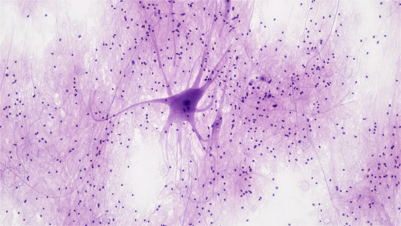
The stellate cell, derived from the Latin term stella meaning star, is a fundamental type of neuron characterized by a symmetrically radiating dendritic arborization that gives the cell body, or soma, its distinctive star-like or polygonal appearance. Unlike pyramidal cells, which possess a single, dominant apical dendrite and are typically classified as principal output neurons, stellate cells function primarily as local circuit neurons, often categorized as interneurons, meaning their axons terminate within the immediate vicinity of the soma. This distinction in morphology directly reflects their role in neural networks, where they are responsible for integrating information across localized regions and modulating the activity of surrounding principal cells, thereby ensuring highly regulated and synchronized signal processing within the central nervous system.
Functionally, stellate cells are crucial components of the brain’s computational architecture, acting as essential relays and inhibitory filters. Their defining characteristic—the multi-branched structure—allows them to receive input from numerous sources simultaneously, facilitating complex integration before transmitting a modulated signal locally. This intensive local processing is vital for cognitive functions, sensory perception, and motor control. The classification of stellate cells is further refined by their neurotransmitter usage and the presence or absence of dendritic spines, which dictates whether they exert an inhibitory (GABAergic) or excitatory (glutamatergic) influence on their targets.
The historical context of the stellate cell’s discovery is deeply tied to the pioneering work of early neuroanatomists who utilized staining techniques, such as the Golgi method, to visualize the intricate cellular landscape of the brain. While their generalized structure—the radiating branches—is consistent, the specific functional subtypes vary significantly across different brain regions, necessitating a comprehensive understanding of their microanatomical distribution. The study of stellate cells is inseparable from the study of cortical organization, as these cells are heavily concentrated within specific layers, playing a definitive role in how the cortex handles incoming sensory information and organizes its output signals.
Morphological Characteristics
The defining characteristic of the stellate cell is the morphology of its dendritic tree, which exhibits a radial symmetry unlike the polarized structure of pyramidal neurons. The dendrites emerge from the soma and extend outwards in all directions, creating a roughly spherical or star-shaped domain. This extensive, multi-directional branching ensures that the neuron can sample inputs equally from a wide, localized area of the surrounding neuropil. This extensive reach allows for sophisticated integration of inputs from various sources, including local axons of principal cells and long-range inputs from subcortical structures such as the thalamus. The complexity of this arborization is a key factor in the cell’s ability to perform rapid, local computational tasks.
The soma of a stellate cell is typically small to medium-sized and polygonal. The axon, conversely, is generally shorter than those of projection neurons, confirming the stellate cell’s status as an interneuron—a neuron whose entire structure, including its axon terminals, is confined to a particular brain region. This short axon often branches profusely near the soma, targeting adjacent neurons, often within the same cortical column. For instance, inhibitory stellate cells frequently target the somata or proximal dendrites of principal cells, exerting powerful control over their firing threshold and timing, a mechanism crucial for generating synchronized oscillations in neural networks.
Morphological features are often used to subtype these cells, particularly the texture of their dendrites. Stellate cells can be broadly categorized based on the presence or absence of dendritic spines:
- Spiny Stellate Cells: These neurons possess dendritic spines, which are typical sites for excitatory synaptic input. They are predominantly glutamatergic (excitatory) and are highly characteristic of the granular layer (Layer IV) of the sensory cortices, where they serve as primary recipients of thalamic afferents.
- Smooth Stellate Cells: These cells lack significant dendritic spines (or have very few). They are typically GABAergic (inhibitory) and function as crucial inhibitory interneurons distributed across various cortical layers, essential for balancing the excitatory drive and regulating network stability.
- Axonal Termination Patterns: Variations in how the axon branches and terminates also define subtypes, such as basket cells (which form ‘baskets’ around the somata of principal cells) and chandelier cells (which target the initial axon segment), many of which fall under the general morphological umbrella of stellate cells.
Classification and Types
The classification of stellate cells is intricate, moving beyond simple morphology to incorporate immunochemical markers, physiological properties, and connectivity patterns. The fundamental division remains the functional role: whether the cell is inhibitory or excitatory. This division dictates the cell’s overall contribution to the circuit dynamics, determining whether it contributes to feed-forward excitation or local feedback inhibition. Recognizing these functional subtypes is critical for understanding pathological conditions where specific interneuron populations are compromised.
Inhibitory Stellate Cells constitute a significant portion of the cortical inhibitory interneuron population. These cells utilize the neurotransmitter GABA (gamma-aminobutyric acid) and are vital for preventing runaway excitation and shaping the temporal precision of neural firing. Further classification within this inhibitory group often relies on the expression of specific calcium-binding proteins or neuropeptides. For example, some inhibitory stellate cells express parvalbumin (PV), rendering them fast-spiking neurons that enforce rapid, synchronous inhibition, often targeting the perisomatic region of principal cells. Others might express somatostatin (SST) or vasoactive intestinal peptide (VIP), each contributing uniquely to different forms of circuit regulation, such as controlling dendrite excitation or mediating disinhibition.
Conversely, Excitatory Stellate Cells are predominantly glutamatergic. The most prominent example is the spiny stellate cell found in cortical Layer IV. These cells are specialized for receiving the primary sensory input relayed from the thalamus. Upon receiving this input, the excitatory stellate cells quickly process and distribute the signal upwards to Layer II/III pyramidal cells, initiating the vertical flow of information through the cortical column. Their highly branched, spiny dendrites allow them to integrate numerous excitatory thalamic inputs efficiently, serving as the essential gateway for sensory information entering the cortex.
Location and Distribution
While stellate cells are ubiquitous throughout the central nervous system, their density and specific function are highly dependent upon their regional location. The instruction to “see cortical layers” underscores their critical role within the cerebral cortex, which is characterized by six distinct horizontal layers (I to VI). Stellate cells are particularly concentrated in the granular layer, Layer IV, often referred to as the primary recipient layer of sensory information. In this layer, the highly concentrated spiny stellate cells act as the first stage of cortical processing for incoming sensory signals, whether visual, auditory, or somatosensory, depending on the specific cortical area.
The distribution is not restricted to Layer IV, however. Inhibitory smooth stellate cells are found dispersed across nearly all cortical layers, where they provide the necessary inhibitory tone to maintain excitability balance. For example, inhibitory stellate cells in Layer II/III play a crucial role in regulating the activity of supragranular pyramidal cells, which are involved in higher-order associative processing. The density and subtype distribution can vary significantly based on the functional requirements of the specific cortical region; for instance, the primary visual cortex (V1) has a particularly high density of Layer IV spiny stellate cells due to the massive influx of visual data from the lateral geniculate nucleus (LGN).
Beyond the neocortex, stellate cells are also fundamental components of subcortical structures. In the cerebellum, for example, inhibitory stellate cells reside in the molecular layer, where they regulate the activity of Purkinje cells—the sole output neurons of the cerebellar cortex. Similarly, in the hippocampus, certain interneurons within the dentate gyrus and CA regions exhibit stellate morphology, contributing to memory formation and spatial navigation by controlling the flow of information through the tri-synaptic circuit. This widespread distribution confirms the stellate cell’s generalized importance as a versatile local processing unit across diverse neural systems.
Functional Roles in Neural Circuits
The primary functional role of the stellate cell is to act as a precision modulator and integrator within local neural circuits. In the context of the cerebral cortex, the spiny stellate cells of Layer IV are responsible for transforming raw thalamic input into a format suitable for complex cortical computations. They receive highly specific, focal input and distribute this excitatory signal widely to the neurons in overlying layers (Layer II/III), initiating the vertical processing stream. Without this critical relay function, incoming sensory information would fail to gain access to the higher-order processing centers of the cortex.
Conversely, the inhibitory smooth stellate cells perform crucial gating and timing functions. By releasing GABA, they hyperpolarize or shunt the membranes of their target cells, effectively controlling when and how strongly those targets can fire. This inhibitory action is critical for several processes:
- Temporal Precision: Inhibitory stellate cells synchronize the firing of large populations of principal cells, leading to the generation of cortical oscillations (e.g., gamma rhythms) that are associated with attention and consciousness.
- Gain Control: They regulate the overall excitability of the network, preventing saturation and ensuring that neural responses remain proportional to the strength of the input signal.
- Feature Selectivity: In sensory cortices, inhibitory stellate cells contribute to sharpening receptive fields, ensuring that neurons respond selectively to specific features (like the orientation of a line in V1) by suppressing responses to irrelevant stimuli.
The strategic placement and symmetric arborization of stellate cells allow them to maintain a highly effective local sphere of influence. They integrate information rapidly and provide immediate feedback or feed-forward control, ensuring that the cortical column operates as a cohesive, finely tuned computational unit capable of handling massive amounts of information without succumbing to excessive noise or instability. Their role is thus foundational to the dynamic equilibrium necessary for conscious processing.
Synaptic Integration and Plasticity
Stellate cells exhibit complex synaptic integration due to their unique morphology. Their radially symmetric dendrites allow them to receive inputs distributed evenly across their domain, leading to highly effective integration of signals arriving from diverse local sources. The synapses formed by stellate cells are highly heterogeneous; excitatory spiny stellate cells primarily receive input from thalamic afferents and local pyramidal axons, forming classical asymmetric synapses, while inhibitory smooth stellate cells receive input from local excitatory neurons and other inhibitory interneurons, forming both axo-dendritic and axo-somatic connections.
A key aspect of their function is their capacity for synaptic plasticity, the ability of synaptic strength to change over time, which underlies learning and memory. While Long-Term Potentiation (LTP) and Long-Term Depression (LTD) are classically studied in principal cells, stellate cells, particularly inhibitory subtypes, also exhibit profound forms of plasticity. This plasticity is crucial for maintaining the balance between excitation and inhibition (E/I balance). For instance, if an excitatory synapse on a principal cell strengthens (LTP), the inhibitory synapse provided by a stellate cell onto that same principal cell must often strengthen commensurately to prevent the circuit from becoming hyperexcitable.
The plasticity of inhibitory stellate cell synapses is often modulated by various neuromodulators, including acetylcholine and dopamine, linking their function to global brain states such as arousal and attention. This regulatory mechanism ensures that the filtering and gating functions of the stellate cells are flexible, adapting quickly to changes in environmental demands or internal processing requirements. For example, during periods of intense learning, the plasticity of inhibitory stellate cells allows for the temporary weakening of inhibitory control, facilitating the strengthening of new excitatory connections necessary for memory encoding.
Clinical Relevance and Pathophysiology
The proper functioning of stellate cells, particularly the inhibitory GABAergic subtypes, is indispensable for neurological health. Dysfunction or loss of specific stellate cell populations is strongly implicated in a wide spectrum of neurological and psychiatric disorders, highlighting their role as sensitive indicators and mediators of brain pathology. Because inhibitory stellate cells are responsible for maintaining the delicate E/I balance, their compromise often leads to a state of network hyperexcitability or disorganized processing.
One of the most heavily researched areas involves Schizophrenia, where post-mortem studies and genetic research point toward significant deficits in GABAergic interneurons, including PV-expressing inhibitory stellate cells. Impairment in these cells leads to reduced synchronization and disrupted gamma oscillations, which are thought to underlie cognitive deficits, such as working memory impairment and disordered thought processes characteristic of the disorder. Similarly, conditions like Epilepsy are fundamentally characterized by recurrent seizures arising from an imbalance favoring excitation. The failure of inhibitory stellate cell circuits to effectively suppress or contain excitatory activity allows runaway depolarization to spread across the cortex, resulting in seizure activity.
Furthermore, stellate cells are studied in the context of neurodegenerative diseases. In Alzheimer’s Disease, specific populations of interneurons show vulnerability to amyloid plaques and tau tangles, potentially leading to early cognitive decline associated with disrupted cortical rhythms. Therapeutic strategies aimed at restoring the function of specific stellate cell subtypes, perhaps through targeted pharmacological agents or cell replacement therapies, represent a promising frontier in treating conditions where E/I balance has been compromised by cellular pathology.
Historical Context and Discovery
The initial identification and characterization of the stellate cell morphology are credited largely to the revolution in neuroanatomy ushered in by the work of Santiago Ramón y Cajal in the late 19th and early 20th centuries. Utilizing the Golgi method—a silver staining technique—Cajal was able to visualize the full extent of individual neurons, revealing their intricate dendritic and axonal structures that had previously been invisible under conventional microscopy. Cajal meticulously described these non-pyramidal, multi-branched cells, recognizing them as distinct elements within the cortical and cerebellar architecture, often referring to them simply as “short-axon cells” or “star-shaped neurons.”
For many decades following Cajal’s discoveries, stellate cells, particularly those acting as interneurons, were viewed collectively as a heterogeneous group of cells whose complexity made functional differentiation challenging. They were broadly understood to provide local modulation, but specific roles and connectivity patterns remained poorly defined. The advancement of physiological recording techniques and, crucially, the development of immunohistochemistry in the latter half of the 20th century allowed scientists to move beyond pure morphology. Researchers could now identify neurotransmitters (like GABA and Glutamate) and specific molecular markers (like PV and SST) expressed by these cells.
This molecular revolution transformed the classification of stellate cells, moving them from a morphological category to functional classes. Modern neuroscience now recognizes that what looks morphologically like a stellate cell might belong to several distinct functional classes based on its output (inhibitory or excitatory) and its specific molecular machinery. This historical progression—from initial morphological observation to detailed molecular and physiological characterization—underscores the stellate cell’s enduring importance as a fundamental, yet highly diverse, building block of the complex brain.