Adie’s Syndrome: Decoding the Mystery of the Tonic Pupil
Introduction to Tonic Pupil of Adie
The Tonic Pupil of Adie, often simply referred to as Adie’s pupil or Adie’s syndrome, represents a fascinating and clinically significant neurological disorder primarily affecting the eye’s pupillary function. It is characterized by a distinctive set of ophthalmic signs, most notably a unilaterally dilated pupil that exhibits an abnormal response to light and near vision. This condition arises from a disruption in the parasympathetic innervation to the intrinsic muscles of the eye, specifically those responsible for pupillary constriction and accommodation. While considered a rare and benign condition, its unique presentation necessitates careful differentiation from other, potentially more serious, neurological or ocular pathologies.
The identification and understanding of this particular pupillary anomaly are rooted in the meticulous observations of British ophthalmologist William John Adie in the early 20th century. His work was instrumental in distinguishing this condition as a distinct clinical entity, separating it from other causes of unequal pupil size or abnormal pupillary reflexes. Despite its benign nature, Tonic Pupil of Adie can present with symptoms such as photophobia, blurred vision, and cosmetic concerns, prompting individuals to seek medical evaluation.
This encyclopedia entry aims to provide a comprehensive overview of the Tonic Pupil of Adie, designed to be accessible to a general audience while maintaining scientific accuracy and depth. We will delve into its core definition, explore the underlying physiological mechanisms, trace its historical discovery, detail its clinical presentation and diagnostic methods, offer practical real-world examples, discuss its significance within the fields of neurology and ophthalmology, and finally, highlight its connections to other related medical concepts. This structured approach will illuminate the multifaceted aspects of this intriguing disorder.
Core Definition and Mechanisms
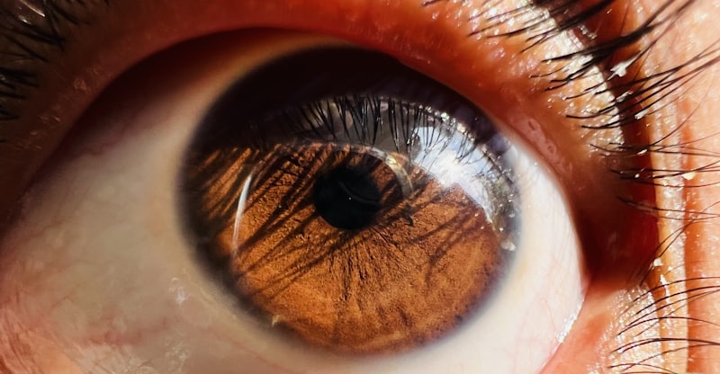
At its core, Tonic Pupil of Adie is defined as a neurological disorder characterized by a unilaterally dilated pupil that reacts sluggishly or not at all to direct light stimulation, but often exhibits a slow, sustained constriction to near vision, followed by an equally slow redilation. This peculiar pupillary behavior is termed “tonic” because the responses, both constriction and redilation, are prolonged and delayed compared to a normal pupil. The condition is almost always benign, meaning it is not associated with life-threatening systemic diseases, and typically affects only one eye, though bilateral cases have been rarely reported.
The fundamental mechanism underlying Adie’s pupil involves a lesion within the
parasympathetic nervous system
pathway that controls pupillary constriction and accommodation. Specifically, the pathology is believed to reside in the
or the short ciliary nerves that emerge from it. The ciliary ganglion is a small, peripheral ganglion located behind the eye, which receives preganglionic parasympathetic fibers from the Edinger-Westphal nucleus in the brainstem. These fibers synapse in the ganglion, and postganglionic fibers then innervate two crucial intrinsic eye muscles: the sphincter pupillae, responsible for constricting the pupil, and the ciliary muscle, essential for accommodation (changing the lens shape for near focus). A lesion or damage to this ganglion or its postganglionic fibers leads to denervation, disrupting the normal signals to these muscles.
The characteristic “tonic” response to near vision, despite impaired light reflex, is attributed to a phenomenon known as denervation supersensitivity. When the postganglionic parasympathetic nerve fibers are damaged, the sphincter pupillae muscle, deprived of its normal neurotransmitter input (acetylcholine), becomes hypersensitive to even small amounts of acetylcholine. This hypersensitivity allows it to respond, albeit slowly and excessively, to the acetylcholine released during the effort of accommodation, even if the light reflex pathway is severely compromised. Over time, there is often a reinnervation by aberrant nerve fibers, which further contributes to the tonicity and light-near dissociation, where the pupil constricts better with accommodation than with light.
Historical Context and Discovery
The condition known as Tonic Pupil of Adie bears the name of the distinguished British ophthalmologist and neurologist,
(1886–1935). Adie meticulously described this peculiar pupillary anomaly in 1931, publishing his seminal observations in the British Journal of Ophthalmology. His work built upon earlier, less comprehensive descriptions of similar pupillary abnormalities, but it was Adie’s detailed clinical characterization that firmly established it as a distinct and recognizable syndrome. He carefully delineated its unique features, distinguishing it from other conditions that might cause unequal pupils or abnormal light reflexes.
Prior to Adie’s comprehensive description, various pupillary abnormalities were often grouped under broader, sometimes ambiguous, diagnostic categories. Adie’s rigorous clinical investigation involved observing numerous patients and systematically documenting the specific characteristics of their pupils, including their responses to light, accommodation, and pharmacological agents. This methodical approach allowed him to identify a consistent pattern: a unilaterally dilated pupil with a sluggish or absent light reflex but a slow, sustained contraction to near vision. He also noted the typically benign course of the condition, which was crucial for differentiating it from more sinister neurological diseases that could present with pupillary changes.
The significance of Adie’s work cannot be overstated. By clearly defining the syndrome, he provided clinicians with a critical diagnostic tool and a framework for understanding the pathophysiology of this particular form of autonomic neuropathy. His contribution not only led to the condition being eponymously named after him but also paved the way for future research into the intricate workings of the autonomic nervous system and its control over ocular functions. His legacy continues to influence the diagnostic approach to pupillary disorders in modern neuro-ophthalmology, ensuring that patients receive accurate diagnoses and appropriate, often conservative, management.
Clinical Presentation and Diagnostic Approaches
The clinical presentation of Tonic Pupil of Adie is quite characteristic, enabling clinicians to identify it through a careful ophthalmic and neurological examination. The most striking feature is the presence of
, or pupillary dilation, in one eye, making it noticeably larger than the pupil in the unaffected eye. This
is typically more pronounced in bright light, as the affected pupil fails to constrict normally, while the healthy pupil reacts appropriately. The affected pupil also exhibits a sluggish or entirely absent direct and consensual pupillary light reflex, meaning it shows little to no constriction when light is shone into either eye.
Another defining feature is the abnormal
. While the pupil may eventually constrict when the patient focuses on a near object, this response is notably slow, sustained, and often described as “tonic.” The pupil remains constricted for an extended period before slowly redilating once the near stimulus is removed. This phenomenon, where the pupil reacts better to near vision than to light, is often referred to as “light-near dissociation” and is a critical diagnostic clue. Patients may also experience mild symptoms such as
(light sensitivity) due to the dilated pupil allowing excess light to enter the eye, or blurred vision, particularly for near tasks, due to the impaired and tonic accommodation.
Diagnosis primarily relies on a thorough
and can be confirmed with a specific pharmacological test. The instillation of a very dilute pilocarpine solution (typically 0.125%) into both eyes is a key diagnostic maneuver. Due to denervation supersensitivity, the Adie pupil, having lost its normal parasympathetic innervation, will constrict significantly in response to this weak cholinergic agent, whereas a normal pupil or one dilated by other causes will show little to no change. This test is highly sensitive and specific for Adie’s pupil. Further objective assessment of pupillary dynamics can be achieved through
, which precisely measures pupil size and reactivity under various lighting conditions, providing quantitative data to support the clinical diagnosis and differentiate it from other causes of anisocoria, such as pharmacologically induced mydriasis or oculomotor nerve palsy.
Practical Implications and Everyday Examples
To illustrate the impact of Tonic Pupil of Adie on an individual’s daily life, consider a common scenario: a person might first notice the condition when they observe that one of their pupils appears consistently larger than the other, especially when looking in a mirror in a brightly lit room. This asymmetry, or
, can be unsettling and might prompt a visit to an eye care specialist or general practitioner. Beyond the cosmetic concern, the dilated pupil’s inability to constrict effectively in bright environments can lead to symptoms like glare sensitivity or
, making outdoor activities on sunny days or driving at night more challenging due to increased light scatter and discomfort.
When this individual attempts to engage in near-vision tasks, such as reading a book, using a smartphone, or threading a needle, they might experience difficulty focusing. This is directly attributable to the impaired
in the affected eye. The ciliary muscle, responsible for changing the lens shape to focus on near objects, does not respond promptly or efficiently. While the pupil might eventually constrict and the eye might achieve some degree of focus, the process is slow and sustained, requiring more effort than usual. This “tonic” nature of accommodation means that shifting focus from a distant object to a near one, and then back again, becomes a deliberate and delayed process, potentially leading to eye strain and frustration.
The practical implications extend beyond mere discomfort. For students, professionals, or anyone whose daily activities heavily rely on precise vision and quick changes in focus, Adie’s pupil can subtly impact performance and quality of life. For instance, a graphic designer might struggle with fine details on a screen, or a musician might find it harder to read sheet music. While not debilitating, these subtle visual disturbances underscore the importance of accurate diagnosis to provide reassurance and appropriate management strategies, such as tinted lenses for photophobia or reading glasses for accommodative difficulties, to help individuals adapt and mitigate these everyday challenges.
Significance, Impact, and Management
The Tonic Pupil of Adie holds significant importance within the fields of ophthalmology and neurology, primarily due to its role as a classic example of a peripheral
neuropathy affecting ocular structures. Its distinct clinical features and predictable pharmacological response offer invaluable insights into the intricate workings of the pupillary light and accommodation pathways. Furthermore, the ability to accurately diagnose Adie’s pupil is crucial for clinicians, as it allows for differentiation from other, potentially more serious, causes of pupillary dilation or anisocoria, such as oculomotor nerve palsy (which can indicate a life-threatening aneurysm or tumor) or pharmacological mydriasis (from accidental exposure to dilating eye drops).
The impact of recognizing Adie’s pupil extends to patient management and reassurance. Once diagnosed, patients can be informed that the condition is generally benign and not indicative of a severe underlying neurological disease. This often alleviates significant anxiety and prevents unnecessary, sometimes invasive, diagnostic tests that might otherwise be performed to rule out more sinister causes of pupillary asymmetry. Thus, Adie’s pupil serves as a critical diagnostic sign, guiding clinical decision-making and ensuring that patients receive appropriate care without undergoing superfluous investigations.
In terms of management, treatment for Tonic Pupil of Adie is generally not medically necessary, as the condition is benign and often remains stable or slowly improves over time. However, symptomatic relief can be provided for patients experiencing significant photophobia or blurred vision. For photophobia, wearing dark glasses or sunglasses can help reduce glare and discomfort. For cosmetic concerns or persistent photophobia, very dilute pilocarpine eye drops (0.125%) can be prescribed. These drops help constrict the pupil, reducing its size and thereby lessening light entry, due to the denervation supersensitivity of the sphincter pupillae muscle. Additionally, patients may benefit from reading glasses if their accommodative difficulties interfere with daily activities, providing a practical solution for near-vision tasks without altering the underlying neurological condition.
Connections to Other Neurological and Ophthalmic Concepts
The Tonic Pupil of Adie is intricately related to, and often compared with, several other key neurological and ophthalmic concepts, particularly other causes of pupillary abnormalities. One of the most important differentiations is from the
, another condition characterized by light-near dissociation. While both show diminished light reflex with preserved or better near response, the Argyll Robertson pupil is typically small, irregular, and bilateral, and crucially, it is strongly associated with neurosyphilis or other midbrain lesions. In contrast, Adie’s pupil is usually large, round, and unilateral, and benign in etiology, highlighting distinct pathologies despite a superficial similarity in pupillary behavior.
Furthermore, Adie’s pupil must be distinguished from other causes of anisocoria (unequal pupil size), such as Horner’s syndrome and third nerve palsy. Horner’s syndrome results from a lesion in the sympathetic pathway to the eye, causing a small pupil (miosis), drooping eyelid (ptosis), and decreased sweating (anhydrosis) on the affected side. A third nerve (oculomotor nerve) palsy, on the other hand, can cause a dilated pupil (mydriasis), often accompanied by severe ptosis and ophthalmoplegia (restricted eye movements), indicating a potentially life-threatening compression of the nerve. Understanding the distinct clinical features and underlying mechanisms of these conditions is paramount in
to ensure accurate diagnosis and appropriate management.
Broadly, Tonic Pupil of Adie falls under the umbrella of
, specifically within the subspecialty of neuro-ophthalmology, which focuses on conditions affecting the visual pathways and ocular motility that have a neurological basis. It is also classified as a form of peripheral autonomic neuropathy, as it involves damage to the peripheral fibers of the parasympathetic nervous system. Its study contributes significantly to our understanding of the complex interplay between the central nervous system, the autonomic nervous system, and their precise control over ocular functions. By examining conditions like Adie’s pupil, researchers and clinicians gain deeper insights into the intricate neural circuits responsible for vision, pupillary reflexes, and accommodation, enhancing the diagnostic and therapeutic capabilities within this specialized field.