Electrophysiology: Mastering Precise Electrode Placement
- Introduction to Body Electrode Placement
- The Core Definition of Body Electrode Placement
- Types of Body Electrodes
- Historical Context of Electrophysiological Measurement
- Optimizing Electrode Location
- The Importance of Electrode Angle and Skin Preparation
- A Practical Example: ECG Monitoring During a Stress Task
- Significance and Impact in Psychology and Research
- Connections to Related Concepts and Broader Fields
- Conclusion
Introduction to Body Electrode Placement
The precise and deliberate placement of body electrodes is an absolutely critical and foundational element within the realm of electrophysiological experimentation. This meticulous process ensures that researchers can reliably capture the subtle yet profound electrical signals generated by biological systems, which are indispensable for understanding various physiological and psychological phenomena. Without careful consideration and adherence to established protocols, the integrity and accuracy of collected data can be severely compromised, leading to erroneous conclusions. Therefore, a comprehensive understanding of factors such as the specific type of electrode employed, its exact anatomical location on the subject’s body, and the optimal angle of placement are paramount for obtaining the highest quality, most accurate, and unequivocally reliable data possible in any given study. This detailed entry aims to elucidate the multifaceted considerations and methodologies that underpin effective electrode positioning for a diverse range of physiological and psychological experiments.
Electrophysiology, as a scientific discipline, relies inherently on the ability to detect and quantify the minute electrical currents produced by cells and tissues, from the rhythmic contractions of the heart to the complex synaptic transmissions within the brain. The quality of these measurements directly influences the validity of scientific findings, making electrode placement not merely a technical step but a crucial determinant of experimental success. An improperly placed electrode can introduce noise, obscure genuine signals, or even lead to the misinterpretation of data, highlighting why this aspect demands such rigorous attention. The principles discussed herein are applicable across various domains, from fundamental neuroscience research to clinical diagnostics and applied psychophysiology, underscoring the universal importance of this methodological cornerstone.
The Core Definition of Body Electrode Placement
At its essence, body electrode placement refers to the strategic positioning of specialized conductive devices on the surface of the skin or, in some invasive procedures, within tissues, with the primary objective of measuring underlying electrical signals generated by biological activity. These signals, typically in the microvolt to millivolt range, are manifestations of physiological processes such as neuronal firing, muscle contraction, or cardiac rhythm. The fundamental mechanism involves establishing a conductive pathway between the body’s electrical activity and an external recording system. Electrodes act as transducers, converting ionic currents within the body into electronic currents that can be amplified, filtered, and recorded by sophisticated instrumentation.
The key idea underpinning effective electrode placement is the principle of minimal impedance and maximal signal fidelity. Biological tissues, including the skin, inherently possess electrical resistance, or impedance, which can attenuate or distort the electrical signals emanating from internal sources. Therefore, electrodes are designed and placed to minimize this impedance, ensuring a clear and robust connection that allows for the faithful capture of the physiological signal of interest. This involves not only selecting the appropriate electrode material and design but also meticulous skin preparation to reduce the barrier posed by the outermost layer of the skin. The precision of placement is also crucial for targeting specific physiological sources and avoiding interference from adjacent structures, thereby isolating the desired signal from background noise or artifact.
Ultimately, the goal is to achieve an optimal interface between the living organism and the electronic measurement device. This interface must be stable, low-noise, and capable of transmitting the full spectrum of relevant electrical activity without significant attenuation or distortion. The success of any electrophysiological study—whether it is an electroencephalography (EEG) study examining brain waves, an electrocardiography (ECG) assessment of heart function, or an electromyography (EMG) recording of muscle activity—hinges directly on the quality and appropriateness of its body electrode placement methodology.
Types of Body Electrodes
Body electrodes are small, intricately designed, metal-based devices specifically engineered to attach to the skin surface or, in some cases, to be implanted, for the purpose of accurately measuring the intrinsic electrical signals of the body. These devices are remarkably diverse, varying significantly in their shapes, sizes, and underlying operational principles to accommodate the wide array of electrophysiological measurements. Common materials include silver/silver chloride (Ag/AgCl), which is favored for its stable electrochemical properties and low noise, as well as stainless steel or gold, each chosen based on specific experimental requirements, signal characteristics, and duration of recording. The physical form factor can range from small disposable adhesive patches to reusable cup electrodes, needle electrodes for intramuscular recordings, or highly specialized arrays for high-density brain mapping.
A primary distinction among electrode types is whether they are active electrodes or passive electrodes. Passive electrodes, which constitute the majority of commonly used surface electrodes, function by directly picking up the body’s electrical activity. They rely entirely on the intrinsic bioelectrical potentials to generate a signal, which is then transmitted to an amplifier. These electrodes are typically connected to a high-input impedance amplifier to minimize current draw and prevent loading the biological source. In contrast, active electrodes incorporate a pre-amplifier directly within the electrode housing. This integrated amplification serves to boost the weak biological signal very close to the source, thereby reducing susceptibility to external electrical noise and interference during transmission over longer cables to the main recording equipment. The choice between active and passive electrodes often depends on the signal strength, the length of cables, and the ambient electromagnetic environment of the recording setup.
The selection of the appropriate electrode type is a critical decision that is intrinsically linked to the specific physiological experiment being conducted and the desired level of accuracy and resolution for the collected data. For instance, in an electrooculography (EOG) study measuring eye movements, small disposable Ag/AgCl electrodes placed around the eyes are often sufficient. However, for high-density EEG recordings aiming to localize brain activity with precision, specialized caps containing dozens or even hundreds of miniature cup electrodes might be employed. Furthermore, the longevity of the recording also dictates electrode choice; long-term monitoring might necessitate more robust, stable electrodes, while short, acute measurements could use simpler, disposable options. Understanding the characteristics and limitations of each electrode type is fundamental to optimizing signal acquisition and ensuring the validity of experimental outcomes.
Historical Context of Electrophysiological Measurement
The scientific journey into understanding the body’s intrinsic electricity, and subsequently developing methods for its measurement, began centuries ago, laying the groundwork for modern body electrode placement techniques. The genesis of electrophysiology can be traced back to the late 18th century with the pioneering work of Italian physician and physicist Luigi Galvani. In the 1780s, Galvani observed that dissected frog legs would twitch when touched by dissimilar metals, even in the absence of an external electrical source. He famously coined the term “animal electricity,” hypothesizing that animals possessed their own inherent electrical fluid. This groundbreaking discovery marked the first recognition of bioelectricity as a fundamental biological phenomenon, challenging prevailing theories about nerve and muscle function and igniting intense scientific curiosity.
Galvani’s work was soon followed and refined by his countryman, Alessandro Volta, who, while initially disagreeing with Galvani’s interpretation of “animal electricity,” ultimately invented the voltaic pile (the precursor to the modern battery) in 1800. Volta’s invention demonstrated that electricity could also be generated from inanimate materials, providing a controllable source of current for further experimentation. Despite their initial disagreements, the work of both Galvani and Volta was mutually foundational, establishing the concept that living organisms generate electricity and that this electricity could be manipulated and measured. These early experiments, though crude by today’s standards, directly led to the development of increasingly sensitive galvanometers and electrometer devices throughout the 19th century, allowing scientists to begin quantifying these subtle biological currents with greater precision.
The leap to human electrophysiological recording, particularly of brain activity, came much later. It was the German psychiatrist Hans Berger who, in 1929, published his seminal work demonstrating the existence of electrical activity in the human brain, which he termed the “Elektrenkephalogramm” or EEG. Berger meticulously developed techniques for placing electrodes on the scalp to record these brain waves, validating his findings by correlating changes in EEG patterns with different mental states, such as wakefulness and sleep. His painstaking efforts to perfect electrode placement and signal acquisition truly ushered in the modern era of non-invasive human electrophysiology. The evolution from Galvani’s twitching frog legs to Berger’s recordings of human brainwaves underscores a continuous thread of innovation in understanding and measuring the body’s electrical language, with careful electrode placement remaining a consistent, central challenge and solution throughout this rich history.
Optimizing Electrode Location
The precise location of electrodes on the body is perhaps the most critical determinant for acquiring accurate and reliable data in any electrophysiological study. The fundamental principle guiding electrode placement is to position them directly over or as close as possible to the physiological source of the electrical signal of interest, while simultaneously minimizing interference from other sources, such as muscle activity or external electrical noise. Generally, electrodes should be situated in areas of the body that are relatively stable, meaning they are free of significant movement artifacts that could contaminate the signal. This often means avoiding bony prominences where movement can cause electrodes to lift, or areas with dense muscle groups that produce competing electrical activity.
Common and well-established locations for electrodes vary significantly depending on the target physiological system. For example, in ECG, electrodes are strategically placed on the limbs (e.g., wrists and ankles) and across the chest to capture the heart’s electrical axis from multiple perspectives, following standardized systems like the 12-lead ECG. For EEG, electrodes are placed on the scalp according to internationally recognized systems, such as the 10-20 system, which uses anatomical landmarks (nasion, inion, preauricular points) to ensure consistent electrode positioning relative to underlying brain regions. For EMG, electrodes are placed over the belly of the specific muscle whose activity is to be measured. Each of these placement systems is designed to optimize the signal-to-noise ratio for the target physiological phenomenon.
A crucial aspect of optimizing electrode location is ensuring strict consistency from subject to subject within an experiment, and ideally across different studies. This standardization is absolutely essential for the comparability and generalizability of research findings. Minor variations in electrode placement can lead to substantial differences in recorded signal morphology, amplitude, and latency, making it challenging to draw valid comparisons or to replicate results. Researchers often use precise anatomical measurements, landmark-based placement protocols, and sometimes even digital mapping tools to ensure this consistency. Furthermore, the placement strategy must also consider potential sources of artifacts, such as sweat glands, hair, or areas prone to movement, and design placements to mitigate their impact, thereby enhancing the overall accuracy and reliability of the collected data.
The Importance of Electrode Angle and Skin Preparation
Beyond the precise anatomical location, the physical orientation, or angle of placement, of the electrode relative to the skin surface is another critical factor influencing data quality. It is paramount to ensure that the electrode makes full, even, and consistent contact with the skin. Ideally, the electrode should be placed parallel to the body surface, facilitating a broad and stable interface. An electrode that is tilted, partially lifted, or making uneven contact can create high and variable impedance pathways, leading to signal attenuation, increased noise, and susceptibility to movement artifacts. This imperfect contact can result in a distorted or weakened signal, making accurate interpretation challenging or impossible. Furthermore, maintaining an appropriate distance between adjacent electrodes is vital to prevent electrical interference or “cross-talk,” especially in dense arrays used for EEG or high-resolution EMG, ensuring that each electrode predominantly captures signals from its intended localized source.
Equally important, and often a precursor to optimal angle and contact, is meticulous skin preparation. The outermost layer of the skin, the stratum corneum, is primarily composed of dead skin cells and acts as a significant electrical insulator, presenting a substantial barrier to the transmission of subtle bioelectrical signals. This high skin impedance can severely degrade signal quality. Therefore, proper skin preparation is essential to reduce this impedance and create a low-resistance pathway for the electrical signals. This process typically involves several steps: first, gently cleaning the skin with an alcohol swab to remove oils, dirt, and dead skin cells; second, and often most critically, lightly abrading the skin surface. Abrasion, usually performed with a mild abrasive paste or a specialized preparation gel, helps to remove a thin layer of the stratum corneum, significantly lowering the impedance without causing discomfort or damage to the subject.
Once the skin is prepared and the electrode is positioned at the correct angle, a conductive medium, such as an electrolyte gel or paste, is applied. This conductive gel serves several vital functions: it further reduces the impedance between the electrode and the skin, ensures an even and stable electrical connection, and prevents air pockets that could otherwise act as insulators. The combination of proper skin preparation, careful electrode placement at the optimal angle, and the use of a suitable conductive medium collectively ensures that the recording system can efficiently capture the true physiological signals with minimal noise and maximal fidelity. Neglecting any of these steps can lead to suboptimal data quality, rendering the entire experimental effort less fruitful and potentially invalidating the research outcomes.
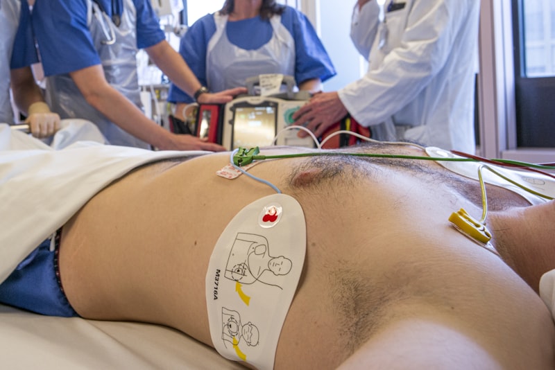
A Practical Example: ECG Monitoring During a Stress Task
To illustrate the practical application and critical importance of meticulous body electrode placement, let us consider a common scenario in psychophysiology research: monitoring a participant’s physiological response, specifically heart rate variability (HRV), during a psychologically stressful task. HRV, which reflects the beat-to-beat variations in heart rate, is a sensitive indicator of autonomic nervous system activity and is often used as a marker for stress, emotional regulation, and cognitive load. To accurately measure HRV, a high-quality electrocardiography (ECG) signal is indispensable, and this hinges entirely on correct electrode placement.
The “how-to” of applying this psychological principle in a real-world scenario begins with preparing the participant. Before any electrodes are attached, the skin at the intended sites must be thoroughly cleaned with an alcohol wipe to remove natural oils and any dirt, followed by gentle abrasion with a mild abrasive gel to reduce skin impedance. This preparation is crucial for obtaining a clear, low-noise signal. For a standard three-lead ECG setup, which is often sufficient for HRV analysis, three disposable Ag/AgCl electrodes would typically be used. The placement generally follows a modified Einthoven’s triangle configuration: one electrode (often the positive lead) is placed on the right infraclavicular fossa (just below the right collarbone), another (negative lead) on the left infraclavicular fossa (below the left collarbone), and a third (ground lead) on the left lower rib cage or abdomen.
During the actual stress task, for instance, a public speaking simulation or a demanding cognitive arithmetic test, the participant’s heart’s electrical activity is continuously recorded via these carefully placed electrodes. The electrodes detect the minute electrical potentials generated by the heart’s depolarization and repolarization, transmitting them through the conductive gel and cables to an amplifier. This amplifier boosts the tiny signals, which are then digitized and processed by a computer. The resulting waveform, known as an electrocardiogram, allows researchers to precisely identify each R-peak (the most prominent spike in the ECG waveform, corresponding to ventricular depolarization). By measuring the time intervals between successive R-peaks (R-R intervals), researchers can calculate various HRV metrics. Incorrect electrode placement—such as poor skin contact, electrodes placed over large muscle groups, or too close to each other—would introduce significant noise and artifacts into the ECG signal, making accurate R-peak detection impossible and thus invalidating the HRV data, ultimately hindering the ability to assess the participant’s stress response effectively.
Significance and Impact in Psychology and Research
The meticulous practice of body electrode placement holds immense significance and impact across various domains within psychology and broader scientific research. Its importance stems from its fundamental role in enabling the non-invasive (or minimally invasive) measurement of physiological processes that are intricately linked to psychological states, cognitive functions, and emotional responses. In the field of psychophysiology, the discipline dedicated to the study of the mind-body connection, electrode placement is the gateway to understanding how mental processes manifest as physiological changes. For instance, accurate EEG recordings, facilitated by precise scalp electrode placement, allow researchers to investigate brain activity patterns associated with attention, memory, emotion, and decision-making, providing crucial insights into the neural underpinnings of cognition.
Beyond basic research, the applications of proper electrode placement extend into clinical psychology, neurorehabilitation, and even consumer science. In clinical settings, electrophysiological measures obtained through carefully placed electrodes are indispensable for diagnosing neurological disorders such as epilepsy (via EEG), sleep disorders (polysomnography, which includes EEG, EOG, and EMG), and cardiac abnormalities (ECG) that might have psychological implications, such as anxiety-induced palpitations. In neurorehabilitation, biofeedback techniques, which rely on electrodes to provide real-time physiological data to patients, empower individuals to gain conscious control over involuntary bodily functions, aiding in stress reduction, pain management, and even stroke recovery by improving muscle control. In marketing and human-computer interaction, researchers use measures like skin conductance (electrodermal activity, EDA) and facial EMG to gauge emotional responses to products or interfaces, all predicated on accurate electrode application.
Moreover, the reliability of data generated through well-executed electrode placement underpins the validity and replicability of scientific findings, which is paramount for the advancement of psychology as an empirical science. Without standardized and precise methods of electrode application, research outcomes would be inconsistent, making it challenging to build robust theories or develop effective interventions. Thus, the seemingly technical act of placing an electrode correctly is, in fact, a cornerstone of methodological rigor, enabling profound insights into the complex interplay between the mind and the body, and driving innovation across diverse scientific and clinical applications.
Connections to Related Concepts and Broader Fields
The practice of body electrode placement is not an isolated technique but is intimately connected to a wide array of related concepts and forms a foundational component of several broader fields within psychology and neuroscience. At its most fundamental, it is the practical methodology that enables the entire field of electrophysiology, which is the study of the electrical properties of biological cells and tissues. This, in turn, is a core component of psychophysiology, a subfield of psychology that investigates the interrelationship between psychological processes and physiological responses, where electrode-based measurements are the primary tools. Techniques like EEG (brain activity), ECG (heart activity), EMG (muscle activity), EOG (eye movements), and electrodermal activity (EDA, or skin conductance) are all distinct but interconnected methodologies that rely entirely on the principles of effective electrode placement to capture their respective electrical signals.
Beyond these specific measurement techniques, electrode placement also facilitates methodologies such as biofeedback, where real-time physiological data (like heart rate, muscle tension, or brain waves) are fed back to an individual to help them gain voluntary control over these bodily functions. It also provides critical data that can be correlated with more advanced neuroimaging techniques, such as functional magnetic resonance imaging (fMRI), within the field of cognitive neuroscience, helping to bridge the gap between brain structure, function, and behavior. The electrical signals captured by electrodes can also be used as inputs for brain-computer interfaces (BCIs), allowing individuals to control external devices using their thoughts, a highly interdisciplinary area drawing from neuroscience, engineering, and computer science.
This essential technique primarily belongs to the broader category of Biological Psychology (also known as Biopsychology or Behavioral Neuroscience), which examines how biological processes, particularly those in the nervous system, influence behavior, thoughts, and emotions. Within this expansive field, psychophysiology is a specialized branch heavily dependent on electrode-based measurements. It also significantly overlaps with Cognitive Neuroscience, as EEG and ERPs (Event-Related Potentials, derived from EEG) are crucial for understanding the temporal dynamics of cognitive processes. Furthermore, its clinical applications place it within areas like clinical neuropsychology and health psychology. Ultimately, the proper placement of body electrodes is a fundamental skill and concept that underpins a vast array of empirical investigations into the biological underpinnings of psychological phenomena, serving as a critical bridge between the physiological and psychological realms.
Conclusion
In conclusion, the meticulous and informed practice of body electrode placement stands as an absolutely essential and foundational component of virtually all electrophysiological experimentation. The integrity and validity of scientific research in psychology, neuroscience, and related biomedical fields are inextricably linked to the quality of the physiological data collected, which in turn is directly dependent on the precision of electrode application. As we have explored, a comprehensive understanding of three pivotal factors—the specific type of electrode selected, its exact anatomical location on the subject’s body, and the optimal angle of placement—is paramount for securing the most accurate, reliable, and interpretable data possible.
From the foundational discoveries of Luigi Galvani and Alessandro Volta regarding bioelectricity to the groundbreaking work of Hans Berger in recording human brain waves, the evolution of electrophysiological measurement has consistently highlighted the critical role of the electrode-skin interface. Modern practices, including systematic skin preparation to minimize impedance and adherence to standardized placement systems like the 10-20 system for EEG, underscore the ongoing commitment to methodological rigor. These careful considerations are not merely technical formalities; they are the bedrock upon which meaningful insights into human cognition, emotion, and behavior are built, allowing researchers to differentiate genuine biological signals from confounding artifacts.
With diligent attention to these multifaceted factors, researchers are empowered to ensure that the data gleaned from their experiments are not only accurate and reliable but also robust enough to contribute significantly to our understanding of the intricate interplay between mind and body. The enduring importance of proper electrode placement thus reinforces its status as a core competency for anyone engaged in the measurement and interpretation of physiological signals in scientific and clinical contexts, serving as a crucial gateway to unlocking the mysteries of biological and psychological function.