Neuroanatomy: How Brain Planes Map Your Mental State
- Introduction: Defining the Coronal Plane
- Historical Perspectives on Anatomical Planes
- Anatomical Divisions and Reference Points
- The Coronal Plane in Medical Visualization
- Clinical and Surgical Applications
- The Role of Anatomical Planes in Human Understanding
- Interconnections with Other Anatomical Frameworks
- Conclusion and Future Directions
Introduction: Defining the Coronal Plane
The coronal plane, also extensively known as the frontal plane, represents one of the three fundamental planes employed in the study of human anatomy to precisely describe the orientation, position, and movement of the body and its constituent parts. This crucial conceptual division is a vertical plane that geometrically bisects the body into distinct anterior (front) and posterior (back) segments. Its orientation is conceived as passing from the right lateral aspect of the body to the left lateral aspect, effectively creating a slice that delineates the front from the back. Understanding the coronal plane is foundational for anyone delving into the intricacies of human structure and function, serving as a universal language for medical professionals, researchers, and educators alike. Its application extends far beyond theoretical discussions, becoming indispensable in clinical diagnostics, surgical procedures, and the broader comprehension of biological systems.
This conceptual framework allows for a standardized way of discussing anatomical locations, ensuring clarity and accuracy across various disciplines. Without such precise spatial definitions, describing the location of organs, the trajectory of a surgical incision, or the precise nature of a physical movement would be fraught with ambiguity. The coronal plane, alongside the transverse plane (horizontal) and the sagittal plane (dividing left and right), forms a three-dimensional coordinate system that underpins much of modern medical and anatomical understanding. These planes are not physical entities within the body but rather conceptual tools, invaluable for visualizing complex anatomical relationships and pathological changes. The ability to mentally segment the body along these planes is a core skill for medical practitioners, enabling them to interpret imaging studies and plan interventions with precision.
The utility of the coronal plane stems from its ability to provide a consistent reference point, irrespective of an individual’s posture or movement. Whether a patient is standing, sitting, or lying down, the intrinsic anatomical relationship defined by the coronal plane remains constant. This consistency is paramount in fields requiring exact spatial descriptions, such as radiology where images are often presented in coronal views, or in physical therapy where movements are analyzed relative to these established planes. The clear demarcation of anterior and posterior portions facilitates the precise identification of structures located towards the front or back of the body, aiding in diagnosis and treatment planning.
Historical Perspectives on Anatomical Planes
The systematic division of the human body into conceptual planes, including the coronal plane, emerged gradually throughout the history of anatomy as a scientific discipline. Early anatomical studies, dating back to ancient Egypt and Greece, involved direct observation and dissection, which naturally led to rudimentary conceptualizations of body orientation. Figures like Herophilus and Erasistratus in Alexandria, around the 3rd century BCE, made significant strides in understanding human structure through dissection, laying groundwork for future anatomical descriptions. However, the formalization of distinct anatomical planes as we understand them today, with specific nomenclature, evolved much later, driven by the need for standardized communication among anatomists and physicians. The detailed mapping of the human body required a common language, and the concept of planes provided this essential framework.
During the Renaissance, the resurgence of anatomical study, spearheaded by figures such as Andreas Vesalius in the 16th century, brought unprecedented detail and accuracy to anatomical descriptions. Vesalius’s groundbreaking work, “De Humani Corporis Fabrica,” revolutionized the field by presenting highly detailed illustrations and descriptions based on direct human dissection. While Vesalius did not explicitly define the modern tripartite system of planes, his meticulous approach to describing spatial relationships within the body undoubtedly contributed to the eventual formalization of such concepts. The increasing complexity of anatomical knowledge necessitated a more structured way to describe relative positions, leading to the gradual adoption of universally accepted terms for directions and planes. The term “coronal” itself has roots in Latin, referring to a “crown,” likely alluding to the plane’s orientation akin to a crown placed on the head, or perhaps referencing the coronal suture of the skull.
The definitive establishment and widespread adoption of the three main anatomical planes—sagittal, transverse, and coronal—became crucial in the 19th and 20th centuries, particularly with advancements in medical education and the rise of diagnostic imaging. As medicine became more sophisticated and globalized, the need for an unequivocal system to describe anatomical locations and pathologies became paramount. This standardization allowed for seamless communication between practitioners across different regions and specialties, significantly improving the accuracy of diagnoses and the efficacy of treatments. The consistent application of these planes provides a robust foundation for visualizing internal structures and their relationships, a principle that remains indispensable in contemporary medical practice and research.
Anatomical Divisions and Reference Points
The defining characteristic of the coronal plane is its action of dividing the body into its anterior (front) and posterior (back) sections. This division is not merely theoretical but has profound implications for understanding the arrangement of organs, muscles, and skeletal structures. The anterior portion, often referred to as the ventral aspect, encompasses a wide array of prominent features and internal systems. This includes the eyes, the entire chest cavity containing the heart and lungs, the abdomen housing digestive organs, and the pelvic area with its reproductive and excretory systems. Conversely, the posterior portion, or dorsal aspect, comprises the back, including the spinal column and associated musculature, the buttocks, and the posterior surfaces of the extremities. This clear anatomical demarcation is fundamental for precise descriptive anatomy.
Crucially, the coronal plane’s perpendicular relationship to the other two primary anatomical planes—the transverse plane and the sagittal plane—establishes a robust three-dimensional coordinate system for the human body. The transverse plane, often called the horizontal or axial plane, divides the body into superior (upper) and inferior (lower) portions. The sagittal plane, on the other hand, divides the body into left and right sections. When these three planes intersect, they create a comprehensive framework that allows for the precise localization of any point or structure within the body. This orthogonal relationship means that a coronal view provides information about height and width, but not depth, which is instead provided by sagittal and transverse views. The interplay between these planes is essential for a holistic understanding of human spatial organization.
Furthermore, understanding the coronal plane is vital when discussing specific anatomical directions and movements. Terms like “medial” (towards the midline) and “lateral” (away from the midline) become clearer when visualized in relation to the sagittal plane, which is often perpendicular to the coronal plane at the body’s center. Similarly, “superior” (above) and “inferior” (below) relate to the transverse plane. The coronal plane itself is instrumental in describing movements that occur along this axis, such as abduction (movement away from the midline in the coronal plane) and adduction (movement towards the midline in the coronal plane), particularly concerning the limbs. This nuanced understanding of anatomical terminology, anchored by the consistent reference of the coronal plane, facilitates accurate communication in medical and scientific contexts.
The Coronal Plane in Medical Visualization
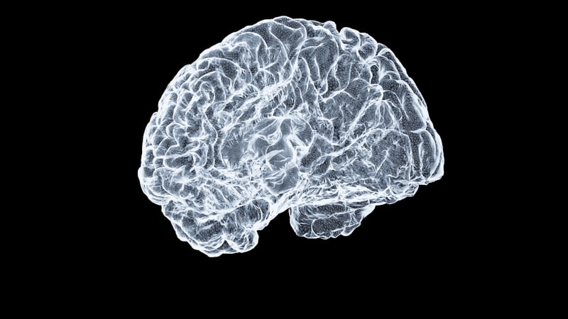
The practical utility of the coronal plane is perhaps most profoundly evident in the realm of medical imaging. Techniques such as CT scans (Computed Tomography), MRI scans (Magnetic Resonance Imaging), and X-rays routinely generate images that are presented in coronal views. These views are indispensable for diagnostic purposes, allowing clinicians to visualize internal structures and pathologies as if the body were sliced vertically from side to side. For instance, a coronal MRI of the brain can reveal the symmetry of the cerebral hemispheres, the extent of a tumor, or the presence of demyelinating lesions, all within a clear anterior-posterior context. This perspective is particularly advantageous for assessing conditions that span across the front and back of the body, offering a comprehensive view that might be obscured in other planes.
In radiology, the ability to reconstruct images along the coronal plane from raw scan data provides critical diagnostic information. For example, when evaluating the lungs, a coronal CT scan can show the extent of pneumonia or the presence of pulmonary nodules across both the anterior and posterior lung fields, allowing for a thorough assessment of their distribution. Similarly, in abdominal imaging, a coronal view can effectively display the relationships between organs like the liver, spleen, and kidneys, along with the major blood vessels, helping to identify abnormalities such as cysts, masses, or fluid collections. The standardized presentation of these images ensures that radiologists and other medical professionals can consistently interpret findings, regardless of where the scans were performed.
Beyond static imaging, the coronal plane is also fundamental in describing the movement of body parts, especially the limbs. When an individual performs an action like raising their arm out to the side (abduction) or bringing it back towards the body (adduction), these movements are occurring predominantly within the coronal plane. Understanding this helps in the analysis of gait, posture, and musculoskeletal function. Physical therapists and sports medicine specialists utilize the concept of the coronal plane to assess range of motion, identify muscular imbalances, and design rehabilitation exercises. By observing movements in relation to this plane, they can accurately diagnose movement dysfunctions and monitor progress during therapy, making the coronal plane a dynamic tool in clinical assessment.
Clinical and Surgical Applications
The significance of the coronal plane extends directly into practical clinical and surgical settings, where its precise definition is critical for patient care. In surgical planning, for instance, surgeons rely heavily on pre-operative imaging (often including coronal views from CT or MRI scans) to meticulously map out the surgical field. This allows them to visualize anatomical structures, identify the exact location of pathologies, and plan the safest and most effective approach for incision and intervention. For complex procedures involving deep-seated organs or structures near vital nerves and blood vessels, a clear understanding of their anterior-posterior relationships, as depicted in the coronal plane, minimizes risks and enhances surgical precision. Without this detailed spatial understanding, surgical outcomes could be compromised, highlighting the plane’s indispensable role.
Furthermore, the coronal plane is instrumental in the visualization of anatomical structures during real-time procedures or post-operative assessments. For example, during endoscopic procedures or arthroscopy, surgeons often orient their instruments and interpret the live video feed based on their understanding of the anatomical planes. This mental mapping allows them to navigate complex internal environments with accuracy. Post-operatively, follow-up imaging in the coronal plane can be used to assess the success of a surgery, monitor for complications, or track the healing process. The consistency of this reference plane across different stages of patient care ensures continuity in evaluation and treatment.
In fields like dentistry and orthodontics, while perhaps less frequently discussed in terms of full body planes, the concept applies to skull and facial anatomy. Coronal views of the skull can be crucial for assessing facial symmetry, the alignment of teeth, or the extent of certain craniofacial anomalies. This detailed perspective helps in planning orthodontic treatments, reconstructive surgeries, or evaluating the progression of developmental conditions. Therefore, the principles derived from the coronal plane’s definition are universally applicable in any medical discipline that requires precise spatial understanding of the body or its parts. The ability to mentally or physically “slice” the body along this plane provides an unparalleled advantage in diagnostic clarity and therapeutic efficacy.
The Role of Anatomical Planes in Human Understanding
While primarily an anatomical concept, the systematic division of the body into planes like the coronal plane profoundly impacts our broader understanding of the human organism, including aspects that border on cognitive and perceptual psychology. The very act of conceptualizing these planes reflects a fundamental human desire to impose order and structure on complex biological forms. This mental framework allows individuals, particularly those in medical fields, to develop a robust internal representation of the human body in three dimensions. This cognitive mapping is essential for diagnosing conditions, planning treatments, and even for understanding how external stimuli interact with internal systems. The ability to mentally rotate and section the body along these planes is a high-level cognitive skill developed through extensive training and practice.
From a cognitive_psychology” target=”_blank”>cognitive psychology perspective, the use of anatomical planes facilitates spatial reasoning and problem-solving within a biological context. When a radiologist interprets a coronal image, they are engaging in a complex process of visual perception, pattern recognition, and spatial transformation, mentally mapping the 2D image back to a 3D reality. This process involves mentally constructing the missing dimension and understanding how structures relate to each other in a cross-sectional view. The standardized nature of these planes reduces cognitive load by providing a predictable reference system, allowing for more efficient and accurate interpretation of complex anatomical data. Therefore, the coronal plane is not just a biological descriptor but a cognitive tool that structures human thought about the body.
Moreover, the understanding of anatomical planes influences how we perceive and describe body posture and movement. When we talk about “frontal” or “back” aspects of the body, we are implicitly referencing the division made by the coronal plane. This linguistic integration demonstrates how deeply ingrained these anatomical concepts are in our everyday language and mental models of the body. In disciplines such as sports psychology or motor learning, while not directly studying the coronal plane, the underlying principles of body orientation and movement in space are crucial. Coaches and athletes often visualize movements in relation to these planes to optimize performance and prevent injury, illustrating a subtle but pervasive psychological impact of these anatomical constructs.
Interconnections with Other Anatomical Frameworks
The coronal plane does not exist in isolation but is intricately connected with other fundamental anatomical frameworks, forming a comprehensive system for spatial orientation. Its relationship with the sagittal plane and the transverse plane is particularly significant, as these three orthogonal planes together define the three-dimensional space of the human body. The coronal plane is perpendicular to both the sagittal and transverse planes, creating a powerful coordinate system. This orthogonality is essential for pinpointing locations with extreme precision and for describing complex movements that involve rotations or translations across multiple axes. For instance, a structure can be described as being “anterior to the sagittal plane and superior to the transverse plane” when viewed in the coronal perspective.
Beyond its relationship with other cardinal planes, the coronal plane also connects to the broader field of kinesiology and biomechanics. These disciplines analyze human movement, often breaking down complex actions into simpler components that occur within or across these anatomical planes. For example, side-bending of the trunk occurs primarily in the coronal plane, while forward flexion occurs in the sagittal plane, and rotation occurs in the transverse plane. Understanding these relationships allows for a sophisticated analysis of muscle actions, joint kinematics, and the forces acting on the body during various activities. This interdisciplinary connection highlights the coronal plane’s foundational role in understanding not just static anatomy but also dynamic human function.
Furthermore, the concept of the coronal plane is integrated into the nomenclature of specific anatomical structures themselves. For instance, the coronal suture of the skull, which runs along the top of the head from ear to ear, is named for its alignment with the coronal plane. This direct naming convention underscores the pervasive influence of these conceptual planes in anatomical description and classification. The consistent use of such terminology across anatomical textbooks, medical literature, and clinical practice ensures a shared understanding and facilitates learning and communication within the vast and intricate domain of human biology. This deep integration demonstrates that the coronal plane is more than just a conceptual slice; it’s a cornerstone of anatomical lexicon and spatial reasoning.
Conclusion and Future Directions
The coronal plane stands as an indispensable concept in the study of human anatomy and its myriad applications in medicine. As one of the three primary planes defining the body’s orientation, it precisely divides the human form into distinct anterior and posterior segments. This fundamental division, running from the right to the left side of the body and maintaining a perpendicular relationship with both the transverse plane and the sagittal plane, provides an unwavering reference system. Its importance cannot be overstated, forming the bedrock upon which complex medical diagnostics, precise surgical interventions, and a comprehensive understanding of human movement are built. The consistent application of this plane ensures clarity and accuracy across diverse clinical and research settings, making it a universal language for describing the human body.
The enduring utility of the coronal plane is particularly evident in advancements in medical imaging, where coronal views from CT scans, MRI scans, and X-rays are vital for visualizing internal structures and pathologies. It is also crucial for sophisticated surgical planning, allowing practitioners to meticulously prepare for procedures by understanding the spatial relationships of organs and tissues. Beyond its direct medical applications, the conceptual framework provided by the coronal plane subtly influences our cognitive understanding of the body, aiding in spatial reasoning and the mental representation of our physical selves. Its integration into anatomical nomenclature and its role in kinesiology further highlight its pervasive influence across the biological sciences.
Looking forward, as medical technology continues to evolve, particularly in areas like augmented reality surgery, 3D printing of anatomical models, and advanced robotics, the foundational principles offered by anatomical planes like the coronal plane will remain central. These technologies will increasingly rely on precise spatial mapping and visualization, where the clear delineation provided by the coronal plane will be more critical than ever. The ability to render and interact with complex anatomical data in virtual or augmented environments, informed by these established spatial frameworks, promises to further enhance diagnostic accuracy, surgical precision, and our overall comprehension of the human body’s intricate architecture. The coronal plane, though a centuries-old concept, will continue to be a cornerstone for future innovations in health and medicine.