Cerebrospinal Fluid: Unlocking Brain Health and Cognition
- Introduction to Dual Definitions of CSF
- Cerebrospinal Fluid (CSF): Core Functions and Production
- Clinical Significance of CSF Dynamics and Pathology
- Introduction to Contrast-Sensitivity Function (CSF)
- Measurement and Assessment of the Contrast-Sensitivity Function
- Clinical Applications of Contrast Sensitivity in Ophthalmology and Psychology
- Interplay of Neuroscience and Functional Assessment
Introduction to Dual Definitions of CSF
The abbreviation CSF is utilized across multiple disciplines within neuroscience, ophthalmology, and psychology, necessitating contextual clarity for precise interpretation. Primarily, CSF stands as the universally accepted abbreviation for Cerebrospinal Fluid, a vital physiological component essential for the mechanical protection and chemical homeostasis of the central nervous system. This fluid plays a critical role in neurobiology, and its status is frequently monitored in clinical settings to diagnose or manage neurological conditions. However, within the domain of visual perception and psychophysics, CSF also represents the Contrast-Sensitivity Function, a complex metric used to evaluate the efficiency and capability of the visual system to discern subtle differences in light intensity across varying spatial scales. Understanding both definitions requires a dual focus on neuroanatomy and sensory processing, respectively, highlighting the diverse applications of this concise abbreviation within academic and clinical literature.
The duality of the term requires careful attention, especially when encountering research findings or clinical reports that do not explicitly define the acronym upon first use. In neurological contexts, discussions involving fluid dynamics, intracranial pressure, or lumbar punctures invariably refer to Cerebrospinal Fluid. Conversely, analyses concerning visual acuity beyond the limitations of the Snellen chart, especially in relation to early-stage ocular disease or environmental visual performance, are almost certainly addressing the Contrast-Sensitivity Function. This initial differentiation is fundamental for maintaining accurate scientific discourse and avoiding significant misinterpretation of results pertaining to either physiological stability or sensory processing capability.
While seemingly disparate, both applications of CSF relate fundamentally to the integrity of the neurological system. Cerebrospinal Fluid ensures the physical and chemical environment necessary for neuronal function, while the Contrast-Sensitivity Function is an output measure reflecting the functional health of the visual pathway, which is an extension of the central nervous system. Therefore, a comprehensive understanding of human physiological and perceptual capabilities necessitates exploring both these critical, yet distinct, concepts represented by the identical three-letter abbreviation.
Cerebrospinal Fluid (CSF): Core Functions and Production
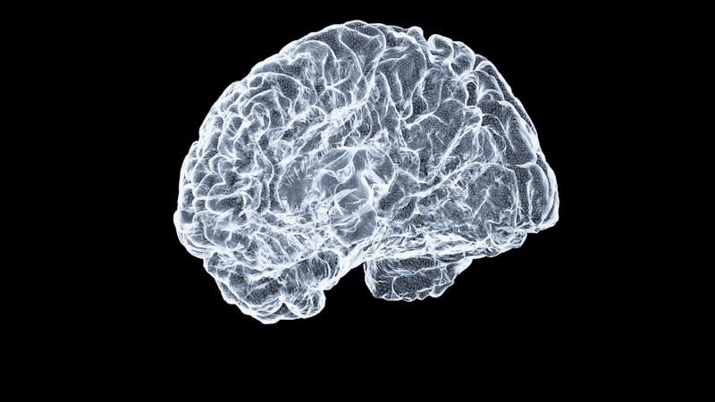
Cerebrospinal Fluid is a clear, colorless liquid that bathes the brain and spinal cord, existing within the cerebral ventricles, the central canal of the spinal cord, and the subarachnoid space. Its existence is crucial for maintaining the delicate biomechanical and biochemical environment required for optimal neural activity. The primary site of CSF production is the specialized epithelial tissue known as the choroid plexus, located within the lateral, third, and fourth ventricles of the brain. This tissue actively filters blood plasma, producing approximately 500 milliliters of fluid per day, though the total volume present in the average adult at any given time is maintained at a relatively stable volume, typically ranging between 125 and 150 milliliters. This constant production and subsequent reabsorption ensures continuous turnover, vital for the removal of metabolic waste products and the maintenance of a stable chemical milieu.
One of the most essential roles of CSF is providing buoyancy to the brain. Because the brain has a density slightly greater than that of the fluid, it is effectively suspended, reducing its net weight from approximately 1,400 grams to nearly 25 grams. This substantial reduction in effective weight prevents the brain from being crushed under its own mass against the rigid structures of the skull base, protecting fragile nerve roots and blood vessels. Furthermore, CSF acts as a primary shock absorber, cushioning the brain against sudden movements or minor trauma. If the head sustains an impact, the fluid dissipates the kinetic energy, minimizing the forces transmitted directly to the delicate neural tissue, thereby preventing contusions and other forms of mechanical injury that could otherwise severely compromise cognitive and motor function.
Beyond physical protection, Cerebrospinal Fluid is indispensable for maintaining the chemical stability of the central nervous system. It helps regulate the concentrations of vital ions, nutrients, and neurotransmitter precursors necessary for synaptic transmission and neuronal metabolism. As a circulating medium, CSF facilitates the transport of hormones and growth factors throughout the neuroaxis. Critically, it serves as the primary drainage route for metabolic byproducts, including harmful molecules and excess proteins produced by active neurons. This waste removal process is increasingly understood to be linked to the glymphatic system, which relies heavily on the bulk flow of CSF to clear interstitial waste, a process particularly active during sleep, highlighting its profound importance in neurological health and disease prevention, such as the clearance of amyloid plaques implicated in neurodegenerative disorders.
Clinical Significance of CSF Dynamics and Pathology
The balance between the production, circulation, and absorption of Cerebrospinal Fluid is tightly regulated, and disturbances in this dynamic equilibrium can lead to severe neurological pathology. The circulation pathway is intricate, moving from the lateral ventricles through the interventricular foramen (of Monro) into the third ventricle, then via the cerebral aqueduct (of Sylvius) into the fourth ventricle, finally exiting into the subarachnoid space through the median and lateral apertures. Reabsorption primarily occurs into the venous sinuses through specialized structures called arachnoid villi or granulations. Any obstruction along this path, or an imbalance in production versus absorption rates, results in a condition known as hydrocephalus, characterized by the abnormal accumulation of CSF within the ventricles, leading to increased intracranial pressure (ICP).
Elevated Intracranial Pressure (ICP) due to issues with CSF dynamics can cause a wide range of symptoms, including headaches, nausea, vomiting, papilledema (swelling of the optic disc), and potentially life-threatening herniation of brain tissue. Treatment for hydrocephalus often involves the surgical insertion of a shunt—a flexible tube system—to divert the excess CSF, typically into another body cavity, such as the peritoneum (ventriculoperitoneal shunt), where it can be safely absorbed. The precise management of CSF pressure and flow is a cornerstone of neurosurgical practice, underlining the profound clinical importance of understanding the fluid’s physics and anatomy.
Furthermore, the composition and pressure of Cerebrospinal Fluid are critical indicators of infectious, inflammatory, and neoplastic processes affecting the central nervous system. Conditions like meningitis (inflammation of the meninges), subarachnoid hemorrhage, or certain autoimmune diseases (e.g., Multiple Sclerosis) often produce characteristic changes in the fluid’s chemistry, cellular content, or protein levels. Therefore, the analysis of CSF obtained via lumbar puncture (spinal tap) remains one of the most powerful diagnostic tools available in neurology. The integrity of the fluid barrier systems, such as the blood-brain barrier, also dictates the passage of substances into the CSF, making its study crucial for pharmacological development and drug delivery strategies targeting the brain.
Introduction to Contrast-Sensitivity Function (CSF)
The second major application of the abbreviation CSF in sensory psychology and visual science is the Contrast-Sensitivity Function. This metric provides a detailed and comprehensive measure of visual performance that goes far beyond the limited scope of standard visual acuity assessments, such as the Snellen chart. Visual acuity typically measures only the ability to resolve fine details—the smallest high-contrast stimulus (usually black letters on a white background) that can be identified—corresponding primarily to high spatial frequencies. In contrast, the Contrast-Sensitivity Function maps the sensitivity of the visual system across a broad spectrum of spatial frequencies, defined as the number of light/dark cycles per degree of visual angle.
The Contrast-Sensitivity Function is fundamentally a graph plotting the threshold contrast required for detection against the spatial frequency of the stimulus. Contrast is defined as the relative difference in luminance between the light and dark parts of the stimulus. A typical CSF curve for a healthy human observer exhibits a characteristic inverted U-shape or band-pass filter profile. Sensitivity is generally low at very low spatial frequencies (large, blurry stimuli) because the visual system is poor at detecting gradual overall luminance changes. Sensitivity peaks around 2 to 5 cycles per degree, where the visual system is most efficient, corresponding roughly to the size of medium-sized objects or features. Sensitivity then rapidly drops off at high spatial frequencies (fine details), eventually reaching the visual acuity limit where even 100% contrast stimuli cannot be resolved.
The utility of the Contrast-Sensitivity Function lies in its ability to predict real-world visual performance more accurately than simple acuity measures. Many daily tasks, such as driving in fog, distinguishing facial features in low light, or navigating low-contrast environments (like stairs with poor lighting), rely heavily on contrast detection across various spatial frequencies, not just the resolution of the smallest letter. Diseases or conditions that impair the visual pathway often affect specific regions of the CSF curve before they impact high-contrast visual acuity, making CSF measurement a highly sensitive tool for early detection and comprehensive visual assessment.
Measurement and Assessment of the Contrast-Sensitivity Function
Measuring the Contrast-Sensitivity Function involves presenting the observer with grating patterns—typically sinusoidal gratings—that vary systematically in both spatial frequency and contrast. These gratings are patterns of light and dark bars whose luminance profile changes sinusoidally across space. The spatial frequency is controlled by the width and spacing of the bars, while the contrast is controlled by the difference between the maximum and minimum luminance levels. Psychophysical methods are used to determine the minimum contrast level (the threshold) at which the observer can reliably detect the presence of the grating for each tested spatial frequency. The reciprocal of this minimum detectable contrast is defined as the sensitivity at that specific frequency.
Standardized testing methodologies often utilize specialized charts or computer-generated displays. Unlike the fixed letter sizes of the Snellen chart, CSF testing requires dynamic control over the stimulus parameters. Common clinical tools include the Vistech chart or the Pelli-Robson contrast sensitivity chart. While the Pelli-Robson chart measures contrast sensitivity at a single, relatively low spatial frequency, full CSF measurement requires testing multiple frequencies to map the entire characteristic curve. The data points derived from these measurements are then plotted to generate the complete function, revealing the overall shape and peak sensitivity of the individual’s visual system.
Detailed analysis of the resulting CSF curve provides crucial insights into the specific deficits an individual may possess. For instance, losses in high spatial frequency sensitivity suggest problems primarily related to fine detail resolution, perhaps due to optical defocus or minor retinal issues. Conversely, a depression in the mid-range or low-range frequencies often indicates significant scattering of light or neurological processing issues, such as those associated with optic neuritis or early-stage cataracts. The slope and shape of the function, therefore, serve as diagnostic signatures for various types of visual impairment, offering precision in diagnosis that standard acuity testing cannot achieve.
Clinical Applications of Contrast Sensitivity in Ophthalmology and Psychology
The clinical application of the Contrast-Sensitivity Function is widespread, particularly in ophthalmology and optometry. It is often employed to assess visual performance in patients who report significant visual difficulties despite having 20/20 or near-normal Snellen acuity. This discrepancy is common in conditions that primarily affect the contrast pathways, such as early cataracts, where scattered light severely reduces contrast before resolution is significantly impaired, or certain corneal diseases. By identifying early losses in contrast sensitivity, clinicians can initiate timely interventions and better counsel patients on their expected functional vision, especially in challenging environments.
Furthermore, CSF testing is highly valuable in monitoring the progression of neuro-ophthalmological diseases. For example, patients with Multiple Sclerosis (MS) often develop optic neuritis, which causes inflammation and demyelination of the optic nerve. This damage frequently results in a marked and early reduction in contrast sensitivity, particularly at mid-to-high spatial frequencies, often preceding or being more pronounced than the loss of visual acuity. Similarly, individuals with glaucoma, a disease causing damage to the optic nerve head, may show specific deficits in their CSF curve, providing a sensitive biomarker for nerve damage progression.
In psychological and human factors research, the Contrast-Sensitivity Function is used to model and predict human performance under various viewing conditions. Understanding how the environment (e.g., glare, low illumination) degrades the effective contrast of objects allows researchers to design safer and more efficient visual displays, cockpit instruments, and signage. The CSF provides the fundamental input sensitivity profile for many computational models of vision, helping to explain phenomena such as camouflage, texture perception, and the visual demands of complex tasks. Thus, the CSF serves not only as a diagnostic tool but also as a foundational component in understanding the limits and capabilities of the human visual processing system across diverse real-world scenarios.
Interplay of Neuroscience and Functional Assessment
While Cerebrospinal Fluid and the Contrast-Sensitivity Function represent distinct physiological and perceptual concepts, their shared abbreviated term, CSF, underscores the interconnected nature of the central nervous system and its functional outputs. The integrity of the brain, maintained biochemically and mechanically by Cerebrospinal Fluid, is a prerequisite for generating any measurable sensory function, including the precise filtering capabilities quantified by the Contrast-Sensitivity Function. If the physical environment of the visual cortex or the optic nerve is compromised—for instance, by increased intracranial pressure due to CSF fluid dynamics issues—the resultant visual performance, as measured by the perceptual CSF, will inevitably degrade.
The clinical observation mentioned in the original entry, regarding the necessity of placing a stent due to the presence of cerebrospinal fluid, is slightly misleading in its simplistic phrasing. The mere presence of CSF is normal; the need for intervention (such as shunt placement or stenting) arises specifically when there is an abnormal accumulation or obstructed flow, leading to pathologies like hydrocephalus or persistent leakage. Such interventions are necessary to restore the normal pressure and flow dynamics, thereby protecting the neural tissues—including the visual pathways—from compressive damage. Thus, ensuring the proper maintenance of the physiological CSF system is essential for preserving the functional integrity measured by the perceptual CSF system.
In summary, both definitions of CSF are cornerstones of high-level scientific inquiry: one pertaining to the foundational environment (Cerebrospinal Fluid) and the other to the resulting measurable performance (Contrast-Sensitivity Function). Researchers and clinicians must remain vigilant regarding context when encountering the abbreviation, ensuring that the critical distinction between the fluid that supports the nervous system and the function that measures its visual output is always maintained for clarity and diagnostic accuracy in psychology, neurology, and ophthalmology.