Nerve Roots: The Biological Map of Your Physical Experience
- Introduction to Nerve Roots
- Anatomical Structure of Nerve Roots
- Physiological Functions and Information Transmission
- Historical Understanding and Discovery
- Radiculopathy: Clinical Implications and Symptoms
- Practical Insights: Understanding Nerve Root Pain in Daily Life
- Broader Significance and Therapeutic Applications
- Interconnections with the Nervous System and Related Conditions
Introduction to Nerve Roots
The nerve root represents a critical anatomical structure within the human spinal cord, serving as the initial segment of a nerve as it branches out from the central nervous system to form the peripheral nervous system. Specifically, these roots are the bundles of nerve fibers emerging directly from the spinal cord, responsible for the vital bidirectional transmission of information between the brain and the rest of the body. They are essentially the conduits through which sensory information from the body’s periphery, such as touch, temperature, and pain, travels to the spinal cord and subsequently to the brain for processing, while also relaying motor commands from the brain and spinal cord to muscles and glands, enabling movement and bodily functions. This intricate network underscores the indispensable role of nerve roots in maintaining physiological homeostasis and facilitating interaction with the external environment.
Each nerve root is meticulously organized into two distinct components: the dorsal root and the ventral root. The dorsal root, also known as the posterior root, exclusively carries afferent, or sensory, nerve fibers, meaning they transmit impulses from the periphery towards the spinal cord. These fibers relay sensations from the skin, muscles, and internal organs, providing the brain with a continuous stream of environmental and internal data. Conversely, the ventral root, or anterior root, is composed solely of efferent, or motor, nerve fibers. These fibers convey impulses from the spinal cord to the skeletal muscles, initiating voluntary movements, and also transmit signals to smooth muscles, cardiac muscle, and glands, thereby controlling involuntary functions. The precise anatomical separation of sensory and motor functions within these roots is a fundamental principle of neuroanatomy, enabling a highly organized and efficient nervous system.
The convergence of these dorsal and ventral roots at each segmental level of the spinal column forms a spinal nerve, which then exits the vertebral column through a specific opening called the intervertebral foramen. This structural arrangement ensures that the nervous system can effectively innervate virtually every part of the body, allowing for complex motor control, sophisticated sensory perception, and the regulation of vital autonomic functions. Understanding the intricate architecture and functional dichotomy of nerve roots is paramount for diagnosing and treating a wide array of neurological conditions that affect sensation, movement, and overall bodily function, highlighting their foundational importance in both healthy physiology and clinical pathology. Their vulnerability to compression or injury due to their passage through bony canals makes them a common site for various debilitating conditions.
Anatomical Structure of Nerve Roots
The spinal cord, a cylindrical bundle of nerve fibers and tissue, is segmented along its length, giving rise to 31 pairs of spinal nerves, each originating from a specific segment. Each pair of spinal nerves begins as two distinct roots—the dorsal (posterior) root and the ventral (anterior) root—which emerge from the lateral aspects of the spinal cord. The dorsal root is characterized by the presence of a dorsal root ganglion, an enlargement containing the cell bodies of all the sensory neurons whose axons comprise the dorsal root. These sensory neurons are pseudounipolar, meaning they have a single process that divides into two, with one branch extending to the periphery (e.g., skin, muscle) and the other entering the spinal cord. This ganglion is crucial for processing sensory input before it reaches the spinal cord’s gray matter.
The ventral root, in contrast, lacks a ganglion because the cell bodies of its motor neurons are located within the gray matter of the spinal cord itself, specifically in the anterior horn. The axons of these motor neurons exit the spinal cord via the ventral root to innervate skeletal muscles and other effector organs. Both the dorsal and ventral roots are covered by extensions of the meninges, the protective membranes that surround the brain and spinal cord, specifically the pia mater, arachnoid mater, and dura mater, which provide structural support and protection. As these roots extend laterally, they converge within the intervertebral foramen, an opening between adjacent vertebrae, where they unite to form a mixed spinal nerve. This nerve then immediately branches into dorsal and ventral rami, which distribute nerve fibers to various parts of the body.
The precise location and trajectory of nerve roots are critically important. They exit the spinal canal at specific levels, corresponding to the vertebral segments. For instance, cervical nerve roots exit above their corresponding vertebrae (e.g., C5 nerve root exits above the C5 vertebra), while thoracic, lumbar, and sacral nerve roots typically exit below their corresponding vertebrae. This anatomical relationship is vital for clinical localization of nerve root pathology. The nerve roots are particularly vulnerable to compression or irritation at the intervertebral foramen due to their proximity to bony structures, intervertebral discs, and ligaments. Any pathological changes in these surrounding structures, such as herniated discs, spinal stenosis, or osteophytes (bone spurs), can impinge upon the delicate nerve roots, leading to a range of neurological symptoms. The detailed understanding of this intricate anatomy is fundamental for accurate diagnosis and effective treatment strategies.
Physiological Functions and Information Transmission
The primary physiological function of nerve roots is to serve as the conduits for the transmission of neural impulses, effectively acting as the communication highways between the central nervous system (CNS) and the peripheral nervous system (PNS). The dorsal roots are exclusively dedicated to afferent transmission, meaning they carry sensory information from various receptors throughout the body towards the CNS. These receptors, located in the skin, muscles, joints, and internal organs, detect stimuli such as touch, pressure, temperature, pain, and proprioception (awareness of body position). This sensory input is then relayed through the dorsal root ganglia, where the cell bodies of the sensory neurons reside, before entering the spinal cord’s posterior horn for further processing and eventual ascent to higher brain centers, allowing for conscious perception and reflex responses.
Conversely, the ventral roots are dedicated to efferent transmission, conveying motor commands from the CNS to effector organs in the periphery. These motor commands originate from motor neurons located in the anterior horn of the spinal cord’s gray matter. Their axons exit via the ventral roots and travel to innervate skeletal muscles, triggering muscle contraction and enabling voluntary movements. Beyond voluntary control, the ventral roots also contain efferent fibers of the autonomic nervous system, which regulate involuntary functions such as heart rate, digestion, respiration, and glandular secretions. This dual role underscores the ventral root’s comprehensive involvement in both somatic (voluntary) and autonomic (involuntary) control mechanisms, illustrating the complexity of neural regulation emanating from the spinal cord.
The integrity of this bidirectional information flow through the nerve roots is absolutely crucial for normal bodily function. Any disruption, whether due to compression, inflammation, or direct injury, can profoundly impair both sensory perception and motor control. For instance, damage to a dorsal root can lead to numbness, altered sensation, or a loss of proprioception in the corresponding dermatome (an area of skin supplied by a single spinal nerve). Conversely, injury to a ventral root can result in muscle weakness, paralysis, or atrophy in the muscles supplied by that root (a myotome). The precise mapping of dermatomes and myotomes to specific nerve roots is a cornerstone of neurological examination, allowing clinicians to pinpoint the exact level of spinal pathology based on the patient’s symptoms. This intricate physiological interplay highlights the nerve roots as indispensable components of the entire nervous system’s operational efficiency.
Historical Understanding and Discovery
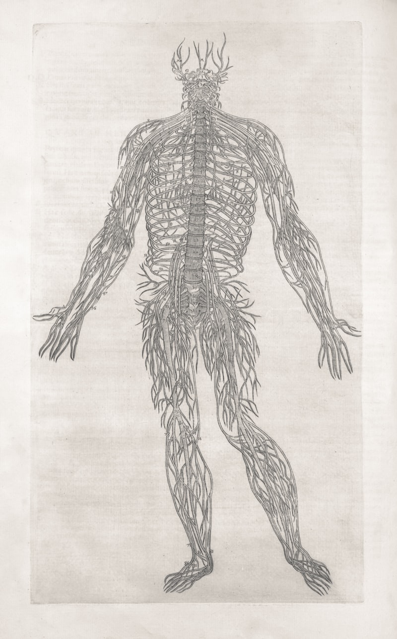
The understanding of nerve roots and their distinct sensory and motor functions evolved significantly over centuries, building upon early anatomical observations. Ancient Greek physicians, notably Galen in the 2nd century AD, made early descriptions of nerves and their connection to the brain and spinal cord, but their understanding of specific functions was rudimentary and often intertwined with philosophical theories. For many centuries, the prevailing view was that nerves were hollow tubes carrying “animal spirits.” It was not until the Renaissance, with figures like Andreas Vesalius in the 16th century, that detailed anatomical dissections began to challenge these older notions, providing more accurate visual representations of the nervous system, though functional insights remained limited.
A pivotal breakthrough in understanding the functional segregation of nerve roots occurred in the early 19th century, primarily through the independent work of two physiologists: Sir Charles Bell in Scotland and François Magendie in France. In 1811, Bell published a pamphlet where he described experiments demonstrating that the anterior (ventral) roots of the spinal nerves were responsible for motor functions, while the posterior (dorsal) roots were associated with sensory functions. However, his initial publication lacked full clarity and definitive proof. A decade later, in 1822, Magendie conducted more conclusive experiments on puppies, directly confirming the distinct roles of the dorsal and ventral roots. He showed that cutting the dorsal roots abolished sensation but not movement, while cutting the ventral roots abolished movement but not sensation. This discovery became known as the Bell-Magendie Law, a cornerstone of modern neurophysiology, firmly establishing the anatomical and functional separation of sensory and motor pathways at the spinal nerve root level.
The establishment of the Bell-Magendie Law profoundly impacted the subsequent study of the nervous system, providing a robust framework for understanding neurological disorders and guiding clinical diagnoses. It marked a significant shift from purely anatomical descriptions to a more functional understanding of neural pathways. This foundational knowledge enabled later researchers to map sensory dermatomes and motor myotomes, linking specific areas of the body to particular spinal nerve roots. This historical progression from speculative theories to rigorous experimental validation underscores the scientific method’s power in unraveling the complexities of the human body. The precise understanding of nerve root function continues to be fundamental in fields ranging from neurosurgery to rehabilitation, influencing diagnostic techniques and therapeutic interventions.
Radiculopathy: Clinical Implications and Symptoms
Radiculopathy is a common and often debilitating condition that arises when a nerve root is compressed, inflamed, or injured. This compression can occur at any level of the spinal column, but it is most frequently observed in the cervical (neck) and lumbar (lower back) regions due to the greater mobility and weight-bearing stresses these areas endure. The symptoms of radiculopathy are directly related to the specific nerve root affected and the type of fibers involved. If a sensory root is compressed, patients typically experience paresthesia, which includes numbness, tingling, or a “pins and needles” sensation in the dermatome supplied by that nerve root. This can also manifest as sharp, shooting pain that radiates along the nerve’s pathway, often described as an electrical sensation.
When a motor nerve root is affected, the primary symptoms involve muscle weakness or impaired motor function in the muscles supplied by that root, known as a myotome. This can range from mild weakness to complete paralysis, making it difficult for individuals to perform everyday tasks. In severe or chronic cases, muscle atrophy (wasting) can occur due to prolonged lack of nerve stimulation. Common causes of nerve root compression include herniated intervertebral discs, where the soft inner material of the disc protrudes and presses on the adjacent nerve root. Other significant causes include spinal stenosis, a narrowing of the spinal canal or intervertebral foramen, often due to degenerative changes like osteophytes (bone spurs) or thickened ligaments. Tumors, infections, and inflammatory conditions can also, albeit less commonly, lead to nerve root compression.
The clinical diagnosis of radiculopathy typically involves a thorough neurological examination, evaluating sensory deficits, muscle weakness, and changes in reflexes. Imaging studies such as Magnetic Resonance Imaging (MRI) are often employed to visualize the spinal structures and identify the source of compression. Electrophysiological studies, including electromyography (EMG) and nerve conduction studies (NCS), can further assess nerve function and confirm nerve root involvement. Treatment strategies vary depending on the severity and cause of the compression. Conservative management often includes rest, anti-inflammatory medications, physical therapy, and epidural steroid injections to reduce inflammation and pain. In cases where conservative treatments fail, or if there is significant neurological deficit, surgical intervention may be necessary to decompress the affected nerve root, such as a discectomy for a herniated disc or a laminectomy for spinal stenosis. Early and accurate diagnosis is crucial for effective management and preventing long-term neurological damage.
Practical Insights: Understanding Nerve Root Pain in Daily Life
To grasp the tangible impact of nerve root issues, consider a common scenario: a person experiencing sciatica. Sciatica is not a diagnosis in itself but rather a symptom complex characterized by pain radiating along the path of the sciatic nerve, which originates from several nerve roots in the lower lumbar and sacral spine (L4, L5, S1, S2, S3). Imagine an individual, let’s call him Mark, who lifts a heavy box incorrectly, causing a herniated disc in his lower back, specifically between the L5 and S1 vertebrae. This herniation means the soft, jelly-like center of the disc protrudes outward, pressing directly onto the S1 nerve root as it exits the spinal column. Mark would immediately feel a sharp, shooting pain in his lower back that quickly travels down his leg, often into his calf and foot, following the distribution of the S1 dermatome. He might also describe numbness or tingling in the affected leg and foot.
The “how-to” of this psychological principle applies as follows: First, the mechanical compression of the S1 nerve root by the herniated disc directly irritates the nerve fibers. This irritation generates abnormal electrical signals that are interpreted by the brain as pain, radiating along the expected path of the S1 nerve root. Second, because the S1 nerve root primarily carries sensory information from the lateral aspect of the foot and motor commands to muscles responsible for plantarflexion (pointing the foot downwards) and ankle eversion, Mark might experience specific symptoms. He would notice weakness when trying to stand on his tiptoes or push off the ground, indicating motor involvement in the S1 myotome. He might also report a diminished sensation or numbness on the outer side of his foot, corresponding to the S1 dermatome. This precise pattern of symptoms allows clinicians to localize the problem to the S1 nerve root, even without immediate imaging.
Furthermore, the inflammation surrounding the compressed nerve root contributes significantly to the pain experience. The body’s inflammatory response releases chemical mediators that sensitize the nerve, making it even more reactive to pressure and movement. Consequently, activities like sitting for prolonged periods, coughing, or sneezing, which increase pressure within the spinal canal, can exacerbate Mark’s pain. Understanding that his symptoms stem from a specific nerve root, rather than a generic “backache,” guides the treatment approach. Physical therapy would focus on exercises that decompress the nerve root, strengthen core muscles, and improve posture. Medications would target inflammation and nerve pain. In severe cases, a surgical procedure like a microdiscectomy would aim to remove the herniated disc material, thereby relieving the pressure on the S1 nerve root directly. This practical example vividly illustrates how a detailed knowledge of nerve root anatomy and function is indispensable for accurate diagnosis, effective treatment, and patient education regarding such common and distressing conditions.
Broader Significance and Therapeutic Applications
The concept of nerve roots holds immense significance in the broader field of neuroscience and clinical medicine, particularly in neurology, neurosurgery, and rehabilitation medicine. Its importance stems from the fact that nerve roots represent the final common pathway for all sensory input from the body to the central nervous system and all motor output from the central nervous system to the skeletal muscles. Therefore, any pathology affecting these structures can have widespread and profound effects on a person’s ability to sense, move, and function autonomously. The detailed mapping of dermatomes (areas of skin supplied by a single spinal nerve root) and myotomes (groups of muscles innervated by a single spinal nerve root) is a foundational element of neurological examination, allowing clinicians to pinpoint the exact level of spinal cord or nerve root injury based on observed sensory deficits, muscle weakness, or reflex changes. This precise localization is critical for guiding diagnostic imaging and therapeutic interventions.
In terms of therapeutic applications, understanding nerve root anatomy and pathology is central to numerous medical procedures and treatment strategies. For instance, in pain management, epidural steroid injections are frequently administered directly around inflamed nerve roots to deliver anti-inflammatory medication, providing targeted relief for conditions like radiculopathy. Surgical interventions for spinal conditions, such as discectomy (removal of a herniated disc) or laminectomy (removal of part of the vertebral bone to decompress the spinal canal), are meticulously planned to relieve pressure on specific nerve roots while preserving their integrity. Furthermore, advancements in neuroimaging techniques, such as high-resolution Magnetic Resonance Imaging (MRI), allow for detailed visualization of nerve roots and surrounding structures, enhancing diagnostic accuracy and informing surgical approaches. These applications underscore how a deep understanding of nerve root biology directly translates into improved patient outcomes and quality of life.
Beyond direct clinical interventions, the study of nerve roots contributes significantly to our comprehension of spinal cord injury and regeneration research. Understanding how nerve roots are damaged and the limited capacity of the adult central nervous system to regenerate axons has driven extensive research into potential therapies, including stem cell transplantation, nerve grafting, and pharmacological interventions aimed at promoting nerve repair. In the field of electrophysiology, techniques like electromyography (EMG) and nerve conduction studies (NCS) are routinely used to assess the functional integrity of nerve roots, helping to differentiate between nerve root compression and peripheral nerve injury. Thus, the concept of nerve roots is not merely an anatomical detail but a dynamic area of study with profound implications for diagnostics, treatment, and the ongoing quest to restore neurological function.
Interconnections with the Nervous System and Related Conditions
Nerve roots serve as the crucial anatomical and functional bridge between the central nervous system (CNS), comprising the brain and spinal cord, and the peripheral nervous system (PNS), which extends throughout the body. This fundamental connection means that nerve roots are intimately involved in nearly all neurological processes, from the simplest reflex arc to complex voluntary movements and sensory perceptions. Each nerve root carries a specific set of sensory and motor fibers, originating from or terminating in distinct areas of the spinal cord’s gray matter. The dorsal roots connect to the posterior horn, where sensory information first synapses, while the ventral roots emerge from the anterior horn, which houses the cell bodies of motor neurons. This direct anatomical and functional continuity ensures seamless communication, allowing the brain to receive a constant stream of information about the body’s internal and external states and to issue appropriate commands.
Several key psychological and neurological concepts are directly related to the function and pathology of nerve roots. Foremost among these is radiculopathy, the clinical syndrome caused by compression or irritation of a nerve root, which we have discussed in detail. This often manifests as sciatica when affecting the lumbar and sacral nerve roots that form the sciatic nerve, or as cervical radiculopathy when nerve roots in the neck are involved, leading to pain, numbness, or weakness in the arm. Another related concept is spinal cord injury, where damage to the spinal cord itself can directly impact the nerve roots emerging from that level, leading to a combination of upper and lower motor neuron signs depending on the extent of the lesion. Conditions such as spinal stenosis and herniated discs are primary causes of nerve root compression, linking them directly to musculoskeletal and neurological health.
The broader category of psychology to which the study of nerve roots most directly belongs is biological psychology or neuropsychology, especially within the subfield of neuroanatomy and neurophysiology. While psychology generally focuses on the mind and behavior, biological psychology specifically examines the biological bases of psychological processes. Understanding nerve roots is fundamental to comprehending how sensory input reaches the brain to influence perception and how motor commands are executed to produce behavior. It provides the anatomical and physiological substrate for sensory experiences (e.g., pain perception, proprioception) and motor actions, both of which are central to human psychology. Furthermore, the study of nerve root pathology is crucial for understanding the physical basis of neurological symptoms that can significantly impact mental health and well-being, highlighting the inextricable link between the physical structure of the nervous system and psychological experience. This interdisciplinary perspective underscores the holistic importance of nerve roots in both human physiology and the broader understanding of behavior and cognition.