Neuroanatomy: The Brain’s Tender Protective Shield
- Introduction and Definition of the Pia Mater
- Anatomical Location and Relationship to the Meninges
- Microscopic Structure and Cellular Composition
- Vascularization and the Role of the Choroid Plexus
- Functional Significance: Protection and Nutrient Exchange
- Clinical Relevance: Pathologies and Disorders
- Development and Embryological Origin
- Comparative Anatomy and Evolutionary Aspects
Introduction and Definition of the Pia Mater
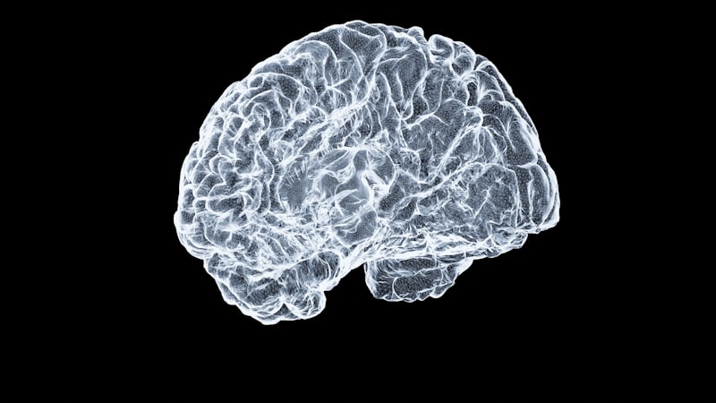
The pia mater, translating literally from Latin as “tender mother” or “soft mother,” constitutes one of the most critical elements of the central nervous system’s protective architecture. It is an exquisitely delicate, highly vascularized connective tissue membrane that intimately follows the contours of the brain and spinal cord, enveloping every gyrus, sulcus, and fissure. This proximity is paramount to its function, as it forms the final, innermost barrier separating the neural tissue itself from the surrounding cerebrospinal fluid (CSF) and the tougher layers of the meninges. Unlike the other meningeal layers, the pia mater is not easily dissectible from the neural surface, a characteristic that underscores its fundamental role in both structural support and physiological regulation.
Serving as the innermost layer among the three meningeal membranes—the dura mater, the arachnoid mater, and the pia mater—its primary function extends beyond mere physical containment. It acts as a selective interface, playing a pivotal role in the diffusion of substances between the CSF and the underlying neural parenchyma. Its agility and thinness allow it to conform precisely to the intricate three-dimensional structure of the CNS, ensuring that even the deepest crevices are protected and supported. Understanding the pia mater is essential for comprehending the overall neuroprotective system, as its integrity is intrinsically linked to the health and functionality of the brain and spinal cord tissue it safeguards.
Historically, the study of the pia mater was instrumental in early neuroanatomy, providing insights into the brain’s vascular supply and its dependency on external protective structures. Modern neuroscience recognizes the pia mater not merely as a passive wrapping but as an active component of the blood-brain barrier system, particularly at the interface with the perivascular spaces. Disturbances or congenital absence of this layer, as illustrated by clinical observations, can lead to severe neurological deficits, highlighting that its seemingly fragile nature belies its immense importance in maintaining homeostatic balance and structural cohesion within the most complex organ system of the body. Clinical evidence suggests that when the pia mater is compromised or entirely absent, it is highly likely to be the primary factor responsible for extensive damage to the underlying spinal cord or brain structure.
Anatomical Location and Relationship to the Meninges
The anatomical positioning of the pia mater dictates its interactions with the other two protective membranes. Superior to the pia mater lies the arachnoid mater, separated by the subarachnoid space, a critical region filled with cerebrospinal fluid and traversed by delicate arachnoid trabeculae. These trabeculae connect the two layers, giving rise to the appearance of a spider web structure, which lends the arachnoid mater its name. The relationship between the pia and the arachnoid is crucial because the subarachnoid space is where major cerebral arteries and veins reside before they penetrate the neural tissue, utilizing the perivascular spaces formed by the pia mater itself.
In contrast to the dura mater, which is a thick, fibrous, and relatively rigid outer layer, and the arachnoid mater, which is avascular and non-adherent to the brain surface, the pia mater is highly vascularized and fundamentally adherent. This adherence is facilitated by astrocytic endfeet that terminate on the pial surface, forming the glia limitans superficialis, which is a specialized basement membrane crucial for barrier function. This intimate bond means that damage penetrating the pia mater immediately affects the neural tissue, emphasizing why its protective layer must remain intact across the entire surface of the central nervous system, including the deep fissures like the longitudinal fissure that divides the cerebral hemispheres.
The continuation of the pia mater along the spinal cord is structurally analogous to its cerebral counterpart, though it exhibits specialized modifications essential for mechanical stability. In the spinal column, the pia mater forms the denticulate ligaments, which are lateral extensions that anchor the spinal cord to the dura mater, preventing excessive longitudinal and lateral displacement within the vertebral canal. Furthermore, at the caudal end of the spinal cord, the pia mater coalesces to form the filum terminale, a slender strand that descends through the lumbar cistern to anchor the spinal cord to the coccyx. These specialized spinal structures confirm the pia mater’s dual role: providing both cellular protection and essential mechanical stabilization of the entire neuroaxis.
Microscopic Structure and Cellular Composition
Microscopically, the pia mater is exceedingly thin, typically measuring only a few micrometers in thickness, and is composed primarily of two layers: an outer layer of collagen fibers and delicate fibroblasts, and an inner layer that integrates seamlessly with the glia limitans. The cellular component consists mainly of flattened, modified fibroblasts known as pial cells, which are characterized by broad, attenuated cytoplasmic processes. These cells are arranged in sheets and are responsible for synthesizing the delicate matrix that allows the membrane to be strong yet pliable, enabling it to accommodate the slight but constant volume changes associated with cerebral blood flow and CSF pulsations, which are critical for preventing undue mechanical stress on the neural tissue.
The innermost boundary of the pia mater, facing the neural parenchyma, is inextricably linked to the underlying astrocytes. The glia limitans is essentially a continuous basement membrane reinforced by the terminal expansions (endfeet) of astrocytes, which tightly abut the pial surface. This composite structure—the pial membrane fused with the glia limitans—is often referred to collectively as the pial-glial membrane. This fusion is critical for establishing the impermeable boundary of the CNS and for regulating the passage of large molecules and inflammatory cells from the CSF into the neural tissue. The integrity of this basement membrane is vital, as its disruption is a common feature in neuroinflammatory conditions and various neurodegenerative diseases, underscoring its importance in chronic neurological health.
Crucially, the pia mater is penetrated by countless blood vessels that supply the cerebral cortex and deep brain structures. As these arteries and arterioles enter the neural tissue, the pia mater accompanies them, forming a sleeve or cuff known as the perivascular space (or Virchow-Robin space). These pial-lined tunnels extend deep into the brain substance, creating channels that are integral to the brain’s unique lymphatic-like waste clearance system, the glymphatic pathway. The precise cellular arrangement and matrix composition of the pia mater thus facilitate not only physical protection but also active participation in fluid dynamics and immunological surveillance within the highly controlled central nervous system environment.
Vascularization and the Role of the Choroid Plexus
The pia mater is distinguished by its rich vascular network, which is essential for nourishing the superficial layers of the CNS and for forming specialized structures involved in cerebrospinal fluid production. Unlike the avascular arachnoid mater, the pial layer contains numerous small arteries and veins that ramify extensively across the neural surface before penetrating the underlying tissue. This intricate plexus of blood vessels ensures a robust and widespread distribution of oxygen and nutrients directly to the cerebral and spinal cortices, which are characterized by exceptionally high metabolic demands. The distribution pattern of these pial vessels is critical for understanding ischemic events, as blockages often follow these specific anatomical routes, leading to predictable patterns of cortical infarction.
A particularly important specialization of the pia mater related to vascularization is its involvement in the formation of the choroid plexus. In specific regions of the cerebral ventricles, the pia mater fuses with the ependyma (the specialized epithelial lining of the ventricles) and highly convoluted capillary loops. This composite structure, known as the tela choroidea, invaginates into the ventricles to form the choroid plexus, which is recognized as the principal site of cerebrospinal fluid (CSF) production. The pial component contributes the supporting connective tissue framework and the necessary vascular supply required for the specialized epithelial cells of the plexus to actively filter plasma and secrete the nutrient-rich cerebrospinal fluid.
The dynamic interplay between the pial vasculature and the CSF environment underscores the pia mater’s role as a physiological mediator and regulator of cerebral blood flow. The vessels within the pial space are subject to strict autoregulatory mechanisms, ensuring constant cerebral perfusion despite acute fluctuations in systemic blood pressure, thereby protecting sensitive neural tissue from ischemia or hyperperfusion. Furthermore, the pia mater acts as a crucial conduit for the venous drainage of the superficial neural tissue. Veins emerging from the cerebral cortex traverse the pial layer, often running alongside arteries in the subarachnoid space before piercing the arachnoid and dura mater to empty into the large dural venous sinuses, thus completing the vascular circuit essential for CNS metabolic homeostasis.
Functional Significance: Protection and Nutrient Exchange
The core functional significance of the pia mater lies in its indispensable multifunctional role as a protective sheath and an active exchange membrane. As a protective sheath, its close adherence to the brain surface provides a crucial mechanical buffer against minor external forces that have already bypassed the robust layers of the skull, dura, and arachnoid mater. More importantly, physiologically, it acts as a selective barrier, meticulously regulating the microenvironment immediately surrounding the neurons and glia. While the most restrictive component of the blood-brain barrier is located at the endothelial cells of the capillaries, the pial-glial membrane provides a secondary, yet critical, physical barrier against large particles, toxins, and migrating immune cells originating from the cerebrospinal fluid.
In terms of nutrient exchange, the pia mater facilitates the necessary diffusion of vital substances such as oxygen, glucose, and other essential metabolites from the highly vascularized subarachnoid space and the CSF into the superficial layers of the neural parenchyma. This highly regulated exchange is absolutely essential for maintaining the precise chemical stability and ion balance required for optimal neuronal signaling and synaptic transmission. Conversely, metabolic waste products generated by the neural tissue are channeled efficiently through the perivascular spaces, which are often lined by pial extensions, before entering the CSF for eventual bulk clearance. This essential role in bidirectional transport highlights the active, rather than passive, nature of this delicate membrane in supporting the brain’s high metabolic turnover.
Furthermore, the pia mater harbors various cell types capable of participating in immunological and inflammatory responses, though its primary function often involves maintaining the immune privilege of the CNS. Fibroblasts and resident macrophages residing within the pial layer are positioned strategically to respond rapidly to pathogens or debris that may enter the subarachnoid space. By contributing to the structural integrity of the perivascular spaces, the pia mater indirectly supports the recently understood glymphatic system, which relies heavily on controlled CSF flow dynamics for the efficient removal of soluble waste. Maintaining the elasticity and impermeability of the pia mater is therefore central to preventing the accumulation of toxic protein aggregates associated with chronic neurological conditions like Alzheimer’s and Parkinson’s diseases.
Clinical Relevance: Pathologies and Disorders
The clinical importance of the pia mater becomes starkly evident when considering acute conditions that compromise its structural integrity or involve inflammation of the meninges. Meningitis, an inflammation of the protective membranes surrounding the brain and spinal cord, involves the pia mater directly and is often life-threatening. In bacterial meningitis, pathogens often proliferate aggressively in the subarachnoid space and incite a severe inflammatory response that rapidly affects the pial layer, potentially leading to critical complications such as cerebral edema, widespread vasculitis, and subsequent irreversible damage to the underlying neural tissue. The intimate contact between the pia mater and the brain makes it the critical point of entry for infection to penetrate the neural parenchyma, transforming a peripheral infection into a central neurological crisis.
Traumatic injuries, such as severe concussions, blast injuries, or penetrating head wounds, can also lead to significant pial damage. Subpial hemorrhages, though often localized and microscopic, can result from intense shear forces that disrupt the delicate pial vessels and tear the glia limitans, leading to localized bleeding and disruption of the crucial microenvironment. More severe trauma may cause the complete tearing or focal absence of the pia mater in specific regions, a scenario that dramatically increases the vulnerability of the spinal cord or brain to further mechanical or infectious insult, consistent with clinical observations that link pial deficiency directly to profound structural damage.
Furthermore, the pia mater is implicated in certain congenital anomalies and neurodevelopmental disorders. Developmental abnormalities affecting the formation or closure of the neural tube can sometimes involve defects in the overlying meningeal layers, including the pia mater. Conditions such as spinal dysraphism or certain types of encephaloceles involve profound malformation of these protective sheaths. Recognizing the structural and protective role of the pia mater is also vital in neurosurgery, where procedures require meticulous handling of this layer to minimize disruption of the superficial vasculature and prevent the introduction of contaminants into the subarachnoid space or neural tissue, emphasizing the surgical precision required when operating near the central nervous system.
Development and Embryological Origin
The development of the pia mater is intricately linked to the early embryogenesis of the central nervous system and the surrounding embryonic tissue. During early development, after the neural tube has formed, it is enveloped by undifferentiated mesenchymal tissue known as the primitive meninx. This mesenchyme subsequently differentiates into the three definitive layers of the meninges. The outermost layer, the dura mater, typically arises from the periosteum of the developing skull and vertebrae (paraxial mesoderm), while the inner two layers—the arachnoid and the pia mater—are believed to derive primarily from the neural crest cells and possibly some components of the paraxial mesoderm, highlighting their shared lineage and close developmental coordination.
Initially, the arachnoid and pia mater exist as a single, combined layer known as the meninx primitiva or the leptomeninx, which closely adheres to the neural tube. Around the fifth to sixth week of human gestation, a crucial process of cavitation begins. Fluid accumulation within the meninx primitiva creates the subarachnoid space, effectively separating the developing arachnoid from the developing pia mater. The pia mater then differentiates into the final, thin, highly vascularized membrane that closely follows the convoluted contours of the rapidly growing brain, a process that must be highly coordinated with the formation of the gyri and sulci to ensure complete, intimate coverage.
The successful differentiation and maturation of the pia mater are necessary prerequisites for establishing the final, secure protective boundaries of the CNS. Defects in the migration or differentiation of the cells contributing to the leptomeninx can lead to severe developmental abnormalities, potentially leaving regions of the brain or spinal cord unprotected, poorly supported, or vulnerable to infection. The tight temporal and spatial relationship between pial development and the establishment of the underlying glia limitans emphasizes the highly coordinated biological processes required to construct the robust, multi-layered protective system essential for mature, higher neurological function and lifelong neurological resilience.
Comparative Anatomy and Evolutionary Aspects
Examining the structure and function of the pia mater across different vertebrate species provides valuable insight into the evolution of centralized nervous system protection. In most higher vertebrates, particularly mammals, the basic three-layered meningeal structure (dura, arachnoid, pia) is structurally and functionally conserved, reflecting the shared adaptive pressures for robust neurological protection. However, significant variations exist in lower vertebrates, particularly in non-mammalian species, where the clear histological distinction between the arachnoid and pia mater may be less pronounced, sometimes resulting in a single composite membrane still referred to as the leptomeninx.
In fish and amphibians, for instance, the protective covering of the CNS is generally simpler, often consisting of only one or two membranes (the primitive meninx). As vertebrates transitioned to terrestrial life and developed progressively more complex, convoluted brains with greater surface area, the mechanical and physiological requirements for protection increased dramatically. This evolutionary pressure led to the refinement and eventual separation of the leptomeninx into the distinct arachnoid and the highly adherent, vascularized pia mater observed consistently in reptiles, birds, and ultimately, mammals. This evolutionary trajectory underscores the functional necessity of the pia mater’s intimate adherence and high vascularity for supporting complex, energy-intensive neural tissue.
The specialization of the pia mater in mammals, particularly its enhanced role in forming the intricate perivascular spaces and contributing significantly to the blood-CSF barrier at the choroid plexus, reflects the adaptation necessary for maintaining a strictly controlled biochemical and immune environment required for endothermy and advanced cognitive function. The high metabolic rate and corresponding vulnerability of the mammalian brain necessitate an exceptionally efficient supply of oxygen and nutrients, a need directly addressed by the dense, regulated vascular network embedded within the pial layer. Thus, the delicate structure of the pia mater is not merely an anatomical detail but a crucial evolutionary adaptation supporting the unparalleled complexity and functional demands of the human brain.