The Spinal Cord: The Neural Bridge Between Mind and Body
Introduction and Definition of the Spinal Cord
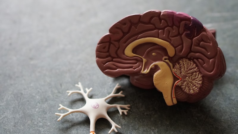
The spinal cord constitutes a vital, elongated component of the central nervous system (CNS), serving as the primary conduit for information exchange between the brain and the rest of the body. Originating continuously from the lower end of the medulla oblongata, situated at the base of the brainstem, this critical structure extends caudally through the protective channel known as the vertebral canal, which is formed by the stacked vertebrae of the spine. Its terminus is typically found in the adult human at the level of the first or second lumbar vertebra (L1 or L2), tapering into a structure called the conus medullaris. This specific anatomical location is crucial, as the lower lumbar and sacral nerves continue downward as the cauda equina (horse’s tail) within the vertebral canal before exiting at their respective levels, a detail essential for clinical procedures such as lumbar punctures.
Functionally and structurally, the spinal cord is inseparable from the brain, together forming the core nervous system responsible for processing, coordinating, and executing all bodily functions. It operates not merely as a passive cable carrying signals but also as a semi-autonomous processing center, capable of initiating simple motor responses through reflex arcs without direct cortical command. The length and diameter of the cord vary slightly throughout its course, featuring two distinct enlargements—the cervical enlargement, which supplies nerves to the upper limbs, and the lumbar enlargement, which supplies nerves to the lower limbs—reflecting the increased density of neural connections required for complex limb movements.
A fundamental characteristic of the spinal cord, critical for its survival and function, is its robust protection. The entirety of the cord is meticulously encased within the spinal column, or vertebral column, a bony structure providing mechanical shielding against external trauma. Furthermore, like the brain, the spinal cord is enveloped by three layers of protective membranes called the meninges, and it is cushioned by cerebrospinal fluid (CSF). This multilayered protection system ensures the delicate neural tissues remain stable and protected from mechanical shock, maintaining the pristine environment required for high-speed electrochemical signaling necessary for movement, sensation, and autonomic control throughout the body.
Anatomy and Protective Structures
The protection afforded to the spinal cord is intricate and multilayered, beginning with the bony framework of the vertebral column. This column is composed of 33 individual bones (vertebrae) separated by intervertebral discs, which provide both flexibility and shock absorption. The central passage through these stacked vertebrae is the vertebral canal, where the spinal cord resides. Beyond this skeletal protection, the cord is shielded by the three meningeal membranes: the outermost dura mater, a thick, fibrous layer that provides durable containment; the middle arachnoid mater, a delicate, web-like structure; and the innermost pia mater, which adheres directly to the surface of the cord itself.
The spaces created by these meningeal layers are clinically significant. The space between the dura mater and the wall of the vertebral canal is the epidural space, filled with adipose tissue and blood vessels, and is the site for administering epidural anesthesia. Immediately beneath the arachnoid mater is the subarachnoid space, which is crucial because it circulates the cerebrospinal fluid (CSF). The CSF provides buoyancy to the cord, effectively reducing its weight and protecting it from minor jolts, while also serving metabolic functions by transporting nutrients and removing waste products. The integrity of these protective layers is paramount; breaches, infections (meningitis), or hemorrhages within these spaces can severely compromise the function and viability of the underlying neural tissue.
Furthermore, the spinal cord is anchored within the vertebral canal by specialized structures to prevent excessive movement during body motion. The denticulate ligaments, which are lateral extensions of the pia mater, pierce the arachnoid and dura mater to attach the cord firmly to the surrounding bone. At the caudal end, the pia mater continues as the filum terminale, a slender, non-neural filament that descends to anchor the cord to the coccyx, thus maintaining longitudinal stability. This complex system of bony casing, resilient membranes, and fluid suspension ensures that the spinal cord remains structurally fixed yet mobile enough to accommodate the bending and flexing movements of the human torso.
Internal Structure: Gray Matter and White Matter
A cross-section of the spinal cord reveals a distinct organization characterized by an inner core of gray matter and a surrounding sheath of white matter, an arrangement that is reversed compared to the cerebral hemispheres. The gray matter is centrally located and shaped like a butterfly or the letter ‘H’, consisting primarily of neuron cell bodies, dendrites, unmyelinated axons, glial cells, and synapses. It is the primary site for synaptic integration and processing of information within the cord. This gray matter is divided into specific regions known as horns: the dorsal (posterior) horns receive sensory information from the body via afferent neurons; the ventral (anterior) horns house the cell bodies of motor neurons that send commands to skeletal muscles; and the lateral horns, present only in the thoracic and upper lumbar segments, contain the cell bodies of autonomic motor neurons.
The white matter encases the gray matter and is composed almost entirely of bundles of myelinated axons, which are organized into tracts or pathways. The myelin sheath, a fatty substance produced by oligodendrocytes, provides insulation and dramatically increases the speed of electrical signal transmission. These tracts run longitudinally up and down the cord, forming the superhighways of the CNS. The white matter is anatomically divided into three major regions, or funiculi: the anterior funiculi, the posterior funiculi, and the lateral funiculi, each containing distinct sets of ascending (sensory) and descending (motor) tracts. The specific location of these tracts is highly organized, meaning that damage to a particular area of the white matter results in predictable losses of sensation or motor control.
The organization within the gray matter follows Rexed’s laminae, a system that divides the gray matter into ten distinct layers (Laminae I-X) based on cellular structure and function. This laminar organization reflects the systematic way in which sensory input is processed, modulated by interneurons, and transformed into motor output. For instance, Laminae IX corresponds to the motor nuclei in the ventral horn, while Laminae I and II are involved in processing pain and temperature sensation. The precise layering underscores the spinal cord’s complexity as an integration center, where intricate neural circuits govern everything from muscle tone to the gating of pain signals before they ascend to higher brain centers.
Major Functions: Conduction and Reflex Arcs
The spinal cord performs two principal functions essential for life and interaction with the environment: serving as a major conduction pathway for nerve impulses traveling between the brain and the periphery, and mediating spinal reflexes. As a conduction pathway, the white matter tracts facilitate bidirectional communication. Ascending tracts carry sensory information—such as touch, pressure, pain, temperature, and proprioception—from sensory receptors throughout the body up to the brain for conscious perception and processing. Conversely, descending tracts carry motor commands from the brain (primarily the cerebral cortex and brainstem nuclei) down to the motor neurons in the ventral horn, which then initiate voluntary and involuntary movements.
The second critical function involves the execution of reflex arcs, which are rapid, automatic, and involuntary responses to specific stimuli. These reflexes are integrated entirely within the gray matter of the spinal cord, bypassing the need for conscious brain involvement, thus ensuring extremely fast reaction times necessary for survival. A typical reflex arc involves five components: a sensory receptor that detects the stimulus; a sensory neuron that transmits the signal to the CNS; an integration center (usually one or more interneurons within the gray matter); a motor neuron that transmits the output signal; and an effector (muscle or gland) that produces the response.
Examples of spinal reflexes include the simple stretch reflex (monosynaptic, like the patellar knee-jerk reflex) and the more complex withdrawal (flexor) reflex (polysynaptic), which causes rapid withdrawal of a limb from a painful stimulus. The autonomy of these reflexes highlights the spinal cord’s capacity for local control. Furthermore, even when reflexes are executed locally, the spinal cord simultaneously transmits information about the event up the ascending tracts to the brain. This ensures that while the body reacts instantly to danger, the brain is eventually informed, allowing for subsequent adjustments or cognitive responses, demonstrating the coordinated operation between the spinal cord and higher cortical centers.
Ascending and Descending Tracts
The white matter is organized into highly specific pathways known as tracts, each dedicated to carrying a particular type of information. Among the most important ascending tracts is the Dorsal Column-Medial Lemniscus (DCML) pathway, which transmits highly discriminative sensory information, including fine touch, vibratory sense, and conscious proprioception. These fibers ascend ipsilaterally (on the same side) in the posterior funiculus, synapse in the brainstem, and then cross over (decussate) before continuing to the thalamus and eventually the somatosensory cortex. In contrast, the Spinothalamic tracts (lateral and anterior) convey crude touch, pain, and temperature sensations. These fibers typically cross over immediately upon entering the spinal cord, ascending contralaterally (on the opposite side) before reaching the thalamus. The differing decussation points of these major sensory tracts are essential for diagnosing the precise location of spinal cord lesions.
The primary motor control pathways are the descending tracts. The most crucial pathway for voluntary, skilled movement is the Corticospinal tract, originating in the motor cortex. Approximately 90% of these fibers cross over in the medulla (forming the lateral corticospinal tract) and descend to innervate motor neurons controlling distal limbs, enabling fine motor skills. The remaining 10% descend ipsilaterally as the anterior corticospinal tract, generally controlling axial and proximal muscles. Damage to the corticospinal tracts results in upper motor neuron signs, characterized by spastic paralysis, hyperreflexia, and pathological reflexes such as the Babinski sign.
In addition to the corticospinal pathway, several other descending tracts collectively form the extrapyramidal system, which modulates posture, balance, and involuntary movements. Key tracts in this system include the Rubrospinal tract, originating in the red nucleus and primarily involved in upper limb motor control; the Vestibulospinal tracts, which originate in the vestibular nuclei and help maintain posture and balance in response to head movements; and the Reticulospinal tracts, originating in the brainstem reticular formation, which influence muscle tone and autonomic functions. The complexity of these interlinked tracts ensures that even the simplest movement is executed with appropriate stability and coordination, demonstrating the highly integrated nature of spinal cord function under the command of various brain centers.
Spinal Nerves and Segmentation
The spinal cord is segmented, although the segments are not physically separated by tissue boundaries; rather, they are defined by the pairs of spinal nerves that emerge from them. There are 31 pairs of spinal nerves, each formed by the fusion of a dorsal (sensory) root and a ventral (motor) root. These nerves are classified according to the region of the vertebral column from which they exit: 8 cervical (C1–C8), 12 thoracic (T1–T12), 5 lumbar (L1–L5), 5 sacral (S1–S5), and 1 coccygeal (Co1). Note that there are 8 cervical nerves but only 7 cervical vertebrae, a discrepancy arising because the C1 nerve exits above the C1 vertebra, while the C8 nerve exits below the C7 vertebra.
Each spinal nerve is a mixed nerve, meaning it carries both sensory (afferent) fibers toward the CNS and motor (efferent) fibers away from the CNS. Upon exiting the intervertebral foramen, the spinal nerve immediately branches into the dorsal ramus (serving deep muscles and skin of the back) and the ventral ramus (serving the limbs and anterior trunk). The distribution of sensory input from these nerves is topographically mapped onto the skin surface in distinct regions called dermatomes. Knowledge of dermatomes is clinically invaluable, as specific patterns of sensory loss allow neurologists to localize the exact segment of the spinal cord or spinal nerve root that has been damaged by disease or injury, such as a herniated disc.
In the cervical, lumbar, and sacral regions, the ventral rami of adjacent spinal nerves intermingle and reorganize extensively, forming complex networks known as nerve plexuses. The four major plexuses are the cervical plexus (C1–C5), which supplies the neck and diaphragm via the critical phrenic nerve; the brachial plexus (C5–T1), which supplies the entire upper limb; the lumbar plexus (L1–L4), supplying the thigh and abdomen; and the sacral plexus (L4–S4), which supplies the lower leg, foot, and pelvic region. This plexal arrangement ensures that multiple segments contribute to the innervation of a single limb muscle, providing a degree of functional redundancy and minimizing the impact of damage to a single spinal segment.
Clinical Significance and Spinal Cord Injury
The clinical significance of the spinal cord cannot be overstated, particularly concerning trauma and disease. A spinal cord injury (SCI), often resulting from vehicular accidents, falls, or violence, leads to disruption of communication pathways, causing varying degrees of paralysis and sensory loss below the level of the injury. The outcome depends heavily on the anatomical level and the extent of the damage. Injuries to the cervical region (C1–C4) are often the most devastating, potentially resulting in quadriplegia (paralysis of all four limbs) and requiring ventilator assistance if the phrenic nerve is affected. Lower injuries (thoracic or lumbar) typically result in paraplegia (paralysis of the lower limbs).
Injuries are categorized as either complete or incomplete. A complete injury involves total loss of motor and sensory function below the lesion, whereas an incomplete injury retains some residual neural function, offering a better prognosis for recovery. Immediately following severe trauma, patients often experience spinal shock, a temporary condition characterized by the loss of all reflexes and muscle tone below the injury site. Recovery from spinal shock is marked by the return of reflexes, though often exaggerated (hyperreflexia), indicative of upper motor neuron damage due to the loss of descending inhibitory control from the brain.
Beyond trauma, the spinal cord is susceptible to various pathological conditions. Poliomyelitis, caused by a virus, specifically targets and destroys the motor neuron cell bodies in the ventral horn, leading to flaccid paralysis. Amyotrophic Lateral Sclerosis (ALS) progressively attacks both upper (descending tracts) and lower (ventral horn) motor neurons. Furthermore, congenital conditions like spina bifida involve incomplete closure of the vertebral column during embryonic development, exposing the spinal cord and often leading to neural deficits. Advances in neurorehabilitation and surgical stabilization continue to improve outcomes, but the profound reliance of the body on the spinal cord makes its injury a major cause of long-term disability.