Psychological Stress: The Hidden Trigger of Blood Clots
- The Core Definition of Thrombus
- Historical Context and Foundational Understanding
- Pathophysiology: The Mechanism of Clot Formation
- Diagnosis of Thrombus in Clinical Practice
- Treatment Strategies and Management
- A Practical Example: Deep Vein Thrombosis (DVT)
- Significance, Impact, and Clinical Relevance
- Connections to Related Physiological Concepts
The Core Definition of Thrombus
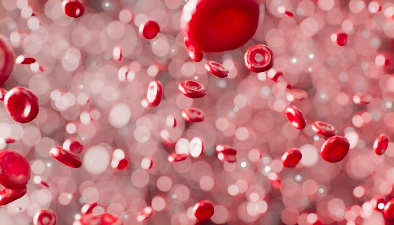
A Thrombus is fundamentally defined as a blood clot that forms and remains attached within a blood vessel or the heart chamber, obstructing the flow of blood. This serious medical condition arises from a pathological process where the normal mechanisms designed to stop bleeding—known as hemostasis—are inappropriately activated within intact vessels. Unlike a normal clot which dissolves after healing, a thrombus persists, growing larger and potentially leading to catastrophic cardiovascular complications such as stroke or heart attack. Its composition is heterogeneous, typically consisting of an intricate meshwork of platelets, trapped red blood cells, white blood cells, and insoluble fibrin, which provides the structural backbone of the clot. The location and specific composition determine whether the thrombus is classified as arterial (often platelet-rich, associated with high flow rates and rupture of atherosclerotic plaque) or venous (often fibrin and red cell-rich, associated with stasis).
The key mechanism underlying the formation of any thrombus relies heavily on factors summarized historically by Virchow’s Triad, which identifies three primary contributing elements: endothelial injury, stasis or turbulent blood flow, and hypercoagulability. Endothelial injury, such as damage to the smooth lining of a blood vessel, exposes subendothelial collagen and tissue factor, triggering immediate platelet activation and adhesion. Abnormal blood flow, whether sluggish (stasis, common in veins) or turbulent (common near atherosclerotic lesions or vessel bifurcations), prevents the dilution of activated clotting factors and hinders the influx of natural anticoagulant inhibitors. Finally, a state of hypercoagulability refers to any alteration in the blood that makes it more prone to clotting, often due to genetic factors, certain medications, or systemic conditions like cancer or severe inflammation. These factors rarely act in isolation; rather, their complex interplay initiates and propagates the thrombotic process, transforming a localized response into a potentially fatal occlusion.
Historical Context and Foundational Understanding
The foundational understanding of thrombosis is inextricably linked to the work of the German physician and pathologist, Rudolf Virchow, in the mid-19th century. Although he did not discover the concept of clotting, Virchow synthesized the observational knowledge of his time into a cohesive theory that remains central to modern hematology and vascular medicine. Around the 1850s, through meticulous autopsy and microscopic examination, Virchow established the critical distinction between a clot formed after death (post-mortem) and a clot formed during life (thrombus). His greatest contribution was the articulation of the aforementioned Virchow’s Triad, formalizing the three key categories of risk factors—vessel wall damage, altered blood flow, and altered blood constituents—that predispose an individual to thrombosis.
Prior to Virchow’s systematic approach, the phenomenon of blood clotting and subsequent vascular occlusion was often attributed vaguely to inflammation or congestion. Virchow’s work provided the critical framework necessary for future research into the intricate biochemical cascade of coagulation. His research not only explained how a thrombus forms but also provided the basis for understanding how pieces of the clot could break off and travel to distant organs, a process he termed embolism. This historical context is vital, as it shifted medical understanding from passive observation to active investigation of the underlying physiological mechanisms, paving the way for targeted preventive and therapeutic strategies centuries later, including the development of modern anticoagulant drugs.
Pathophysiology: The Mechanism of Clot Formation
The pathophysiology of thrombus formation is a complex, multi-factorial process involving the interplay of cellular elements, biochemical pathways, and the vascular environment. The initial trigger often involves damage to the protective layer of endothelial cells lining the vessel, which immediately initiates platelet activation. Activated platelets rapidly adhere to the exposed collagen and begin releasing a variety of potent pro-coagulant and vasoconstrictive molecules, including adenosine diphosphate (ADP) and thromboxane A2. This release amplifies the response, recruiting more platelets to form a primary hemostatic plug—a localized aggregation that attempts to seal the injury.
Simultaneously, the extrinsic pathway of the coagulation cascade is activated by tissue factor released from the injured vessel wall. This initiates a complex sequence of enzymatic reactions involving various clotting factors (proteins denoted by Roman numerals) that ultimately leads to the generation of thrombin. Thrombin is arguably the most crucial enzyme in this cascade, as it converts soluble fibrinogen into insoluble fibrin strands. This fibrin mesh acts like molecular glue, stabilizing the initial platelet plug by trapping additional platelets, red blood cells, and white blood cells, thereby transforming the temporary soft plug into a robust, definitive thrombus.
Furthermore, acute inflammation is increasingly recognized as a significant factor promoting thrombus formation. Inflammatory cytokines and mediators can directly enhance the expression of pro-coagulant factors on the surface of endothelial cells and leukocytes, thereby linking the body’s defensive inflammatory response directly to the risk of thrombosis. This inflammatory state creates a pro-thrombotic environment, contributing to the cycle of clot growth and persistence, particularly in conditions like sepsis, major surgery, or chronic inflammatory diseases. The stability and eventual resolution (or lack thereof) of the resulting fibrin-platelet plug are critical determinants of patient outcomes.
Diagnosis of Thrombus in Clinical Practice
Diagnosing a thrombus, particularly those in deep veins or smaller arteries, can be challenging due to non-specific symptoms, necessitating the use of advanced imaging and laboratory techniques. The diagnostic approach typically begins with a thorough clinical assessment, evaluating patient risk factors based on Virchow’s Triad. However, definitive confirmation relies heavily on specialized imaging techniques. Ultrasonography, specifically Doppler ultrasound, is a cornerstone of diagnosis for deep vein thrombosis (DVT). This non-invasive technique uses sound waves to visualize the blood vessel, detect the presence of a clot (which appears incompressible), and assess blood flow dynamics, providing a rapid and generally reliable method for identifying venous clots.
For more complex or centrally located thrombi, such as those in the pulmonary arteries or cerebral vessels, more sophisticated imaging is required. Computed Tomography (CT) scanning, often enhanced with contrast agents (CT angiography or venography), provides detailed, cross-sectional images of the vessels and the surrounding anatomy. CT scanning is particularly useful for visualizing pulmonary embolism (PE), where it can pinpoint the exact location and extent of the occlusion. Magnetic Resonance Imaging (MRI) is another powerful tool, offering excellent soft tissue contrast, although it is less frequently used than CT or ultrasound for acute diagnosis due to accessibility and time constraints.
Laboratory tests complement imaging studies by assessing the state of the patient’s coagulation system. The D-dimer test is a crucial initial screening tool; it measures the levels of fibrin degradation products that are released when fibrin is broken down by the body’s natural processes. An elevated D-dimer level suggests that significant clot formation and breakdown are occurring, although it is non-specific (meaning it can be elevated in many conditions, not just thrombosis). Other tests, such as Prothrombin Time (PT) and Activated Partial Thromboplastin Time (aPTT), assess the speed and efficiency of the clotting cascade, which is essential both for initial diagnosis and for monitoring the efficacy of anticoagulant therapy.
Treatment Strategies and Management
The treatment of a thrombus is highly dependent on its location, size, and the patient’s overall clinical presentation, but the overarching goal is twofold: to prevent the existing clot from growing or migrating (leading to pulmonary embolism or stroke) and to manage the underlying risk factors contributing to the hypercoagulable state. Anticoagulant medications are the mainstay of treatment. These drugs, often referred to as “blood thinners,” do not dissolve the existing clot but rather inhibit the formation of new fibrin strands and prevent the existing thrombus from expanding. Examples include heparin, warfarin, and the newer direct oral anticoagulants (DOACs). The choice and duration of therapy are individualized, often extending for three to six months or indefinitely for patients with recurrent thrombosis or high inherent risk.
In cases where the thrombus poses an immediate threat to life or limb—such as massive pulmonary embolism or acute arterial occlusion—more aggressive interventions are required. Thrombolytic agents, sometimes called “clot busters,” are powerful pharmacological drugs designed to actively dissolve the thrombus. The most well-known example is tissue plasminogen activator (tPA). These agents work by converting plasminogen into plasmin, an enzyme that rapidly breaks down the fibrin mesh. Due to their high risk of bleeding complications, thrombolytics are reserved for severe cases and must be administered under strict medical supervision, usually within a short therapeutic window following the thrombotic event.
In specific clinical scenarios, surgical or interventional procedures may be necessary. For large, proximal arterial thrombi causing acute ischemia, surgical thrombectomy (physical removal of the clot) might be performed. For venous clots, especially in patients who cannot tolerate anticoagulant therapy, insertion of an inferior vena cava (IVC) filter can be considered; this device is designed to catch large pieces of clot originating from the legs before they can travel to the lungs and cause a pulmonary embolism. Furthermore, catheter-directed thrombolysis or mechanical clot retrieval systems offer less invasive alternatives to traditional surgery, allowing physicians to target the clot directly with thrombolytic agents or mechanical devices.
A Practical Example: Deep Vein Thrombosis (DVT)
A common and clinically significant example of thrombosis is Deep Vein Thrombosis (DVT), which typically occurs in the large veins of the legs. Consider a scenario involving an individual, Mr. J, who has recently undergone major orthopedic surgery. Due to the trauma of the surgery itself (endothelial injury), prolonged immobility during recovery (blood stasis), and the hypercoagulable state often induced by surgical stress, Mr. J is at high risk. Several days post-operation, he notices swelling, pain, and warmth in one calf—classic signs indicating the formation of a thrombus.
The “How-To” application of the principle in this scenario follows a clear sequence. First, the surgical trauma and subsequent inflammation activate platelets and release tissue factor in the affected vein. Second, the enforced stasis of blood in the deep leg veins prevents the natural washout of activated clotting factors, allowing them to accumulate. Third, the resulting high concentration of active clotting factors, particularly thrombin, rapidly drives the coagulation cascade, forming a large fibrin-rich clot.
If Mr. J were to ignore these symptoms, the most significant danger would be fragmentation of the thrombus. A piece of the clot, now termed an embolus, could detach and travel through the circulatory system, passing through the right side of the heart and lodging in the pulmonary arteries, resulting in a life-threatening pulmonary embolism. Therefore, upon suspicion of DVT, immediate diagnostic imaging (ultrasound) and subsequent initiation of anticoagulant therapy are crucial steps to prevent this potentially fatal migration.
Significance, Impact, and Clinical Relevance
The study and management of the thrombus hold immense significance because thrombotic events—myocardial infarction (heart attack), ischemic stroke, and venous thromboembolism (VTE)—are among the leading causes of morbidity and mortality globally. The formation of a thrombus in a coronary artery is the proximate cause of most heart attacks, while arterial thrombi in cerebral vessels account for the majority of ischemic strokes. These events place a tremendous burden on healthcare systems and result in significant long-term disability for survivors.
The application of this knowledge permeates nearly every subfield of modern medicine, extending far beyond the emergency room. In preventative medicine, understanding the risk factors laid out by Virchow’s Triad allows clinicians to identify high-risk individuals and implement prophylactic measures, such as prescribing low-dose anticoagulants for patients undergoing major surgery or those with atrial fibrillation. In pharmacology, the development of novel anti-platelet and anticoagulant drugs continues to be a major area of research, aimed at finding agents that prevent thrombosis without causing undue bleeding risk. Furthermore, in critical care and oncology, recognizing that cancer and severe infections induce a hypercoagulable state is vital for managing complex patient populations and preventing VTE.
Connections to Related Physiological Concepts
The concept of the thrombus is intimately connected to several other key physiological and pathological processes. Most notably, it is linked to Atherosclerosis, the chronic disease characterized by the hardening and narrowing of arteries due to plaque buildup. Arterial thrombosis often occurs when an unstable atherosclerotic plaque ruptures, exposing the highly thrombogenic core material to the blood flow, which instantly triggers massive platelet activation and rapid thrombus formation, leading to acute coronary syndrome or stroke.
Another critical relationship is the distinction between a thrombus and an embolus. While a thrombus is a stationary clot, an embolus is any material (clot fragment, fat globule, air bubble) that travels through the bloodstream and lodges in a distant vessel. When a piece of a venous thrombus breaks off, it becomes a thromboembolus, most commonly resulting in a pulmonary embolism. The distinction is crucial for diagnosis and treatment, as managing the source thrombus is key to preventing recurrent embolic events.
The broader category of study for thrombosis falls primarily under **Hematology** (the study of blood, including clotting factors and platelets), **Cardiology** (when affecting the heart), and **Vascular Medicine**. However, given its systemic implications and the involvement of inflammatory responses, it also has deep ties to **Pathophysiology** and **Immunology**. The ongoing discovery of new genetic and biochemical factors that influence the delicate balance between bleeding and clotting continues to expand our understanding of how the body regulates hemostasis and why this regulation sometimes fails, leading to life-threatening thrombotic disorders.