Corpus Callosum: The Neural Bridge Uniting Your Mind
Scientifically Reviewed · Dr. Marwa Abd-Alazim · September 8, 2026
Review Criteria & Clinical Standards
This content undergoes rigorous scientific peer-review and medical editorial standards at Arab Psychology Network to ensure clinical accuracy, validity, and compliance with evidence-based guidelines from leading psychological and healthcare authorities (APA / WHO).
Introduction and Definition of the Corpus Callosum
The Corpus Callosum stands as the largest and most critical structure for interhemispheric communication in the human brain, functioning as an immense pathway of nerve fibers traveling superiorly over the longitudinal fissure. This massive white matter tract serves as the primary linkage for the two opposing cerebral hemispheres, ensuring seamless integration of cognitive, sensory, and motor information. Without this essential bridge, the two hemispheres, specialized in their respective functions through the process of lateralization, would operate largely in isolation, severely compromising unified consciousness and coordinated action. It is composed of dense bundles of axons—estimated to contain between 200 and 300 million fibers—that project contralaterally to homologous cortical regions, allowing complex processing to occur across the entire neocortex rather than being confined to the site of initial processing.
In essence, the Corpus Callosum is the neurological foundation upon which the brain achieves holistic function. While the left hemisphere is often dominant for language processing and analytical thought, and the right hemisphere excels in spatial reasoning and emotional context, these specializations must be synthesized into a coherent whole. The efficiency and integrity of the Corpus Callosum directly correlate with the brain’s ability to execute high-level cognitive tasks, requiring rapid data transfer and immediate synchronization. The sheer volume and myelination status of these fibers underscore its importance, distinguishing it as the paramount commissural pathway dedicated to the integration necessary for complex human behavior and adaptability.
The structure acts dynamically, facilitating both excitatory and inhibitory signals. It not only transfers information from one side to the other but also plays a crucial role in balancing hemispheric activity, often inhibiting activity in the opposing hemisphere to prevent interference when one side is heavily engaged in a specific task. This carefully modulated interplay of excitation and inhibition is fundamental to maintaining focus and executing coordinated actions, highlighting that the Corpus Callosum is not merely a conduit, but a sophisticated regulator of bilateral cerebral activity.
Anatomical Structure and Composition
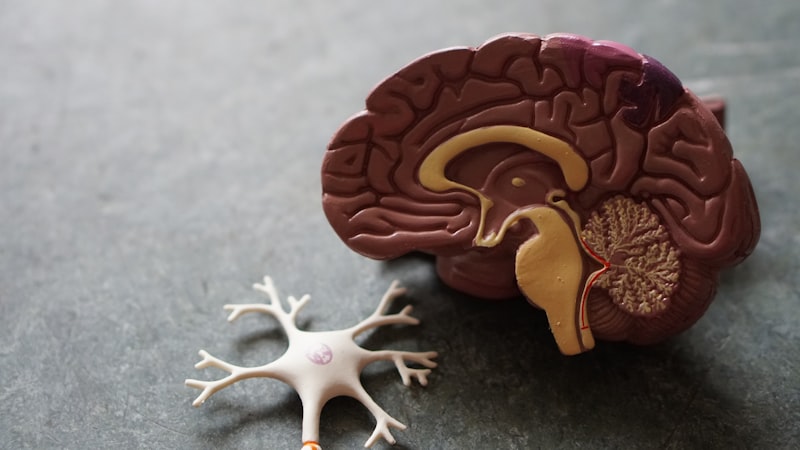
Anatomically, the Corpus Callosum is divided into four main segments, each distinguished by its location and the specific cortical areas whose fibers it carries. These segments, moving from anterior to posterior, are the Rostrum, the Genu, the Body (or Trunk), and the Splenium. The Rostrum and the Genu constitute the anterior curved portion, with the Genu projecting fibers primarily connecting the frontal lobes. These connections are vital for executive functions, planning, working memory, and complex motor control. The fibers originating here spread widely in the frontal white matter, forming the frontal forceps (or forceps minor).
The Body or Trunk comprises the extensive central portion of the structure, connecting the massive expanses of the parietal and temporal lobes. These connections are instrumental in coordinating somatosensory processing, spatial awareness, and auditory functions. The fibers originating from the Body project posteriorly, forming the superior longitudinal fasciculus. The dense organization within the Body reflects the continuous need for coordination of ongoing sensory input and motor output across the midline, ensuring that the brain accurately maps the external world and controls bilateral movements.
The posterior-most segment is the Splenium, which is typically the thickest part of the Corpus Callosum. The Splenium primarily connects the occipital lobes and the posterior parietal cortices, carrying crucial information related to visual perception. The fibers projecting from the Splenium fan out to form the occipital forceps (or forceps major). This connection is particularly important for integrating the visual fields; for example, allowing an image perceived initially by the right visual field (processed by the left hemisphere) to be immediately shared with the right hemisphere for holistic spatial interpretation. The topographic organization ensures that neighboring regions of cortex are connected via adjacent fibers within the Corpus Callosum, maintaining a precise map of connectivity across the cerebral hemispheres.
Development and Myelination Timeline
The formation of the Corpus Callosum is a highly orchestrated process beginning early in prenatal development, specifically around the 12th week of gestation. This development relies heavily on the presence of specialized glial structures, known as the glial sling, which guide the pioneering axons migrating across the midline. The initial establishment of the pathway is critical; if genetic or environmental factors disrupt this process during the second trimester, it can result in structural anomalies such as partial or complete Agenesis of the Corpus Callosum (ACC). The subsequent growth involves the massive proliferation of axons until the structure reaches its full complement of fibers.
While the axons are largely in place by birth, the functional maturation of the Corpus Callosum is a protracted process defined by myelination—the wrapping of axons in a fatty sheath that dramatically increases the speed and efficiency of signal transmission. Myelination begins shortly after birth and proceeds in a distinct sequence, starting in the posterior Splenium and moving forward toward the Genu and Rostrum. Crucially, this process does not conclude in childhood; the anterior fibers, connecting the frontal lobes responsible for complex executive functions, continue to myelinate well into late adolescence and early adulthood (often extending into the third decade of life).
This prolonged myelination timeline has profound implications for cognitive development. The gradual refinement of callosal conductivity parallels the increasing sophistication of skills such as abstract reasoning, complex problem-solving, and social cognition, all of which require rapid, high-bandwidth communication between the frontal lobes. Disruptions to myelination, whether due to nutritional deficits, injury, or neurological conditions, can impede processing speed and interhemispheric transfer time, leading to subtle but significant cognitive challenges that manifest as the individual attempts to master increasingly demanding cognitive tasks.
Function: Interhemispheric Communication and Integration
The overarching function of the Corpus Callosum is to facilitate the rapid, high-fidelity transfer of information, ensuring that the brain acts as a cohesive unit rather than two separate processing centers. This communication is essential for tasks requiring the simultaneous involvement of both specialized hemispheres. For instance, reading requires the left hemisphere to decode linguistic symbols, while the right hemisphere processes the spatial layout of the text and the emotional tone implied by the context; the Corpus Callosum must integrate these streams instantaneously to achieve comprehension.
Beyond simple data transfer, the Corpus Callosum is vital for coordinating bimanual motor skills. Activities such as tying shoelaces, playing a musical instrument, or catching a ball necessitate precise, time-locked motor commands issued by both hemispheres. The fibers connecting the motor cortices transmit timing and force information to ensure that movements performed by one hand are smoothly coordinated with the other. Disruptions in this pathway often result in motor apraxia or difficulty performing complex sequences of movements that cross the midline, illustrating the critical role of the Corpus Callosum in bilateral motor execution.
Furthermore, the Corpus Callosum serves a crucial inhibitory role, a function often overlooked but essential for mental clarity. When one hemisphere is dedicated to a specific task (e.g., the left hemisphere analyzing language structure), the Corpus Callosum inhibits competing activity in the homologous region of the opposite hemisphere. This mechanism prevents cognitive interference, allowing the focused hemisphere to operate efficiently without distraction from unnecessary or conflicting neural chatter. This inhibitory modulation is particularly important for tasks involving focused attention and swift decision-making.
Clinical Significance: Agenesis of the Corpus Callosum (ACC)
Agenesis of the Corpus Callosum (ACC) refers to the complete or partial absence of this critical white matter tract, a congenital condition that occurs when the structure fails to develop properly during gestation. The clinical presentation of ACC is highly variable, ranging from severe intellectual disability and pervasive developmental delay to cases where individuals exhibit only subtle neurocognitive deficits, often attributed to the remarkable compensatory ability (neuroplasticity) of the developing brain. However, the absence of the primary linkage results often in delayed development across multiple domains.
A core set of symptoms associated with ACC involves difficulties with motor skills and coordination. Individuals with ACC frequently exhibit clumsy gait, challenges with balance, and significant trouble integrating complex bilateral movements. The lack of direct connectivity between the motor cortices necessitates reliance on less efficient, longer subcortical pathways, slowing down response times and reducing the precision of coordinated actions. This difficulty often persists throughout life, requiring extensive physical and occupational therapy.
Beyond motor difficulties, ACC is associated with distinct cognitive and social challenges. Affected individuals frequently display deficits in abstract reasoning, problem-solving that requires rapid integration of disparate information, and subtle impairments in social cognition, such as difficulty interpreting nuanced social cues or understanding non-literal language. Processing speed is characteristically slow, as information must traverse longer, indirect routes. While the brain compensates by developing abnormal but functional pathways (probst bundles), these routes are fundamentally less efficient than the direct callosal connection, underscoring the vital nature of the Corpus Callosum for optimal cognitive performance.
Surgical Interventions: Callosotomy and Split-Brain Studies
The Corpus Callosum became the focus of intense clinical and research scrutiny following the development of callosotomy, a radical surgical procedure performed primarily to manage intractable, generalized epilepsy. In this procedure, the Corpus Callosum is partially or completely severed (a commissurotomy), preventing epileptic seizures originating in one hemisphere from spreading uncontrollably to the other. While callosotomy is highly effective in reducing seizure severity, it functionally separates the cognitive processing streams of the two hemispheres, leading to the well-documented phenomenon of the “split brain.”
The study of split-brain patients, pioneered by Nobel laureates Roger Sperry and Michael Gazzaniga, offered unprecedented insight into hemispheric specialization and the necessity of interhemispheric communication for consciousness. These studies revealed that when visual or tactile information was presented exclusively to one hemisphere (e.g., showing a word only to the right visual field, which projects to the language-dominant left hemisphere), the patient could verbalize the stimulus. However, if the stimulus was presented only to the left visual field (right hemisphere), the patient often could not name the object, yet could correctly point to it with the left hand, demonstrating that the information was processed but inaccessible to the verbal center in the separated left hemisphere.
The clinical consequences of a callosotomy, while manageable, highlight the Corpus Callosum’s role in unifying subjective experience. Patients may exhibit behavioral phenomena such as intermanual conflict, where the left hand seems to act autonomously against the conscious will of the right hemisphere. Modern surgical techniques often favor partial callosotomies, focusing on severing only the anterior or posterior two-thirds of the structure, as this minimizes the profound cognitive and behavioral side effects while often providing sufficient control over seizure generalization, demonstrating a refined understanding of which sections carry the most clinically relevant fibers for seizure spread.
Corpus Callosum and Cognitive Functions
The integrity of the Corpus Callosum is intrinsically linked to the efficiency of various high-level cognitive functions, particularly those requiring the rapid synthesis of data from disparate cortical regions. In the domain of attention, the callosal pathway ensures that attentional resources allocated by the prefrontal cortex in one hemisphere are consistently aligned with the processing demands of the other, enabling smooth shifts in focus and sustained concentration across the visual and spatial fields.
For memory function, the Corpus Callosum is essential for both encoding and retrieval. While memory traces may be distributed across both hemispheres, the CC facilitates the cross-cueing necessary to access stored information, particularly regarding contextual and spatial memories often lateralized to the right hemisphere. Furthermore, the efficiency of the pathway influences processing speed, which is a fundamental measure of overall cognitive capability. Slower callosal transfer times, often seen in aging or neurodevelopmental disorders, correlate directly with reduced performance on tasks requiring quick mental manipulation or simultaneous information handling.
Research has also revealed subtle morphological differences in the Corpus Callosum in various neurodevelopmental and psychiatric conditions. Individuals diagnosed with Autism Spectrum Disorder (ASD), for example, often exhibit atypical callosal development, sometimes manifesting as reduced overall size or altered microstructure, particularly in the anterior portions. Similar findings of altered connectivity have been noted in schizophrenia, suggesting that compromised interhemispheric communication may contribute to the fragmented thought processes and perceptual disturbances characteristic of these disorders, cementing the structure’s role as a biomarker for large-scale brain network health.
Modern Research and Imaging Techniques
Advancements in neuroimaging have revolutionized the study of the Corpus Callosum, moving beyond simple structural observation to detailed analysis of its microstructural integrity and functional connectivity. Diffusion Tensor Imaging (DTI) is the principal technique employed today, allowing researchers to map the directionality and organization of the white matter fibers. DTI measures water diffusion anisotropy; highly aligned, dense, and myelinated fibers (like those in the Corpus Callosum) restrict water movement, resulting in high fractional anisotropy (FA) values. Deviations from normal FA values can indicate demyelination, axonal damage, or developmental disorganization, providing quantitative data on subtle structural deficits not visible through standard MRI.
Complementary to structural mapping, Functional Magnetic Resonance Imaging (fMRI) allows researchers to observe the synchronized activity of the hemispheres during specific cognitive tasks. Studies using fMRI have confirmed that the Corpus Callosum is not merely a passive cable but actively modulates its connectivity based on task demands. For instance, during complex tasks requiring spatial and linguistic integration, fMRI shows heightened interhemispheric synchronization across the callosal projection areas, confirming the structure’s dynamic role in coordinating neural resources.
Ongoing research continues to explore the relationship between callosal microarchitecture and individual differences in cognition and behavior. Longitudinal studies utilizing these advanced imaging methods are crucial for understanding how developmental changes in myelination throughout adolescence impact the emergence of sophisticated executive functions. The Corpus Callosum remains a central focus in systems neuroscience, serving as a critical model for understanding general principles of long-range neural connectivity and the etiology of disorders rooted in disrupted brain network organization.